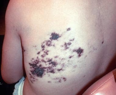
Continually Updated Clinical Reference
 
 
  All Sources     eMedicine     Medscape     Drug Reference     MEDLINE
 
eMedicine - Cobb Syndrome : Article by

Quick Find
Authors & Editors
Introduction
Clinical
Differentials
Workup
Treatment
Follow-up
Miscellaneous
Multimedia
References

Related Articles
Angiokeratoma Corporis Diffusum (Fabry Syndrome)

Herpes Zoster

Infantile Hemangioma


Patient Education
Click here for patient education.


Author: Christopher Norwood, MD, MS, Assistant Clinical Professor, Department of Dermatology, University of Connecticut Health Center

Christopher Norwood is a member of the following medical societies: American Academy of Dermatology, American Society for Dermatologic Surgery, and American Society for Laser Medicine and Surgery

Coauthor(s): Stephen J Krivda, MD, Assistant Professor of Dermatology, Uniformed Services University of the Health Sciences; Chief of the Integrated Department of Dermatology, Chief of Dermatology Service, Director of Dermatopathology, Staff Dermatopathologist, Walter Reed Army Medical Center; Head, Department of Dermatology, Staff Dermatologist and Dermatopathologist, National Naval Medical

Editors: Abby S Van Voorhees, MD, Assistant Professor, Director of Psoriasis Services and Phototherapy Units, Department of Dermatology, University of Pennsylvania School of Medicine, Hospital of the University of Pennsylvania; David F Butler, MD, Professor of Dermatology, Texas A&M University College of Medicine; Director, Division of Dermatology, Scott and White Clinic; Director Dermatology Residency Training Program, Scott and White Clinic; Rosalie Elenitsas, MD, Associate Professor of Dermatology, University of Pennsylvania School of Medicine; Director, Penn Cutaneous Pathology Services, Department of Dermatology, University of Pennsylvania Health System; Catherine Quirk, MD, Clinical Assistant Professor, Department of Dermatology, Brown University; Dirk M Elston, MD, Director, Department of Dermatology, Geisinger Medical Center

Author and Editor Disclosure

Synonyms and related keywords: cutaneomeningospinal angiomatosis

Background

Cobb syndrome is a rare, noninherited disorder that involves the association of spinal angiomas or arteriovenous malformations (AVM) with congenital, cutaneous vascular lesions in the same dermatome. Berenbruch first described the disorder in 1890, but it was not widely known until Cobb's report in 1915.1 The importance of this syndrome is the recognition that cutaneous vascular lesions may hint at an accompanying spinal cord angioma or AVM that may result in weakness or paralysis.

Cobb's patient initially had been presumed to have poliomyelitis; however, Cushing deduced that a spinal cord lesion was present by the complete paralysis with definite upper level of anesthesia, priapism, visceral paralysis, and exaggerated reflexes. He even suggested that an angioma was the cause by noting a similarity to meningeal angioma with facial port-wine stain (Sturge-Weber syndrome).

Patients originally were treated with laminectomy/decompression as attempted ligation of the vascular malformations resulted in death by hemorrhage. Therapeutic radiation later was attempted with moderate success.

Pathophysiology

The cutaneous manifestations of Cobb syndrome typically are present as port-wine stains (PWS) or angiomas, but reports exist of angiokeratomas,2 angiolipomas, and lymphangioma circumscriptum.3 The intraspinal lesions may be angiomas or AVMs. One case report exists of a patient with Cobb syndrome who had brain angiomas in addition to the classic lesions.

Frequency

International

In the world literature, only 35 cases of Cobb syndrome are reported. The actual incidence may be higher as only symptomatic cases are diagnosed. In a 1927 study, autopsy findings showed that approximately 12% of cadavers had angiomas; these angiomas had been asymptomatic during life.

Mortality/Morbidity

The major debility from Cobb syndrome is the onset of weakness, paresis, sensory loss, and loss of bowel and bladder control. Patients generally experience the sudden onset of pain and weakness as children or young adults. These symptoms may remit or remain stable; however, they do tend to worsen over time either by discrete steps or continuously.

  • Paralysis may lead to early reporting; treatment can lessen the degree of impairment.
  • Early development of weakness may portend a more aggressive course.
  • A possible complication if treatment is delayed is Foix-Alajouanine disease4 or subacute necrotic myelopathy due to thrombosis in the spinal angioma.

Race

No racial predilection is known, although most reported cases have been in whites.

Sex

Cobb syndrome has a slight male predominance.

Age

Disease onset is in childhood or adolescence. Recently, a report described a 5-month-old child with a T5-T12 hemangioma and paraparesis.


History

Patients typically present with sudden onset of back or lower extremity radicular pain associated with numbness that can be localized below a specific dermatome. Less commonly, weakness or bowel/bladder dysfunction may be presenting symptoms.

Physical

  • Cutaneous lesions may be distributed anywhere in the dermatome, from midline back to abdomen.
  • Midline back lesions, on rare occasions, are associated with spina bifida.
  • Unilateral lesions provide a clue to the location of the feeding artery in the spinal canal.
  • The cutaneous lesion may be very faint and may be more pronounced when the patient performs a Valsalva maneuver. The increased abdominal pressure causes preferential filling of the cutaneous angioma.
  • Neurological examination reveals weakness or paralysis and numbness or decreased sensation with a sharp upper cutoff.

Causes

Though Cobb syndrome is thought to be noninherited, two case reports exist of inherited cutaneous angiomas in patients with Cobb syndrome. The forebears had no clinical evidence of spinal lesions.


Angiokeratoma Corporis Diffusum (Fabry Syndrome)
Herpes Zoster
Infantile Hemangioma


Imaging Studies

  • MRI, radiography, and angiography all may be ordered in consultation with a neurologist and radiologist.
  • MRI probably is the most effective study, though CT, plain radiography, and angiography all provide useful information.
  • Criteria that are significant in the evaluation of cord compression are the following:
    • Localization of a lesion between T3 and T9
    • Involvement of the entire vertebral body
    • Extension to the neural arch
    • An expanded cortex with indistinct margins
    • Irregular honeycomb pattern
    • A soft tissue mass
  • Prior to the advent of CT and MRI, the classic finding on plain film was vertical striations of the vertebral body.

Procedures

  • Perform lumbar puncture to rule out infectious etiology.


Surgical Care

Patients should be referred to a neurosurgeon and an interventional radiologist for embolization5, 6 and decompression.

Consultations

  • Neurosurgeon
  • Interventional radiologist
  • Neurologist


Complications

A possible complication if treatment is delayed is Foix-Alajouanine disease4 or subacute necrotic myelopathy due to thrombosis in the spinal angioma.

Prognosis

The prognosis was grim with paralysis inevitable and death from infection likely a century ago. Current intervention provides some hope for minimizing permanent neurological damage. The key is early diagnosis.


Medical/Legal Pitfalls

In the past, patients were initially diagnosed with atypical poliomyelitis. Since early decompression can minimize symptoms, correct diagnosis is key. Patients with lower extremity weakness or pain should have a thorough skin examination and be examined with the proper imaging modalities promptly.


Media file 1:  Cutaneous angioma. Courtesy of L. Cooke, MD.
Click to see larger pictureClick to see detailView Full Size Image
Media type:  Photo

Media file 2:  Cutaneous angioma. Courtesy of L. Cooke, MD.
Click to see larger pictureClick to see detailView Full Size Image
Media type:  Photo

Media file 3:  Cutaneous angioma. Courtesy of L. Cooke, MD.
Click to see larger pictureClick to see detailView Full Size Image
Media type:  Photo

Media file 4:  MRI of spinal angioma. Courtesy of L. Cooke, MD.
Click to see larger pictureClick to see detailView Full Size Image
Media type:  MRI


  1. Cobb S. Haemangioma of the spinal cord associated with skin naevi of the same metamere. Annals Surgery. 1915;62:641-9.
  2. Clinton TS, Cooke LM, Graham BS. Cobb syndrome associated with a verrucous (angiokeratomalike) vascular malformation. Cutis. Apr 2003;71(4):283-7. [Medline].
  3. Shim JH, Lee DW, Cho BK. A case of Cobb syndrome associated with lymphangioma circumscriptum. Dermatology. 1996;193(1):45-7. [Medline].
  4. Wirth FP, Post KD, Di Chiro G. Foix-Alajouanine disease. Spontaneous thrombosis of a spinal cord arteriovenous malformation: a case report. Neurology. 1970;20:1114-18.
  5. Miyatake S, Kikuchi H, Koide T, Yamagata S, Nagata I, Minami S, et al. Cobb's syndrome and its treatment with embolization. Case report. J Neurosurg. Mar 1990;72(3):497-9. [Medline].
  6. Soeda A, Sakai N, Iihara K, Nagata I. Cobb syndrome in an infant: treatment with endovascular embolization and corticosteroid therapy: case report. Neurosurgery. Mar 2003;52(3):711-5; discussion 714-5. [Medline].
  7. Aminoff MJ, Logue V. Clinical features of spinal vascular malformations. Brain. Mar 1974;97(1):197-210. [Medline].
  8. Aminoff MJ, Logue V. The prognosis of patients with spinal vascular malformations. Brain. Mar 1974;97(1):211-8. [Medline].
  9. Brant AJ, James HE, Tung H. Cutaneomeningospinal angiomatosis (Cobb syndrome) with tethered cord. Pediatr Neurosurg. Feb 1999;30(2):93-5. [Medline].
  10. Doppman JL, Wirth FP Jr, Di Chiro G, Ommaya AK. Value of cutaneous angiomas in the arteriographic localization of spinal-cord arteriovenous malformations. N Engl J Med. Dec 25 1969;281(26):1440-4. [Medline].
  11. Jessen RT, Thompson S, Smith EB. Cobb syndrome. Arch Dermatol. Nov 1977;113(11):1587-90. [Medline].
  12. Kaplan P, Hollenberg RD, Fraser FC. A spinal arteriovenous malformation with hereditary cutaneous hemangiomas. Am J Dis Child. Dec 1976;130(12):1329-31. [Medline].
  13. Karshner R, Rand C, Reeves D. Epidural hemangioma associated with hemangioma of the vertebrae. Report of a case. Arch Surgery. 1939;39:942-51.
  14. Laredo JD, Reizine D, Bard M, Merland JJ. Vertebral hemangiomas: radiologic evaluation. Radiology. Oct 1986;161(1):183-9. [Medline].
  15. Mercer RD, Rothner AD, Cook SA, Alfidi RJ. The Cobb syndrome: association with hereditary cutaneous hemangiomas. Cleve Clin Q. 1978;45(2):237-40. [Medline].
  16. Wakabayashi Y, Isono M, Shimomura T, Tajima A, Terashi H, Asada Y, et al. Neurocutaneous vascular hamartomas mimicking Cobb syndrome. Case report. J Neurosurg. Jul 2000;93(1 Suppl):133-6. [Medline].

Cobb Syndrome excerpt

Article Last Updated: May 9, 2006