Background
Venous insufficiency is caused by a refluxing circuit that results from failure of the primary valves at the saphenofemoral junction and typically leads to superficial varicose veins. Varicose veins that branch off an incompetent saphenous vein are called branch veins or secondary varicosities. [1] The incidence of varicose veins is estimated to be 25% of the White population. The incidence is higher with age and with female hormonal environment.
Histologic specimens of removed varicose vein typically demonstrate features of veins that have had a dynamic response to venous hypertension. Varicose veins are dilated and tortuous veins with significantly larger wall areas and higher amounts of collagen. They have a higher content of smooth muscle and elastin.
The typical signs and symptoms of venous insufficiency, including ankle edema, stasis dermatitis, and possibly ulceration, may occur when varicose veins are untreated. The most important aspect of pathophysiology is the origin point of reflux and its elimination. Only then can branch varicosities be treated.
Ambulatory phlebectomy permits removal of incompetent veins below the saphenofemoral and saphenopopliteal junctions, not including the proximal great saphenous vein (GSV) or small saphenous vein (SSV). [2] The junctions themselves cannot be treated with simple phlebectomy, because junctional reflux must be addressed with endovenous ablation methods, which allow saphenous reflux to be treated.
Cornelius Celsus first described phlebectomy in 45 CE. The earliest phlebectomy hooks were described in 1545 in the Textbook of Surgery authored by WH Ryff. Dr Robert Muller, a Swiss dermatologist in private practice in Neuchâtel, Switzerland, rediscovered the technique in 1956. He developed his own technique and instruments and taught the technique to hundreds of physicians. [3, 4, 5] Dr Albert-Adrien Ramelet, one of Dr Muller's students and a former president of the Swiss Society of Phlebology, further advanced the technique for smaller reticular veins with his own hooks. [6, 7, 8]
Indications
Although any branch varicosity can be removed by means of hook extraction, inexperienced physicians should be careful to avoid the popliteal fold, the dorsum of the foot, and the prepatellar and pretibial areas. These regions are more susceptible to injury, and they contain veins that can be more difficult to extract.
Veins most readily treated with phlebectomy include branch varicosities of the GSV and SSV, pudendal veins in the groin, and reticular varices in the popliteal fold or lateral part of the thigh. Phlebectomy can also be used as an immediate treatment for small segments of superficial phlebitis because the intravascular coagulum is expressed and the involved vein segment can be extracted through the same incision.
Large, tortuous distal branch varicosities are typically treated by means of ambulatory phlebectomy, but some large branch varicosities may rarely be treated by means of endovenous ablation. Ambulatory phlebectomy is best for tortuous varicosities. Radiofrequency ablation (RFA) catheters or optical laser fibers cannot easily be passed along a tortuous vein.
Large, tortuous varicosities can also be treated by means of foam sclerotherapy, in which a detergent sclerosant, such as 1-3% sodium tetradecyl sulfate, is agitated with air. The physician's assessment of the thickness of the vein wall can be the determining factor in the decision to use ambulatory phlebectomy or foam sclerotherapy, with the latter procedure being reserved for thinner-walled veins.
Clinical practice guidelines published by the European Society of Vascular Surgery (ESVS) stated that phlebectomy can be considered either as an adjunctive treatment in association with stripping or endovenous ablation of the main refluxing truncal vein or as the sole treatment of varicose veins. [9] Guidelines for the management of varicose veins, including the use of phlebectomy, have also been published by the Society for Vascular Surgery (SVS), the American Venous Forum (AVF), and the American Vein and Lymphatic Society (AVLS). [10]
Contraindications
The main contraindication for ambulatory phlebectomy is reflux at the saphenofemoral or saphenopopliteal junction. These junctions must be treated by other means, such as endovenous RFA or endovenous laser ablation (EVLA).
Technical Considerations
Veins that may be removed by means of ambulatory phlebectomy include major tributaries such as the anterolateral vein, pudendal vein, and branches of the saphenous vein around and below the knee (see the image below). Perforators and reticular veins may also be addressed, rarely including small reticular veins associated with telangiectasias.
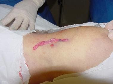 This vein on calf represents major varicose tributary of small saphenous vein that was removed by means of ambulatory phlebectomy.
Skin incisions as small as 1 mm or needle punctures with an 18-gauge or larger needle are used to extract veins with a phlebectomy hook. The procedure is well tolerated by patients under local anesthesia and typically produces good cosmetic results. Long-term results from the authors’ experience are excellent, as long as the most proximal source of reflux is eliminated via endovenous ablation.
In contrast to sclerotherapy of large varicose veins, ambulatory phlebectomy minimizes the risks of intra-arterial injection, skin necrosis, and residual hyperpigmentation. The source vein is extracted by the procedure.
Traditional venous ligation is no longer considered an acceptable method, because the vein is interrupted rather than removed, and this leads to relatively high recurrence rates. With ambulatory phlebectomy, the small size of the skin incision or puncture usually results in little or no scarring. This procedure, performed with only local anesthesia, leads to greatly reduced surgical risks as compared with traditional surgery for truncal (axial) reticular varicose veins and incompetent perforators.
Outcomes
Long-term results after phlebectomy are excellent when the procedure is performed for the appropriate indications. For the main indication—an incompetent primary or secondary branch of the GSV or the SSV—long-term success rates of 90% or greater are reported. Long-term success is typically associated with the elimination of high-grade junctional reflux before or immediately prior to phlebectomy. It is common practice to perform endovenous ablation of saphenous reflux and then perform ambulatory phlebectomy of varicose branches arising from the saphenous system.
A randomized trial involving 50 patients undergoing EVLA for GSV insufficiency, in which 25 underwent ambulatory phlebectomy concomitantly with EVLA and 25 underwent EVLA alone with subsequent phlebectomy as needed a minimum of 6 weeks later, found that the former approach yielded better results with regard to disease severity and quality of life. [11]
The AVULS (Ambulatory Varicosity avUlsion Later or Synchronized) trial, in which 101 patients undergoing endovenous truncal ablation received either simultaneous phlebectomy (n = 51) or delayed varicosity treatment (n = 50), found that the patients in the simultaneous group had improved clinical outcomes and less need for further procedures, as well as early improvements in quality of life. [12]
As with any therapy, new varicose veins may develop over time, and patients must be informed about the likely evolution and progression of venous insufficiency and the associated genetic predisposition.
In the randomized controlled SAPTAP trial (N = 464), Scheerders et al addressed the question of whether isolated ambulatory phlebectomy with or without delayed endovenous truncal ablation (SAP; n = 227) is noninferior to thermal endovenous ablation with concomitant phlebectomy (TAP; n = 237). [13] The primary outcome was the VEnous INsufficiency Epidemiological and Economic Study Quality of Life/Symptoms (VEINES-QOL/Sym) score at 12 months. SAP was found to be noninferior to TAP, as well as less costly.
A prospective study by Kishore et al compared ambulatory phlebectomy with foam sclerotherapy in patients with isolated perforator incompetence with regard to evaluating clinical (return to normal activity, primary symptom relief), functional (procedural time, change in disease severity, course of venous ulcer), and duplex ultrasonography (US) parameters (recurrence in treated veins, complete occlusion of treated veins). [14] Whereas the procedural time was shorter with foam sclerotherapy, the other parameters of primary symptom relief were better with ambulatory phlebectomy. The authors concluded that ambulatory phlebectomy was superior for treating patients with isolated perforator incompetence.
Zolotukhin et al studied short-term outcomes of isolated phlebectomy with preservation of an incompetent GSV (ambulatory selective varices ablation under local anesthesia [ASVAL] procedure) in 67 patients (51 women, 16 men; 75 limbs; age range, 17-71 y; mean age, 46.8 y) with primary varicose veins and GSV incompetence. [15] The ASVAL procedure led to disappearance of reflux in most cases and significantly decreased vein diameter in all. The authors suggested that ASVAL could be considered in selected cases as a less aggressive and less expensive option but noted that no clear indications for the procedure had yet been established.
Garavello et al retrospectively studied ASVAL with a mixed phlebectomy-foam technique in 30 consecutive patients with minimal GSV insufficiency (main GSV ≤ 1 cm). [16] They found this approach to be safe, simple, inexpensive, and minimally traumatic, with no need for US guidance and less risk of foam migrating in the GSV.
Albernaz et al, in a study (N = 171; mean age, 52.3 y; 270 feet) comparing the outcomes and complications of ambulatory phlebectomy (157 feet) and EVLA (113 feet) in the treatment of varicose veins of the foot, found that the two procedures were similar with respect to treatment complications. [17] In both patient groups, dysesthesia was the most common complication; the only complication that occurred at a significantly different rate with two procedures was transient induration (phlebectomy, 0.0%; EVLA, 7.1%).
-
Tumescent anesthesia placed subcutaneously pushes vein closer to skin for easier removal.
-
Various hooks are used in ambulatory phlebectomy (eg, Ramelet, Muller, Oesch, and Varady hooks).
-
Before and 2 months after ambulatory phlebectomy. Reflux at saphenofemoral junction was treated with radiofrequency endoluminal ablation during same procedure.
-
This vein on calf represents major varicose tributary of small saphenous vein that was removed by means of ambulatory phlebectomy.
-
Extraction of veins by means of ambulatory phlebectomy.


