Background
Neurofibroma is an uncommon benign tumor of the oral cavity derived from the cells that constitute the nerve sheath. [1] See the images below.
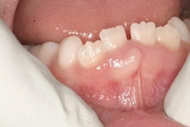 An 11-year-old girl with an asymptomatic raised lesion on the anterior mandibular gingiva.
Neurofibroma is seen either as a solitary lesion or as part of the generalized syndrome of neurofibromatosis (usually neurofibromatosis type 1 [NF-1], also called von Recklinghausen disease of the skin). The solitary form does not differ from the disseminated form or the multiple form of the disease, except that systemic and hereditary factors present in the disseminated form are absent in the solitary type. Multiple neurofibromas have also been strongly associated with the polyglandular syndrome multiple endocrine neoplasia type 3 (MEN-3). [2]
Oral cavity involvement by a solitary and peripheral plexiform neurofibroma in patients with no other signs of neurofibromatosis is uncommon. Sporadic cases have been reported in the submandibular gland, tongue, and on the periosteum at the mental foramen. This sporadic syndromic occurrence has also been seen in the cutaneous region, and several authors have suggested that these isolated neurofibromas may represent a hamartomatous growth.
The World Health Organization (WHO) has subdivided neurofibromas into 2 broad categories: dermal and plexiform. Dermal neurofibromas arise from a single peripheral nerve, while plexiform neurofibromas are associated with multiple nerve bundles. [3] Other clinicopathologic subtypes include localized neurofibroma (sporadic neurofibroma), diffuse neurofibroma, plexiform neurofibroma, and epithelioid neurofibroma.
Localized or solitary neurofibroma is the most frequent manifestation and develops along a peripheral nerve as a focal mass with well-defined margins but not encapsulated. Localized or solitary neurofibroma is rare in infancy and typically appears in late childhood or during teenage years. The majority of isolated or solitary neurofibromas are sporadic, and a small minority may be associated with the NF-1 syndrome. Most of these arise in the third to fourth decades of life. Neurofibroma involving a major nerve, especially those encased in bone, such as the inferior alveolar nerve in the mandible, results in a fusiform expansion of the nerve canal—the so-called blunderbuss canal formation. Soft tissue growths are noted when smaller peripheral nerves are involved. [4, 5]
Plexiform neurofibroma arises along peripheral nerves and tends to involve the smaller branches of the nerves, producing a poorly circumscribed and locally invasive tumor. Approximately 21% of patients with NF-I present with plexiform neurofibromas. [6] These lesions can result in substantial morbidity, mainly due to their size and ability to cause disfigurement. Plexiform neurofibromas are considered to be pathognomonic of for NF-1. These lesions produce the classic "bag-of-worms" appearance. Occasionally, malignant transformation of plexiform neurofibroma is reported (< 5%). This tumor is better designated as a malignant peripheral nerve sheath tumor (MPNST), which tends to exhibit a poor prognosis compared with nonsyndromic MPNSTs.
An uncommon variant of neurofibroma is the diffuse neurofibroma, which typically involves the skin and subcutaneous tissues, resulting in enlargement of affected tissues. Diffuse neurofibromas are most often seen in the head and neck region and may involve the oral cavity. [7, 8] These lesions are more common in adolescents or young adults. Malignant transformation of diffuse neurofibromas is rare, and rapid growth and careful microscopic examination of the biopsy sample is essential to rule out such a change. A minority of diffuse neurofibromas (< 10%) are seen in the setting of NF-1.
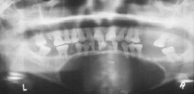 Intrabony presentation of neurofibroma. Note the extensive bone destruction caused by the lesion.
Pathophysiology
The cell of origin for neurofibroma has not been definitively identified. Some believe it arises from the Schwann cell. Perineural fibroblasts are neuroectodermal tissue cells that synthesize collagen and create a network that envelops the individual axis cylinders of the nerves with their associated Schwann cells. Some investigators believe that perineural fibroblasts give rise to the neurofibroma.
The cause of solitary neurofibroma is unknown. However, neurofibromatosis is inherited as an autosomal dominant trait with a high degree of penetrance but variable expressivity. As many as 50% of cases are reported to be the result of spontaneous mutation. Two subsets have been defined: one is associated with the NF-1 (NF1) gene, and the other is associated with the neurofibromatosis type 2 (NF2) gene. [9]
Etiology
The cause of these lesions is unknown; however, neurofibromatosis syndrome or the disseminated form is inherited as an autosomal dominant trait and may present with a variety of lesions, including a highly variable number of neurofibromas.
Epidemiology
US frequency
Little information is available regarding the relative frequency of solitary oral neurofibromas and oral manifestations of neurofibromatosis. A survey of series on oral neurofibromas has reported that approximately 20-60% of cases are associated with neurofibromatosis. Intraoral neurofibromas may be seen in as many as 25% of patients with neurofibromatosis. Another review of head and neck neurofibromas reported that approximately 25% of all neurofibromas are seen in the head and neck region and 6.5% occur in the oral cavity as solitary or multiple lesions associated with NF-1.
Although these lesions may be seen anywhere in the oral cavity, the most common location of this tumor is the tongue. They have also been described on the gingiva, palate, major salivary glands, and maxillary bones.
Race
No racial predilection is recognized for oral neurofibromas.
Sex
No sexual predilection has been described for oral neurofibromas.
Age
The average age of patients presenting with solitary neurofibromas ranges from 10 months to 70 years, with an average of approximately 45 years; however, for intraoral neurofibromas associated with neurofibromatosis, the lesions may present at any age, with no specific bias. [10]
Prognosis
Solitary neurofibromas have a good prognosis, with only rare instances of local recurrence after excision. However, in neurofibromatosis, a larger proportion of patients develop recurrence after excision, and multiple recurrences are associated with malignant transformation. Spontaneous malignant transformation of 1 or more lesions is also reported. The rate of transformation is estimated to be 5-15%. This neurofibrosarcomatous change has an extremely poor prognosis, and distant metastasis is common. The average 5-year survival rate is dismal and ranges around 10-15%. A study involving 66 cases of head and neck neurofibromas reported that no recurrence of the neurofibromas was detected after surgical removal in approximately 33% of the patients during follow-up ranging from 3-230 months.
-
Intrabony presentation of neurofibroma. Note the extensive bone destruction caused by the lesion.
-
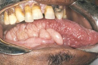
Multiple neurofibromas on the tongue.
-
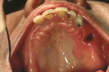
Solitary neurofibroma on the hard palate.
-
An 11-year-old girl with an asymptomatic raised lesion on the anterior mandibular gingiva.
-
Isolated palatal lesion in a 27-year-old African American woman.


