Background
Geographic tongue (benign migratory glossitis) is a benign condition that occurs in up to 3% of the general population. Most often, patients are asymptomatic; however, some patients report increased sensitivity to hot and spicy foods. The etiology and pathogenesis of geographic tongue are still poorly understood. Geographic tongue affects males and females and is noted to be more prominent in adults than in children. [1, 2]
The classic manifestation of geographic tongue is an area of erythema, with atrophy of the filiform papillae of the tongue, surrounded by a serpiginous, white, hyperkeratotic border. The patient often reports spontaneous resolution of the lesion in one area, with the return of normal tongue architecture, only to have another lesion appear in a different location of the tongue. Lesion activity in geographic tongue may wax and wane over time, and patients are occasionally free of lesions.
See the image below.
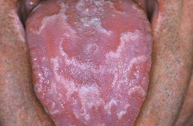 Geographic tongue. Courtesy of Dimitrios Malamos (own work), via Wikimedia Commons.
Pathophysiology
The most commonly affected site is the tongue; however, other oral mucosal soft tissue sites may be affected. Geographic tongue has been reported with increased frequency in patients with psoriasis [3, 4, 5, 6, 7] and in patients with fissured tongue. [8, 9] Geographic tongue and fissured tongue have been reported in association with chronic granulomatous disease. [10] Although geographic tongue is an inflammatory condition histologically, a polygenic mode of inheritance has been suggested because it is seen clustering in families. Associations with human leukocyte antigen (HLA)–DR5, HLA-DRW6, and HLA-Cw6 have also been reported. [11, 12]
Etiology
A definitive cause has not been elucidated, but lesions are seen with increased frequency in patients with psoriasis. In a study of patients with psoriasis, geographic tongue occurred in 10% of the patients, in contrast to only 2.5% of age- and sex-matched controls. [13] A polygenic mode of inheritance has been suggested for geographic tongue. [14] No increased incidence of geographic tongue has been noted with medication use or environmental agents. Immunologic and psychologic parameters have been associated with geographic tongue. [15]
Epidemiology
Frequency
Geographic tongue has reportedly occurred in up to 3% of the general population in the United States. International frequency rates for geographic tongue are similar to those reported in the United States.
Race
No racial or ethnic predilection is reported for geographic tongue.
Sex
Females have been reported to be affected twice as often as males. [16] Exacerbations have been suggested to be related to hormonal factors.
Age
Geographic tongue can affect all age groups; however, it is more predominant in adults than in children.
Prognosis
Geographic tongue is a benign condition.
Patient Education
Defining geographic tongue, describing its clinical appearance, and reinforcing its benign nature is usually all that is needed to educate patients and allay any concerns they may have about geographic tongue.
-
Geographic tongue. Courtesy of Dimitrios Malamos (own work), via Wikimedia Commons.


