Practice Essentials
Necrotizing sialometaplasia (NS) is a nonneoplastic inflammatory condition of the salivary glands. In 1973, Abrams et al first reported this condition. [1] The clinical and histopathologic features of necrotizing sialometaplasia often simulate those of malignancies such as squamous cell carcinoma or salivary gland malignancy. [2] All subsequent reports of necrotizing sialometaplasia stress the importance of correct diagnosis. Familiarity with necrotizing sialometaplasia and correct diagnosis are paramount in avoiding misdiagnosis and inappropriate treatment. Ischemia of salivary gland tissue leading to infarction (trauma) is the most likely cause.
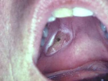
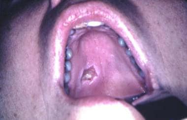
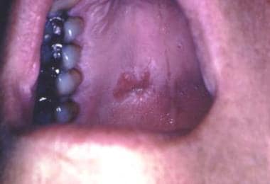
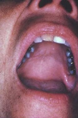
See Prognosis for images.
A related Medscape Reference article that may be of interest is Cancers of the Oral Mucosa.
Signs and symptoms
Necrotizing sialometaplasia manifests as a swelling with or without ulceration in anatomic sites that have mucous or serous glandular tissue.
The typical clinical presentation of necrotizing sialometaplasia is that of a crateriform ulcer of the palate that simulates a malignant process. These ulcerated lesions are 1-3 cm and are usually unilateral, but bilateral synchronous lesions and metachronous lesions can occur. [3, 4, 5]
Some lesions of necrotizing sialometaplasia may present as a submucosal swelling, without ulceration of the overlying mucosa. An intact surface mucosa may be noted in an evolving lesion at the time of diagnosis, although most cases are accompanied by mucosal ulceration. Erosion of the palatal bone may occur in either ulcerated or nonulcerated lesions.
Examination of a biopsy specimen is usually required to establish the correct diagnosis and to exclude a malignant or infectious process or an inflammatory condition such as granulomatosis with polyangiitis (formerly Wegener granulomatosis). Extranodal lymphoma also may be considered in the clinical differential diagnosis of a palatal swelling or ulceration.
Case reports
In an example of a typical case, a 57-year-old male smoker presented with oral pain. Right posterolateral hard palate NS was diagnosed based on clinical features and cone beam CT imaging. The lesion resolved after 3 days with no treatment, and no biopsy was performed. [6]
Another case involved a 19-year-old female with ulcerative lesions of the oral mucosa and bilateral facial swelling. The lesions' appearance was interspersed by several weeks. A culture grew S anginosus. A diagnosis of asynchronous bilateral NS with superinfection was made. [7]
The case of a 26-year-old female illustrates the importance of accurate diagnosis with NS. The patient underwent surgery for a left palatal bone defect. An erythematous area was noted, and the lesion was mistaken for a malignant neoplasm. After an unnecessary left maxillectomy, an accurate diagnosis of NS was made. [8]
In the case of a non-ulcerated but painful, erythematous lesion on the hard palate of a 32-year-old male present for 1 month, clinicians initially suspected a salivary gland tumor. However, histopathologic and immunohistochemical investigations proved the lesion to be NS. [9]
Diagnostics
A definitive tissue diagnosis of necrotizing sialometaplasia should exclude the need for radiographic imaging. If erosion of the palatal bone occurs with or without perforation, radiologic examination may be performed. [10]
Incisional biopsy is necessary to establish the diagnosis of necrotizing sialometaplasia. An inadequate biopsy specimen may lead to the misdiagnosis of squamous cell carcinoma or mucoepidermoid carcinoma. Findings in a superficial or limited biopsy specimen may be misinterpreted as a nonspecific ulcer or pseudoepitheliomatous hyperplasia of the surface mucosa.
Also see Histologic Findings.
Management
Necrotizing sialometaplasia (NS) resolves spontaneously. No treatment is necessary.
Surgical care for NS consists of incisional biopsy for diagnostic purposes.
Periodic evaluation of the affected site is recommended until spontaneous resolution occurs.
Pathophysiology
Necrotizing sialometaplasia was first reported to involve the minor salivary glands of the oral cavity, particularly those of the palate. Seventy-five percent of all cases occur on the posterior palate. [11] Most are unilateral, with one third occurring in a bilateral or midpalatal location. Reports of this entity in the minor glands of the retromolar pad area, buccal mucosa, tongue, incisive canal, and labial mucosa followed. In addition, necrotizing sialometaplasia is recognized in the parotid and submandibular salivary glands, [12] minor mucous glands in the lung, [13] nasal cavity, [14, 15] larynx, [16, 17] trachea, [18] nasopharynx, and maxillary sinus. [19] Similar lesions are identified in the breast; the condition is referred to as posttraumatic lobular metaplasia of the breast. [20]
Etiology
In most cases of necrotizing sialometaplasia, the etiology is believed to be related to vascular ischemia. Cases are reported in which vascular compression is caused by a necrotic myocutaneous reconstruction flap, embolization from carotid endarterectomy, sickle cell anemia, [21] Buerger disease, [22] or Raynaud phenomenon. [22]
The association of adjacent neoplasia that results in ischemic necrosis of the glandular elements and the histologic features of necrotizing sialometaplasia supports this pathogenic mechanism. In an experimental study in a rat model, local anesthetic injections induced necrotizing sialometaplasia. [23] Tobacco use is suggested as a possible etiologic risk factor for necrotizing sialometaplasia.
Epidemiology
Frequency
United States
Mesa and colleagues reported an incidence of 0.03% based on findings in 10,000 oral biopsy specimens. [24] However, they state that this percentage does not account for cases of necrotizing sialometaplasia that heal spontaneously without biopsy.
International
Necrotizing sialometaplasia is reported worldwide. Isolated cases and reviews from Europe, North America, South America, and Asia are reported in the literature.
Race-, sex-, and age-related information
Brannon and colleagues [25] reported that cases of necrotizing sialometaplasia in whites outnumbered cases in blacks by a ratio of 4.9:1. Given the ratio of Whites to Blacks in the United States, a significant racial predilection does not appear to exist.
The male-to-female ratio is approximately 2:1.
The average age of patients with necrotizing sialometaplasia in the Armed Forces Institute of Pathology (AFIP) registry is 47.9 years, with a range of 17-80 years. The average age is 43.1 years for female patients and 50.3 years for male patients. A case of necrotizing sialometaplasia in an 18-month-old infant is reported.
Prognosis
The prognosis for necrotizing sialometaplasia (NS) is excellent. Necrotizing sialometaplasia is a completely benign lesion that is most likely caused by ischemia secondary to trauma (examples of causes could be dental injection or hot liquid burn). The lesions heal on their own with or without biopsy. The reason for biopsy is to rule out cancer from their worrisome clinical presentation.
The average healing time for necrotizing sialometaplasia of the minor salivary glands of the hard and soft palates is approximately 5 weeks. The size of the lesion and whether or not bony perforation has occurred are clinical parameters that may influence the healing time.
The lesions of necrotizing sialometaplasia often are painless; less frequently, they cause pain and numbness. The clinical appearance that suggests cancer is the significant feature of this lesion. The clinical pictures show a patient with a lesion thought to be cancer who underwent biopsy and was monitored for 9 weeks. Over that time, regression of the lesion can be seen (see the images below).
Patient Education
Necrotizing sialometaplasia is a completely benign condition that heals on its own following biopsy.
-
Initial presentation.
-
Three weeks later after biopsy.
-
At 6 weeks.
-
Nine weeks. Salivary gland infarction.


