Continually Updated Clinical Reference
 
 
  All Sources     eMedicine     Medscape     Drug Reference     MEDLINE
 
eMedicine - Epulis Fissuratum : Article by

Quick Find
Authors & Editors
Introduction
Clinical
Differentials
Workup
Treatment
Follow-up
Miscellaneous
Multimedia
References

Related Articles
Metastatic Neoplasms to the Oral Cavity

Pyogenic Granuloma (Lobular Capillary Hemangioma)

Squamous Cell Carcinoma


Patient Education
Click here for patient education.


Author: Diane Stern, DDS, Clinical Professor, Department of Surgery, Section of Oral and Maxillofacial Surgery, University of Miami; Clinical Professor, Nova Southeast University School of Dental Medicine

Editors: Marjan Garmyn, MD, PhD, Professor, Faculty of Medicine, Katholieke Universiteit Leuven, Belgium; Chair and Adjunct Head, Department of Dermatology, University of Leuven, Belgium; David F Butler, MD, Professor of Dermatology, Texas A&M University College of Medicine; Director, Division of Dermatology, Scott and White Clinic; Director Dermatology Residency Training Program, Scott and White Clinic; Drore Eisen, MD, DDS, Consulting Staff, Department of Dermatology, Dermatology Research Associates of Cincinnati; Glen H Crawford, MD, Assistant Clinical Professor, Department of Dermatology, University of Pennsylvania School of Medicine; Chief, Division of Dermatology, The Pennsylvania Hospital; William D James, MD, Paul R Gross Professor of Dermatology, University of Pennsylvania School of Medicine; Vice-Chair, Program Director, Department of Dermatology, University of Pennsylvania Health System

Author and Editor Disclosure

Synonyms and related keywords: denture-induced hyperplasia, inflammatory hyperplasia, denture hyperplasia, denture-induced fibrous hyperplasia

Background

This common lesion is a mucosal hyperplasia that results from chronic low-grade trauma induced by a denture flange.

Pathophysiology

Epulis fissuratum arises in association with denture flanges. Consequently, it is usually observed in the maxillary or mandibular vestibule.

Mortality/Morbidity

Significant morbidity does not occur with epulis fissuratum.

Race

Most cases are observed in whites. This, no doubt, relates to the predominance of whites as denture wearers.

Sex

Most studies indicate a clear predilection for females. The fact that women are more likely than men to wear their dentures for prolonged periods because of their reluctance to be seen without them probably plays a significant role. In addition, more women than men wear dentures and are more likely to seek treatment. Possibly, atrophic epithelial changes secondary to menopause may influence an increased reaction to trauma in older females.

Age

Epulis fissuratum occurs in greatest numbers in the fifth, sixth, and seventh decades, but it can be observed at almost any age. Epulis fissuratum has been described in children. The fact that the lesions are related to denture wear and chronicity of an irritative process explains the higher incidence in older individuals.


History

  • Epulis fissuratum develops slowly over a prolonged period of time in patients with ill-fitting dentures. It is associated with a denture flange that may be either a full or partial denture.
  • Typically, patients with this condition are asymptomatic.

Physical

  • Examination of the patient typically reveals folds of hyperplastic mucosa, which encompass the border of the denture flange. The edge of the denture usually fits in a groove between the folds. The lesions are most frequently observed at the facial aspect of the denture. The occurrence of this on the lingual surface is unusual. They are more often observed in the anterior portion of the jaws; however, a predilection for the maxilla or the mandible does not seem to exist.
  • The surface of the mass tends to be smooth; however, occasionally, it is ulcerated (most often within the depth of the groove) or papillary.
  • The size of the lesion is variable; some lesions are small, but they can be extensive and involve the entire length of the vestibule.
  • Although frequently of normal mucosal color, erythema may be associated with inflammation. Some lesions have a more pyogenic granuloma–like appearance because of capillary proliferation.

Causes

The cause of epulis fissuratum is chronic low-grade irritation from an ill-fitting denture. Frequently, this is the consequence of resorption of the alveolar ridge so that the denture moves further into the vestibular mucosa, creating an inflammatory fibrous hyperplasia that proliferates over the flange.


Metastatic Neoplasms to the Oral Cavity
Pyogenic Granuloma (Lobular Capillary Hemangioma)
Squamous Cell Carcinoma


Procedures

  • Surgically excise and microscopically examine the epulis fissuratum.

Histologic Findings

Epulis fissuratum is a hyperplastic reactive lesion, often with inflammatory and reparative phases. The histologic picture can be variable. Most frequently, a dense fibrous hyperplasia occurs, often with varying degrees of inflammation and vascularity. Because capillary proliferation is considerable, an overlap with pyogenic granuloma occurs. Mucous glands are often present in the specimen and may show a chronic sialadenitis. Occasionally, the glands may have an associated lymphoid hyperplasia and papillary ductal hyperplasia. The epithelium may be atrophic or hyperplastic and occasionally shows a pseudoepitheliomatous hyperplasia. Ulceration can occur. Infrequently, chondroid or osseous metaplasia can develop within the mass.


Surgical Care

Surgically excise the epulis fissuratum because even removal of the offending stimulus (ie, denture) will not result in complete resolution. In addition, correct the denture; otherwise, the lesion will recur. Either make a new denture or reline the old denture.

Consultations

  • Oral and maxillofacial surgeon for excision
  • General dentist or prosthodontist for correction of the denture


Deterrence/Prevention

  • Regular dental care can prevent epulis fissuratum. Patients who wear dentures frequently believe that they no longer require care, and, under these circumstances, dentures lose their correct fit and become the source of irritation.

Prognosis

  • With correction of the poorly fitting denture, the prognosis is excellent.

Patient Education

  • Instruct the patient that regular dental care is necessary and that the oral tissues are changing constantly. This means that dentures are not permanent and need adjustments over time.


Medical/Legal Pitfalls

  • The potential problem is that masses that occur in the vestibular area and are associated with dentures tend to be dismissed as epulis fissuratum. Unfortunately, on rare occasions, these masses prove to be squamous cell carcinomas or metastatic disease. Therefore, do not ignore these lesions, and always examine them histologically.


Media file 1:  
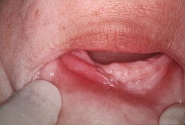
An epulis fissuratum in the anterior part of the mandible shows a central groove where the denture flange rests. Note the inflammatory erythema. The surface of the lesion is usually smooth as shown in the image.
Click to see larger pictureClick to see detailView Full Size Image
Media type:  Photo

Media file 2:  The mass in the posterior part of the maxillary vestibule is associated with a full denture; however, in this patient, the mass represented a squamous cell carcinoma. The surface is more granular in appearance, although this is not always the case.
Click to see larger pictureClick to see detailView Full Size Image
Media type:  Photo

Media file 3:  A view of a whole mount of a tissue section taken from an epulis fissuratum shows that it is essentially a fibrous hyperplasia. The central groove can be observed, and, in this patient, papillary hyperplasia is present in some areas.
Click to see larger pictureClick to see detailView Full Size Image
Media type:  Photo


  • Buchner A, Begleiter A, Hansen LS. The predominance of epulis fissuratum in females. Quintessence Int. Jul 1984;15(7):699-702. [Medline].
  • Coelho CM, Zucoloto S, Lopes RA. Denture-induced fibrous inflammatory hyperplasia: a retrospective study in a school of dentistry. Int J Prosthodont. Mar-Apr 2000;13(2):148-51. [Medline].
  • Cutright DE. The histopathologic findings in 583 cases of epulis fissuratum. Oral Surg Oral Med Oral Pathol. Mar 1974;37(3):401-11. [Medline].
  • Keng SB, Loh HS. Clinical presentation of denture hyperplasia of oral tissues. Ann Acad Med Singapore. Sep 1989;18(5):537-40. [Medline].
  • Ralph JP, Stenhouse D. Denture-induced hyperplasia of the oral soft tissues. Vestibular lesions, their characteristics and treatment. Br Dent J. Jan 18 1972;132(2):68-70. [Medline].

Epulis Fissuratum excerpt

Article Last Updated: Nov 8, 2006