Continually Updated Clinical Reference
 
 
  All Sources     eMedicine     Medscape     Drug Reference     MEDLINE
 
eMedicine - Mucocele and Ranula : Article by

Quick Find
Authors & Editors
Introduction
Clinical
Differentials
Workup
Treatment
Follow-up
Miscellaneous
Multimedia
References

Related Articles
Branchial Cleft Cyst

Dermoid Cyst

Lipomas

Lymphangioma

Oral Hemangiomas

Oral Lymphangiomas

Oral Pyogenic Granuloma

Venous Lakes


Patient Education
Click here for patient education.


Author: Catherine M Flaitz, DDS, MS, Dean and Co-director of Oral and Maxillofacial Pathology Laboratory, Professor of Oral and Maxillofacial Pathology and Pediatric Dentistry, Department of Diagnostic Sciences, University of Texas Health Sciences Center at Houston, Dental Branch

Catherine M Flaitz is a member of the following medical societies: American Academy of Oral and Maxillofacial Pathology, American Academy of Oral Medicine, American Dental Association, International Association for Dental Research, and International Association of Oral Pathologists

Coauthor(s): M John Hicks, DDS, MS, PhD, MD, Professor with Tenure, Department of Pathology, Baylor College of Medicine; Medical Director of Surgical Pathology, Department of Pathology, Texas Children's Hospital

Editors: Timothy McCalmont, MD, Director, UCSF Dermatopathology Service, Professor of Clinical Pathology and Dermatology, Departments of Pathology and Dermatology, University of California at San Francisco; David F Butler, MD, Professor of Dermatology, Texas A&M University College of Medicine; Director, Division of Dermatology, Scott and White Clinic; Director Dermatology Residency Training Program, Scott and White Clinic; Drore Eisen, MD, DDS, Consulting Staff, Department of Dermatology, Dermatology Research Associates of Cincinnati; Glen H Crawford, MD, Assistant Clinical Professor, Department of Dermatology, University of Pennsylvania School of Medicine; Chief, Division of Dermatology, The Pennsylvania Hospital; William D James, MD, Paul R Gross Professor of Dermatology, University of Pennsylvania School of Medicine; Vice-Chair, Program Director, Department of Dermatology, University of Pennsylvania Health System

Author and Editor Disclosure

Synonyms and related keywords: mucus retention phenomenon, mucus escape reaction, mucous cyst, cervical (plunging) ranula, oral ranula, anterior lingual mucocele, Blandin-Nuhn mucocele

Background

Collectively, the mucocele, the ranula, and the cervical, or plunging, ranula are clinical terms for a pseudocyst that is associated with mucus extravasation into the surrounding soft tissues. These lesions occur as the result of trauma to the salivary gland excretory duct, although obstruction of salivary flow is implicated in some instances.

Mucoceles, which are of minor salivary gland origin, are also referred to as mucus retention phenomenon and mucus escape reaction. The superficial mucocele, a special variant, has features that resemble a mucocutaneous disease. At times, the mucus retention cyst, also referred to as the sialocyst or the salivary duct cyst, is included in this group of lesions but appears to represent a separate entity on the basis of its clinical and histopathologic features. Although the mucus retention cyst is discussed in this article, its features are differentiated from the features of the pseudocysts. The lesions of the sinus, such as sinus mucoceles, pseudocysts, and retention cysts, are not included in this discussion.

Ranulas, which involve the major salivary glands, are divided into 2 types: oral ranulas and cervical ranulas. Oral ranulas are secondary to mucus extravasation that pools superior to the mylohyoid muscle, whereas cervical ranulas are associated with mucus extravasation along the fascial planes of the neck.

Pathophysiology

The development of mucoceles and ranulas depend on the disruption of the flow of saliva from the secretory apparatus of the salivary glands. The lesions are most often associated with mucus extravasation into the adjacent soft tissues caused by a traumatic ductal insult; the insults include a crush-type injury and severance of the excretory duct of the minor salivary gland. The disruption of the excretory duct results in extravasation of mucus from the gland into the surrounding soft tissue. The rupture of an acinar structure caused by hypertension from the ductal obstruction is another possible mechanism for the development of such lesions. Furthermore, trauma that results in damage to the glandular parenchymal cells in the salivary gland lobules is another potential mechanism.

Recent research studies have revealed increased levels of matrix metalloproteins, tumor necrosis factor-alpha, type IV collagenase, and plasminogen activators in mucoceles compared with that of whole saliva. These factors are further hypothesized to enhance the accumulation of proteolytic enzymes that are responsible for the invasive character of extravasated mucus.

Besides ductal disruption, partial or total excretory duct obstruction is involved in the pathogenesis of ranulas in some instances. The duct may become occluded by a sialolith, congenital malformation, stenosis, periductal fibrosis, periductal scarring due to prior trauma, excretory duct agenesis, or even a tumor. Although most oral ranulas originate from the secretions of the sublingual gland, they may develop from the secretions of the submandibular gland duct or the minor salivary glands on the floor of the mouth. The mucus extravasation of the sublingual gland almost exclusively causes cervical ranulas. The mucus escapes through openings or dehiscence in the underlying mylohyoid muscle.

Occasionally, ectopic sublingual glands may be responsible for the problem. When mucus secretions escape into the neck through the mylohyoid muscle, they extend into the fascial tissue planes and cause a diffuse swelling of the lateral or submental region of the neck. The continuous secretions from the sublingual gland allow for relatively rapid accumulation of mucus in the neck and a constantly expanding cervical mass.

The mucus retention cyst may also develop because of ductal obstruction; however, many of these lesions actually represent a distinct cystic entity of unknown cause. When ductal occlusion is involved, it is usually caused by a sialolith or an inspissated secretion that results in ductal dilatation and focal containment of the mucoid material.

Frequency

United States

In the Minnesota Oral Prevalence Study that included 23,616 white adults older than age 35 years, mucoceles represented the 17th most common oral mucosal lesion, with a prevalence of 2.4 cases per 1000 people. Data from the Third National Health and Nutrition Examination Survey (NHANES III) that included 17,235 adults aged 17 years or older documented an overall prevalence ranking of 44 for the mucocele and a point prevalence of 0.02%. In the same study, which consisted of 10,030 children aged 2-17 years, the mucocele had a point prevalence of 0.04%. Congenital mucoceles in newborns are rare, with sporadic case reports and small case series appearing in the literature.

Mucoceles of the anterior lingual salivary glands (glands of Blandin and Nuhn) are relatively uncommon. In the Minnesota Oral Disease Prevalence Study, Blandin and Nuhn mucoceles had a lower prevalence than mucoceles at other locations, or 0.1 cases per 1000 persons. This type of mucocele represents an estimated 2-10% of all mucoceles.

Superficial mucoceles are typically located in the soft palate, the retromolar region, and the posterior buccal mucosa. They represent approximately 6% of all mucoceles. Multiple superficial mucoceles have been reported in a small number of patients.

In an 11-year retrospective review of oral mucoceles and sialocysts from a university-based oral and maxillofacial pathology laboratory, most lesions were found to be mucous retention phenomenon (mucoceles, 91%). In descending order, the other diagnoses included ranulas (6%), and mucous retention cysts (5%). Mucocele outnumbered mucous retention cysts by a ratio of 15.3:1.0. More limited histopathologic studies document that the mucus retention cyst (those lesions with an epithelial lining) accounts for 3-18% of all oral mucoceles.

Ranulas have a prevalence of 0.2 cases per 1000 persons and are ranked 41st in the Minnesota Oral Disease Prevalence Study. As noted previously, ranulas accounted for 6% of all oral sialocysts in a university-based oral and maxillofacial biopsy service. The prevalence of cervical (plunging) ranulas is not known; however, these lesions are considered uncommon. The number of ranulas that represents a true retention cyst ranges from less than 1% to 10%.

International

Large international population studies comparable to those undertaken in the United States are not available for oral diseases, except in Sweden. In a study of 30,000 Swedish individuals aged 15 years or older, the prevalence of mucoceles was 0.11%. In a Brazilian study of 1200 children seen at pediatric hospital clinic, the prevalence of mucoceles was 0.08%.

Mortality/Morbidity

  • Mucoceles and ranulas tend to be relatively painless or asymptomatic lesions with little or no associated morbidity or mortality.
  • Oral and plunging ranulas, if large, may affect swallowing, speech, mastication, or respiratory function.

Race

  • No racial predilection is reported for any of the lesions.

Sex

  • Although no sexual predilection is usually associated with mucoceles, the prevalence of the lesions in the Minnesota Oral Disease Prevalence Study was 1.9 cases per 1000 males compared with 2.6 cases per 1000 females. Other authors have shown that mucoceles are more common in males than in females, with a male-to-female ratio of 1.3:1.
  • In the reported cases, superficial mucoceles and mucoceles of Blandin and Nuhn have a predilection for females.
  • The sexual predilection for oral ranulas slightly favors females, with a male-to-female ratio of 1:1.4, while cervical ranulas have a predilection for males.

Age

  • Most mucoceles occur in young individuals, with 70% of individuals being younger than 20 years. The peak prevalence occurs in persons aged 10-20 years. Although not well studied, superficial mucoceles tend to occur in individuals older than 30 years.
  • Ranulas usually occur in children and young adults, with the peak frequency in the second decade. The cervical variant tends to occur a little later in the third decade.
  • Mucus retention cysts occur in older individuals; the peak prevalence occurs in persons aged 50-60 years.


History

  • Mucoceles are painless, asymptomatic swellings that have a relatively rapid onset and fluctuate in size.
    • They may rapidly enlarge and then appear to involute because of the rupture of the contents into the oral cavity or resorption of the extravasated mucus.
    • The patient may relate a history of recent or remote trauma to the mouth or face, or the patient may have a habit of biting the lip. However, in many cases no insult can be identified.
    • When lesions occur on the anterior ventral surface of the tongue, tongue thrusting may be the aggravating habit, in addition to trauma.
    • The duration of the lesion is usually 3-6 weeks; however, it may vary from a few days to several years in exceptional instances.
  • Patients with superficial mucoceles report small fluid-filled vesicles on the soft palate, the retromolar pad, the posterior buccal mucosa, and, occasionally, the lower labial mucosa.
    • These vesicles spontaneously rupture and leave an ulcerated mucosal surface that heals within a few days.
    • Several lesions may be present, and they are mildly tender.
    • Often, the patient may rupture or unroof the vesicles by suction alone.
    • Typically, patients report a chronic and recurrent history.
  • Individuals with an oral ranula may complain of swelling of the floor of the mouth that is usually painless. The mass may interfere with speech, mastication, respiration, and swallowing because of the upward and medial displacement of the tongue. When oral ranulas are large, the tongue may place pressure on the lesion, which may interfere with submandibular salivary flow. As a result, obstructive salivary gland signs and symptoms may develop, such as pain or discomfort when eating, a feeling of fullness at that site, and increased swelling of the submandibular gland.
  • In patients with a cervical ranula, an enlarging asymptomatic neck mass is reported.
    • Although trauma is not typically associated with the development of a ranula, the patient may have a prior history of a previously removed sialolith, other oral surgical procedures at the floor of the mouth, or transposition of the submandibular ducts for the management of severe drooling.
    • In large oral and cervical ranulas, dysphagia and respiratory distress may be the chief complaints.
    • The patient may have a history of a preceding oral swelling (45%) or a concurrent oral mass at presentation (34%). One fifth of patients with cervical ranula have only a cervical swelling, lacking an oral ranula or a history of an oral ranula.
  • The mucus retention cyst appears as a superficial asymptomatic swelling that is usually not associated with a history of trauma.
    • These cysts tend to have variable growth rates, and they do not fluctuate in size.
    • When the mucus retention cyst involves the submandibular gland, Wharton duct, or Stensen duct, obstructive disease may occur and a pattern of gustatory swelling and pain may be reported.

Physical

The clinical features associated with mucoceles include a nontender, mobile, dome-shaped enlargement with intact epithelium that lies over it. Superficial lesions take on a bluish to translucent hue, whereas deep lesions have normal mucosal coloration. Bleeding into the swelling may impart a bright red and vascular appearance. The mucosa lining is usually intact; however, repeated sucking on the lesion may result in a white, rough, keratotic surface. Occasionally, a punctate sinus tract is observed from which mucoid material is expressed. Palpation reveals a fluctuant mass that does not blanch on compression. An inflammatory response is usually not detected at clinical examination unless it has been irritated recently. Most are less than 1.5 cm in diameter. Although the mucocele can occur anywhere in the oral cavity where minor salivary glands are present, approximately 75% of the cases occur on the lower lip, followed by the buccal mucosa.

  • The Blandin and Nuhn mucocele occurs exclusively on the anterior ventral surface of the tongue at the midline. Although the lesions may have clinical features similar to those of the mucocele, which is found elsewhere, they tend to be more polypoid with a pedunculated base. Because of repeated trauma against the lower teeth, the surface may be red and granular or white and keratotic.
  • Superficial mucoceles appear as single or multiple tense vesicles with intact delicate mucosa. They are transparent, mucous filled, and dome shaped. The lesions tend to persist for several days, rupture spontaneously, and heal a few days after they rupture. Usually, only mild discomfort occurs. Some cases of biopsy-proven concurrent lichen planus and multiple superficial mucoceles have been reported. These lesions are typically 1-4 mm in diameter.
  • The oral ranula is a relatively large unilateral blue to translucent mass in the floor of the mouth that remotely resembles the belly of a frog (Rana species). The lesion may cross the midline when especially large, making the offending salivary gland difficult to localize. Large oral ranulas superiorly and medially displace the tongue. The consistency of the lesion is that of mucus, and the lesion does not blanch on compression. If the mass is located in the deeper aspect of the floor of the mouth, it loses its bluish translucent color. Most commonly, ranulas arise from the sublingual gland and, infrequently, from the submandibular gland.
  • The cervical ranula appears as an asymptomatic, continuously enlarging mass that may fluctuate in size. The overlying skin is usually intact. The mass is fluctuant, freely movable, and nontender. The mass is not associated with the thyroid gland or lymph node chains. In some instances, detecting salivary gland herniation of a portion of the sublingual gland through the mylohyoid muscle into the neck may be possible. The mass may not be well defined but follows the fascial planes of the neck and may extend into the mediastinum. Similar to the oral ranula, the mass tends to cause a lateral swelling; however, it may cross the midline.
  • The mucus retention cyst has a presentation similar to that of a mucocele and a ranula, except that it does not fluctuate in size. The fluid-filled lesions tend to slowly enlarge with well-defined margins that are freely movable. The dome-shaped nodule has a smooth intact surface that imparts a pink, yellow, blue, or red color. The oral floor is the primary site, especially in the area of the orifices of the Wharton's duct and the caruncles, followed by the buccal mucosa. The lesions are usually 5-15 mm in diameter, but they may be much larger when they involve the sublingual or submandibular gland.

Causes

  • The most frequently injured glands are the minor salivary glands of the lower lip.
    • The mechanism of injury is mechanical, with the tissue of the lower lip becoming caught between the maxillary anterior teeth and the mandibular anterior teeth during mastication or with the habit of biting one's lip. This trauma results in a crush-type injury and severance of the excretory duct of the minor salivary gland. In the palate, low-grade chronic irritation (eg, from heat and noxious tobacco products) may cause these lesions to develop.
    • Mucoceles occur when injury to the minor salivary glands occurs usually secondary to trauma; biting one's lip; chronic inflammation with periductal scarring; excretory duct fibrosis; prior surgery; trauma from oral intubation; or rarely, minor salivary gland sialolithiasis.
    • Most mucoceles occur because of severance of the excretory duct and extravasation of mucus into the adjacent tissue.
  • Birth trauma that affects the oral cavity is believed to cause congenital mucoceles in some newborns.
    • Potential causes include the baby sucking his or her fingers in utero or the baby passing through the birth canal.
    • Other causes include the use of forceps during delivery or suctioning of the baby's mouth after birth.
  • Most ranulas are the result of escaped mucus from an injured excretory duct, while ductal obstruction of primarily the sublingual gland and (less often) the submandibular gland is a less common cause.
    • This obstruction is often due to a sialolith or mucus plug; however, chronic inflammation or infection with periductal scarring, trauma, ductal stenosis, ductal hypoplasia or agenesis, and neoplasia are other causes of ranula formation.
    • Isolated case reports have identified Sjögren syndrome and sarcoidosis as contributing to the development of these reactive lesions. In addition, HIV infection may increase the risk of developing a ranula, especially in children.
  • Cervical ranulas are associated with a discontinuity of the mylohyoid muscle.
    • The mylohyoid muscle is regarded as the diaphragm of the floor of the mouth; however, it is not a strict anatomical barrier from entry into the neck. A dehiscence or hiatus in the mylohyoid muscle has been noted in 36-45% of individuals in cadaver studies. This defect is observed along the lateral aspect of the anterior two thirds of the muscle.
    • Projections of sublingual glandular tissue or ectopic glandular tissue may also extend into the neck; these projections facilitate cervical ranula formation.
    • Approximately 45% of plunging ranulas occur after surgery to remove oral ranulas.
  • An obstruction of the excretory duct, with pooling and dilatation of the affected duct, causes the mucus retention cyst. A mucus plug appears to be the cause in most instances, although a sialolith accounts for some of these cysts.
  • With superficial mucoceles, the salivary composition of the minor glands, rather than trauma, induces these lesions.
  • Both lichen planus and chronic graft versus host disease can trigger the formation of superficial mucoceles.
  • Tartar-control toothpaste may be the inciting factor in a few cases of superficial mucoceles.


Branchial Cleft Cyst
Dermoid Cyst
Lipomas
Lymphangioma
Oral Hemangiomas
Oral Lymphangiomas
Oral Pyogenic Granuloma
Venous Lakes

Other Problems to be Considered

Mucoceles may mimic the following:

Benign or malignant salivary gland neoplasms
Hemangioma
Lymphangioma
Venous varix or venous lake
Lipoma
Soft irritation fibroma
Oral lymphoepithelial cyst
Gingival cyst in adults
Soft tissue abscess
Cysticercosis (parasitic infection)

Anterior lingual mucoceles (Blandin-Nuhn mucoceles) may resemble the following:

Hemangioma
Pyogenic granuloma
Squamous papilloma
Fibroepithelial polyp

Superficial mucoceles may be confused with the following:

Cicatricial pemphigoid
Bullous lichen planus
Minor aphthous ulcers

Oral ranula may resemble the following:

Benign or malignant salivary gland neoplasm
Dermoid cyst
Soft tissue space abscess
Hemangioma
Lymphangioma
Lipoma
Plexiform neurofibroma

Cervical ranula may appear similar to the following:

Thyroglossal duct cyst
Branchial cleft cyst
Cystic hygroma
Submandibular sialadenitis
Intramuscular hemangioma
Cystic or neoplastic thyroid disease
Infectious cervical lymphadenopathy (Epstein-Barr virus, cat scratch disease, tuberculosis)
Hematoma
Lipoma
Laryngocele
Dermoid cyst

A mucus retention cyst has a similar differential diagnosis as a mucocele and a ranula, depending on its location.


Imaging Studies

  • In general, imaging studies are not indicated in the evaluation of this disorder. Radiographic evaluation can be considered if sialoliths are considered a contributing factor in the formation of oral and cervical ranulas.
  • Radiologic examination of the head and neck and mediastinum by CT scanning or MRI to define the extent of a cervical ranula and to eliminate other disease processes is prudent prior to surgical intervention.
  • Ultrasonography has also been used to evaluate the lesions.
  • Conventional mucoceles and mucus retention cysts do not require imaging studies.

Procedures

  • Mucoceles usually require excisional biopsy and removal of the servicing minor salivary glands. If a vascular lesion cannot be excluded from the differential diagnosis, then aspiration of the lesion is prudent for evaluation of the fluid contents. Large mucoceles may be best treated by marsupialization because of the risk of traumatizing the labial branch of the mental nerve. Dissection of the lesion along with the adjacent salivary glands is indicated for moderate-sized lesions.
  • Superficial mucoceles may require biopsy, in addition to direct immunofluorescence studies for immunoglobulins and complement, if a mucocutaneous disease is suspected in the differential diagnosis. Laser vaporization may be useful when multiple lesions are present and a diagnosis has been established.
  • Fine needle aspiration of the contents of oral and cervical ranulas may be helpful in the diagnosis prior to excision and subsequent surgery. The fluid consists of mucus with muciphages (macrophages with engulfed mucin), as demonstrated by mucicarmine staining, and other inflammatory cells. Analysis of the aspirated fluid shows increased amylase and protein content. The recurrence of other fluid types or a solid mass with the failure to aspirate fluid indicates that a mass other than a ranula may have been encountered.
  • Oral and cervical ranulas require complete excision of the oral portion of the ranula, in addition to the responsible gland. Usually, the sublingual gland is the origin of the ranula; however, occasionally, the submandibular gland may be the source. Frequently, decompression of the oral ranula or the oral portion of a cervical ranula is necessary. Some authors advocate marsupialization with packing of the pseudocyst with gauze. This technique allows removal of a smaller amount of tissue with better-defined interfaces and less likelihood of injury to the nerve and the Wharton duct. Likewise, some individuals advocate aspiration of the cervical portion of the plunging ranula.
  • Mucus retention cysts require excisional biopsy. If the retention cyst is overlying the Wharton or Stensen duct, the cyst is unroofed, a lacrimal probe is inserted into the duct, and sialodochoplasty is performed.

Histologic Findings

The mucocele and the oral ranula have a well-delineated cavity that contains free mucinous material; this material is characteristic of these entities. The cavity wall lacks an epithelial lining and is considered a pseudocyst. The pseudocyst wall is composed of granulation tissue with fibroblasts, proliferating small-caliber vessels, and a mixed acute and chronic inflammatory reaction. Muciphages are usually present in the fibroconnective wall of the pseudocyst. The muciphage cells may be demonstrated by using mucicarmine staining.

The cyst wall also has free mucin in the connective tissue stroma. The adjacent salivary gland tissue possesses dilated ducts, fibrosis, acinar atrophy, and chronic inflammation. Occasionally, a ruptured salivary duct that is feeding into the area may be identified. The mucosal surface may be atrophic with focal ulceration, or it may show epithelial hyperplasia with hyperkeratosis. Special stains, mucicarmine and Alcian blue, are useful in identifying mucin that is free in the tissue or in the foamy macrophages.

Extravasation of mucin along the mucosal-submucosal interface characterizes the superficial mucocele. The extravasation results in separation of the epithelium from its underlying submucosa and the formation of a subepithelial mucus-filled vesicle.

The cervical ranula appears identical to the mucus extravasation phenomenon. Biopsy of the lateral part of the neck may reveal only amorphous material with rare inflammatory cells, which stains positive for mucin.

The mucus retention cyst is a true cystic entity. It usually demonstrates a unicystic pattern; rarely, a multicystic appearance is identified. The cystic lining is composed of primarily cuboidal to columnar cells; however, mucous cells and squamous cells may be interspersed. Occasionally, oncocytoid and papillary changes of the epithelium lining are found. Typically, free mucin fills the central cavity, but mucus plugs and concentric layers of acellular calcification (sialolith) may be present. The cyst wall is composed of connective tissue with minimal inflammation, and it lacks the granular appearance of the mucus extravasation phenomenon. Atrophic sialadenitis with fibrosis and ductal ectasia may be found in the surrounding tissues.


Medical Care

Multiple superficial mucoceles may be treated with gamma-linolenic acid (oil of evening primrose), which is a prostaglandin E precursor, with some degree of success in limited patients. With cessation of the treatment, the superficial mucoceles recur within a few months. This agent also may improve salivary function and reduce inflammation in patients with Sjögren syndrome.

Surgical Care

  • Mucus extravasation phenomenon: Surgical excision of the mucocele along with the adjacent associated minor salivary glands is recommended. The risk for recurrence is minimal when appropriate surgical excision has been performed. Aspiration of the mucocele contents often results in recurrence and is not appropriate therapy, except to exclude other entities prior to surgical excision. Large lesions may be marsupialized to prevent significant loss of tissue or to decrease the risk for significantly traumatizing the labial branch of the mental nerve. If the fibrous wall is thick, moderate-sized lesions may be treated by dissection. If this surgical approach is used, the adjacent minor salivary glands must be removed.
    • The use of a micromarsupialization technique for mucoceles in pediatric patients has been reported. This technique involves the placement of a 4.0 silk suture through the widest diameter of the lesion (dome of the lesion) without engaging the underlying tissue. A surgical knot is made, and the suture is left in place for 7 days. Patients need to be educated about suture replacement; they must return to have the suture replaced if it should be lost during the 7-day period. The idea behind this alternative treatment for mucoceles of minor salivary glands is that re-epithelization of the severed duct occurs or a new epithelial-lined duct forms, allowing egress of saliva from the minor salivary gland. The recurrence rate after a short follow-up period has been 14% in pediatric patients. This technique is not indicated for lesions larger than 1 cm in diameter.
    • Laser ablation, cryosurgery, and electrocautery are approaches that have also been used for the treatment of the conventional mucocele with variable success.
  • Superficial mucoceles: No surgical treatment is necessary unless the lesion frequently recurs and is problematic to the patient. If treatment is desired, the options include surgical excision, cryotherapy, and laser vaporization. To prevent recurrences when the lesion is associated with an underlying mucocutaneous disease, management of the causative disease is necessary.
  • Oral ranula: With most oral ranulas, surgical management is preferred. Isolated reports demonstrate that oral ranulas have been successfully treated with intracystic injection of the streptococcal preparation, OK-432. Lesion resolution or marked reduction was documented in almost all of the patients following this sclerotherapy. Local pain at the injection site and fever were noted in about 50% of the patients. Currently, the use of this sclerosing agent for the treatment of oral ranulas is considered experimental.
    • Some clinicians use a tiered approach to the management of oral ranulas. The first attempt at management may be marsupialization of the ranula with packing of the entire pseudocyst with gauze for 7-10 days. The entire ranula is unroofed, and the packing material is firmly placed into the entire cavity of the pseudocyst. This technique allows for re-epithelialization of the pseudocyst cavity; seals the mucinous leak; and provokes a foreign body inflammatory reaction, leading to fibrosis and atrophy of the involved acini. The procedure may be effective with the sublingual gland because it has multiple draining excretory ducts. If this does not eliminate the ranula, additional surgical therapy is initiated with removal of the ranula and the offending major salivary gland.
    • The more traditional method of surgery for an oral ranula is complete excision of the ranula and associated major salivary gland. Laser ablation and cryosurgery, either alone or after marsupialization, have been used for some patients with oral ranula.
    • The recurrence rates of an oral ranula with various surgical treatment methods are as follows:

      • Incision and drainage, 71-100%

      • Ranula excision only, 0-25%

      • Marsupialization only, 61-89%

      • Marsupialization with packing, 0-12% (limited studies)

      • Complete excision of the ranula with the sublingual gland, 0-2%
  • Cervical ranula: The elimination of cervical ranulas depends on the complete surgical excision of the oral portion of the ranula with the associated sublingual salivary gland or, rarely, the submandibular gland.

    • When this procedure is performed, the cervical ranula resolves and has a low risk of recurrence. With drainage of the cervical ranula alone, the recurrence rate is greater than 85%. When the sublingual gland is intraorally excised along with drainage of the cervical pseudocyst, no recurrences are observed. A cervical approach to excision of the neck pseudocyst and the sublingual gland has a low recurrence rate (approximately 4%).

    • The most important factor in surgical management for cervical ranulas is removal of the responsible major salivary gland.

    • Besides surgical management, intracystic injection of the streptococcal preparation, OK-432, has been used to treat this lesion in a few reported cases. The use of this sclerosing agent as a treatment approach for the cervical ranula is considered experimental.
  • Mucus retention cyst: These cysts are treated with conservative surgical excision. When they involve the major glands, partial or total removal of the affected gland may be necessary.

Consultations

  • Consultation with a radiologist may be required to determine the tissue extension of oral and cervical ranulas.
  • Consultation with an anesthesiologist is recommended when airway obstruction is a possibility.

Diet

Diet modifications depend on the extent of surgery.

  • After many oral surgical procedures, a liquid or soft and bland diet is usually recommended for the first couple of days.
  • More invasive surgeries that involve the removal of a major salivary gland may require a modified diet for a longer period.
  • Use of tobacco products is not recommended until healing has occurred.

Activity

Depending on the extent of the procedure, strenuous physical and recreational activities are discouraged for several days to several weeks after surgery.


Further Inpatient Care

  • Routine postsurgical care is required for patients who undergo the surgical procedure under general anesthesia.

Further Outpatient Care

  • Typical wound care after surgical management is required.
  • Patients who receive marsupialization with gauze packing should be informed that the dressing is spontaneously expelled in 7-14 days.

Complications

  • A low risk of bleeding and peripheral nerve damage exists after excision of a mucocele.
  • No complications are associated with superficial mucoceles.
  • Complications are more common with surgical intervention in oral and cervical ranulas than other treatments.
    • Possible surgical complications include the following: injury to the Wharton duct, leading to stenosis, obstructive sialadenitis, and leakage of saliva; injury to the lingual nerve with paresthesia; and injury to the marginal mandibular branch of the facial nerve with paresthesia. Postoperative hematoma, infection, or dehiscence of the wound may occur.
    • In addition, incomplete removal of the oral ranula increases the risk for developing a cervical ranula, while a cervical ranula may extend into the mediastinum. Approximately 45% of plunging ranulas occur after attempts to remove oral ranulas, which can result in a compromised airway. Cervical ranulas can extend into the mediastinum and provoke a sterile mediastinitis that may be life threatening.
  • The complications of a mucus retention cyst are the same as those for a mucus retention phenomenon and an oral ranula, depending on the location.

Prognosis

  • If adequate and complete surgical excision is accomplished, the patient should expect no recurrence of mucoceles. If the adjacent minor salivary glands are not removed or are transected, the risk for recurrence increases. In the case of the anterior lingual mucocele, the offending glands of Blandin and Nuhn are deep within the musculature of the tongue and require knowledge of tongue anatomy and adequate resection to prevent recurrences.
  • Superficial mucoceles are likely to recur periodically, and new lesions may develop over time.
  • Inadequate surgical therapy for oral ranulas may result in the creation of cervical ranulas. As noted previously, almost one half of cervical ranulas are those occurring after surgical attempts to eliminate oral ranulas. When these lesions are managed by marsupialization alone, the recurrence rate is high, and the lesions usually develop 6-8 weeks after surgery.
  • With adequate surgical excision, mucus retention cysts are not likely to recur.

Patient Education

  • Educate the patient regarding early recognition of a mucocele, an oral ranula, or a cervical ranula recurrence.
  • Educate the patient to recognize signs and symptoms of wound infection after surgical intervention and to seek the care of a dentist or physician if necessary.


Medical/Legal Pitfalls

  • Failure to differentiate the clinical features of mucoceles, oral ranulas, cervical ranulas, and mucus retention cysts may be a medicolegal pitfall. Because these lesions may be difficult to clinically differentiate from benign and malignant salivary gland tumors, especially cystadenoma and mucoepidermoid carcinoma, submitting excised tissues for histopathologic evaluation is important.
  • At least one report of a squamous cell carcinoma arising in a cervical ranula exists. This finding emphasizes the need for excluding a neoplastic cause for the development of these reactive lesions.

Special Concerns

  • Although a rare complication, the physician should be able to recognize mediastinitis secondary to a plunging ranula, which can be life threatening.


Media file 1:  Classic example of a mucocele in a child. The fluctuant, translucent-blue nodule on the lower labial mucosa has been present for 6 weeks. Trauma from sucking on the lower lip was suspected to be the cause.
Click to see larger pictureClick to see detailView Full Size Image
Media type:  Photo

Media file 2:  Fluctuant submucosal nodule of the lower lip consistent with a mucocele.
Click to see larger pictureClick to see detailView Full Size Image
Media type:  Photo

Media file 3:  Surgical excision of the mucocele in Image 1.
Click to see larger pictureClick to see detailView Full Size Image
Media type:  Photo

Media file 4:  Mucocele on the midline ventral surface of the tongue involving the glands of Blandin and Nuhn.
Click to see larger pictureClick to see detailView Full Size Image
Media type:  Photo

Media file 5:  Example of 2 superficial mucoceles of the soft palate in a 50-year-old woman. The red lesion represents a recently ruptured mucocele, and the translucent papular lesion represents an intact mucocele.
Click to see larger pictureClick to see detailView Full Size Image
Media type:  Photo

Media file 6:  Unilateral oral ranula in a young adult manifesting as a purple swelling.
Click to see larger pictureClick to see detailView Full Size Image
Media type:  Photo

Media file 7:  Ranula on the floor of the mouth with focal ulceration.
Click to see larger pictureClick to see detailView Full Size Image
Media type:  Photo

Media file 8:  
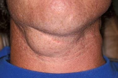
Example of a cervical ranula with no oral involvement in an adult. The swelling developed after a car accident in which the individual had trauma to the face and neck.
Click to see larger pictureClick to see detailView Full Size Image
Media type:  Photo

Media file 9:  Low-power photomicrograph of a mucocele with attenuation of the mucosal surface and pooling of mucus (hematoxylin-eosin, original magnification X40).
Click to see larger pictureClick to see detailView Full Size Image
Media type:  Image

Media file 10:  High-power photomicrograph of a mucocele with pooling of mucus and numerous foamy histiocytes (hematoxylin-eosin, original magnification X400).
Click to see larger pictureClick to see detailView Full Size Image
Media type:  Image

Media file 11:  Intermediate-power photomicrograph of an affected minor salivary gland lobule with atrophy of the acinar structures, ductal ectasia, and fibrosis (hematoxylin-eosin, original magnification X100).
Click to see larger pictureClick to see detailView Full Size Image
Media type:  Image


  • Ali MK, Chiancone G, Knox GW. Squamous cell carcinoma arising in a plunging ranula. J Oral Maxillofac Surg. Mar 1990;48(3):305-8. [Medline].
  • Anastassov GE, Haiavy J, Solodnik P, Lee H, Lumerman H. Submandibular gland mucocele: diagnosis and management. Oral Surg Oral Med Oral Pathol Oral Radiol Endod. Feb 2000;89(2):159-63. [Medline].
  • Axell T. A prevalence study of oral mucosal lesions in an adult Swedish population. Odontol Revy. 1976;27(36):1-103. [Medline].
  • Azuma M, Tamatani T, Fukui K, Yuki T, Hoque MO, Yoshida H, et al. Proteolytic enzymes in salivary extravasation mucoceles. J Oral Pathol Med. Aug 1995;24(7):299-302. [Medline].
  • Balakrishnan A, Ford GR, Bailey CM. Plunging ranula following bilateral submandibular duct transposition. J Laryngol Otol. Aug 1991;105(8):667-9. [Medline].
  • Baurmash HD. Marsupialization for treatment of oral ranula: a second look at the procedure. J Oral Maxillofac Surg. Dec 1992;50(12):1274-9. [Medline].
  • Baurmash HD. Mucoceles and ranulas. J Oral Maxillofac Surg. Mar 2003;61(3):369-78. [Medline].
  • Bermejo A, Aguirre JM, Lopez P, Saez MR. Superficial mucocele: report of 4 cases. Oral Surg Oral Med Oral Pathol Oral Radiol Endod. Oct 1999;88(4):469-72. [Medline].
  • Bessa CFN, Santos PJB, Aguiar MCF, do Carmo MAV. Prevalence of oral mucosal alterations in children from 0-12 years old. J Oral Pathol Med. 2004;33:17-22. [Medline].
  • Bouquot JE. Common oral lesions found during a mass screening examination. J Am Dent Assoc. Jan 1986;112(1):50-7. [Medline].
  • Chidzonga MM, Rusakaniko S. Ranula: another HIV/AIDS associated oral lesion in Zimbabwe?. Oral Dis. Jul 2004;10(4):229-32. [Medline].
  • Crean SJ, Connor C. Congenital mucoceles: report of two cases. Int J Paediatr Dent. Dec 1996;6(4):271-5. [Medline].
  • Crysdale WS, Mendelsohn JD, Conley S. Ranulas--mucoceles of the oral cavity: experience in 26 children. Laryngoscope. Mar 1988;98(3):296-8. [Medline].
  • de Visscher JG, van der Wal KG, de Vogel PL. The plunging ranula. Pathogenesis, diagnosis and management. J Craniomaxillofac Surg. May 1989;17(4):182-5. [Medline].
  • Delbem AC, Cunha RF, Vieira AE, Ribeiro LL. Treatment of mucus retention phenomena in children by the micro-marsupialization technique: case reports. Pediatr Dent. Mar-Apr 2000;22(2):155-8. [Medline].
  • Eversole LR. Oral sialocysts. Arch Otolaryngol Head Neck Surg. Jan 1987;113(1):51-6. [Medline].
  • Fukase S, Ohta N, Inamura K, Aoyagi M. Treatment of ranula with intracystic injection of the streptococcal preparation OK-432. Ann Otol Rhinol Laryngol. 2003;112:214-20. [Medline].
  • Garcia-F-Villalta MJ, Pascual-Lopez M, Elices M, Dauden E, Garcia-Diez A, Fraga J. Superficial mucoceles and lichenoid graft versus host disease: report of three cases. Acta Derm Venereol. 2002;82:453-5. [Medline].
  • Gatti AF, Moreti MM, Cardoso SV, Loyola AM. Mucus extravasation phenomenon in newborn babies: report of two cases. Int J Paediatr Dent. Jan 2001;11(1):74-7. [Medline].
  • Harrison HD. Sublingual gland is origin of cervical extravasation mucocele. Oral Surg Oral Med Oral Pathol Oral Radiol Endod. Oct 2000;90(4):404-5. [Medline].
  • Hoque MO, Azuma M, Sato M. Significant correlation between matrix metalloproteinase activity and tumor necrosis factor-alpha in salivary extravasation mucoceles. J Oral Pathol Med. Jan 1998;27(1):30-3. [Medline].
  • Jinbu Y, Kusama M, Itoh H, Matsumoto K, Wang J, Noguchi T. Mucocele of the glands of Blandin-Nuhn: clinical and histopathologic analysis of 26 cases. Oral Surg Oral Med Oral Pathol Oral Radiol Endod. Apr 2003;95(4):467-70. [Medline].
  • Jinbu Y, Tsukinoki K, Kusama M, Watanabe Y. Recurrent multiple superficial mucocele on the palate: Histopathology and laser vaporization. Oral Surg Oral Med Oral Pathol Oral Radiol Endod. Feb 2003;95(2):193-7. [Medline].
  • Macdonald AJ, Salzman KL, Harnsberger HR. Giant ranula of the neck: differentiation from cystic hygroma. AJNR Am J Neuroradiol. Apr 2003;24(4):757-61. [Medline].
  • McCaul JA, Lamey PJ. Multiple oral mucoceles treated with gamma-linolenic acid: report of a case. Br J Oral Maxillofac Surg. Dec 1994;32(6):392-3. [Medline].
  • Mintz S, Barak S, Horowitz I. Carbon dioxide laser excision and vaporization of nonplunging ranulas: a comparison of two treatment protocols. J Oral Maxillofac Surg. Apr 1994;52(4):370-2. [Medline].
  • Morton RP, Bartley JR. Simple sublingual ranulas: pathogenesis and management. J Otolaryngol. Aug 1995;24(4):253-4. [Medline].
  • Neumann RA, Knobler RM. Treatment of oral mucous cysts with an argon laser. Arch Dermatol. Jun 1990;126(6):829-30. [Medline].
  • Oliveira DT, Consolaro A, Freitas FJ. Histopathological spectrum of 112 cases of mucocele. Braz Dent J. 1993;4(1):29-36. [Medline].
  • Osborne TE, Haller JA, Levin LS, Little BJ, King KE. Submandibular cystic hygroma resembling a plunging ranula in a neonate. Review and report of a case. Oral Surg Oral Med Oral Pathol. Jan 1991;71(1):16-20. [Medline].
  • Pang CE, Lee TS, Pang KP, Pang YT. Thoracic ranula: an extremely rare case. J Laryngol Otol. Mar 2005;119(3):233-4. [Medline].
  • Pownell PH, Brown OE, Pransky SM, Manning SC. Congenital abnormalities of the submandibular duct. Int J Pediatr Otorhinolaryngol. Sep 1992;24(2):161-9. [Medline].
  • Praetorius F, Hammarstrom L. A new concept of the pathogenesis of oral mucous cysts based on a study of 200 cases. J Dent Assoc S Afr. May 1992;47(5):226-31. [Medline].
  • Sugerman PB, Savage NW, Young WG. Mucocele of the anterior lingual salivary glands (glands of Blandin and Nuhn): report of 5 cases. Oral Surg Oral Med Oral Pathol Oral Radiol Endod. Oct 2000;90(4):478-82. [Medline].
  • Takimoto T. Radiographic technique for preoperative diagnosis of plunging ranula. J Oral Maxillofac Surg. Jun 1991;49(6):659. [Medline].
  • Tamaki H, Taniguchi S, Kondo S, et al. Blandin-Nuhn cysts: report of six cases. Int J Oral Maxillofac Surg. 1997;26(Suppl No 1):55-6.
  • Woo JS, Hwang SJ, Lee HM. Recurrent plunging ranula treated with OK-432. Eur Arch Otorhinolaryngol. Apr 2003;260(4):226-8. [Medline].
  • Yamasoba T, Tayama N, Syoji M, Fukuta M. Clinicostatistical study of lower lip mucoceles. Head Neck. Jul-Aug 1990;12(4):316-20. [Medline].
  • Yasumoto M, Nakagawa T, Shibuya H, Suzuki S, Satoh T. Ultrasonography of the sublingual space. J Ultrasound Med. Dec 1993;12(12):723-9. [Medline].
  • Yoshimura Y, Obara S, Kondoh T, Naitoh S. A comparison of three methods used for treatment of ranula. J Oral Maxillofac Surg. Mar 1995;53(3):280-2; discussion 283. [Medline].
  • Zhao YF, Jia J, Jia Y. Complications associated with surgical management of ranulas. J Oral Maxillofac Surg. Jan 2005;63(1):51-4. [Medline].
  • Zhao YF, Jia Y, Chen XM, Zhang WF. Clinical review of 580 ranulas. Oral Surg Oral Med Oral Pathol Oral Radiol Endod. 2004;98:281-7. [Medline].

Mucocele and Ranula excerpt

Article Last Updated: Oct 19, 2006