Practice Essentials
Oral hyperkeratinization is defined as the excessive formation of tenaciously attached keratin in the mouth, which can result from friction. (See the image below.) The diagnosis of oral frictional hyperkeratosisis is typically based on a detailed clinical examination and the finding of an oral habit or some other agent that has produced chronic, low-grade irritation of the mucosa. Treatment consists of removal of the frictional irritant.
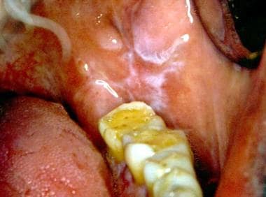 The white line observed on the cheek is level with the biting plane of the teeth. The wear on the occlusal surfaces of the molar teeth suggests that the patient had a habit of bruxism. Courtesy of Catherine M. Flaitz, DDS and Alfredo Aguirre, DDS.
Signs and symptoms
Most patients with frictional keratosis have no symptoms, with the exception of those with aggressive cheek and lip biting habits. In some individuals who repeatedly traumatize the tissues, tenderness, swelling, and a burning sensation may be present.
Typically, the lesions of frictional keratosis appear as distinct, focal, and translucent-to-opaque white asymptomatic patches with sharply delineated borders. One of the more common presentations is the linea alba (white line).
See Presentation for more detail.
Diagnosis
The first step in the identification of white patches suspected of being associated with physical trauma is to use a 2 × 2-inch sterile gauze to wipe off the lesion or lesions. If the patch is not easily wiped off, this finding suggests hyperkeratinization. If any doubt exists about a particular lesion or if residual keratotic foci persist despite the removal of the causative factor, then a biopsy is indicated.
See Workup for more detail.
Treatment
Any frictional irritant should be removed. Biting, sucking, or chewing habits should be discontinued, and fractured or rough tooth surfaces or irregularly fitting dentures or other appliances should be corrected.
See Treatment for more detail.
Background
The oral mucosa is lined by stratified squamous epithelium and has topographic differences that correlate with physical demands or a higher degree of specialization. For example, the epithelium lining the floor of the mouth, the ventral side of the tongue, the buccal mucosa, and the soft palate is nonkeratinized; however, the epithelium associated with the gingiva and hard palate is usually keratinized. The dorsal surface of the tongue is also keratinized, but it is referred to as specialized mucosa because of the presence of papillae. The dorsum of the tongue, the hard palate, and the gingival tissues are keratinized to better respond to masticatory demands.
Hyperkeratinization (excessive formation of tenaciously attached keratin) may be present in a variety of clinical conditions, including genetic, physiologic, inflammatory, immunologic, premalignant, and malignant conditions. The change may result from a local insult, including chemical, thermal, or physical irritants. This article focuses on the oral hyperkeratinization that results from friction. Friction (the constant rubbing of 2 surfaces against one another) in the oral cavity may result in the development of clinically observable white patches.
Various names have been used to describe particular examples of frictional keratosis (FK). These include frictional keratosis arising from excessive force while brushing the teeth (toothbrush keratosis); the constant rubbing of the tongue against the teeth (tongue thrust keratosis); the constant sucking, pressure, and irritation of the teeth against the buccal mucosa along the plane of occlusion (linea alba); and the habit of chronic cheek, tongue, or lip biting (cheek- or lip-bite keratosis). [1] Injuries to the oral mucosa, using items such as a pen, toothpicks, or fingernails, may result in frictional keratosis.
Pathophysiology
The white patches of frictional keratosis that develop in the oral cavity represent a chronic, low-grade, mechanical process that is analogous to the formation of a callus on the skin. The most common local factors involved in this process are tissue chewing (mainly on the buccal mucosa or lips), ill-fitting or irregularly surfaced removable dental prostheses (dentures), fractured or malposed teeth, poorly adapted dental restorations, orthodontic appliances, improper toothbrushing, and constant mastication on edentulous alveolar ridges. The constant irritation stimulates the production of excessive keratin, with a subsequent change in the thickness and the color of the involved mucosa. [2]
Etiology
In most patients with frictional keratosis, the cause is easily identified. An oral habit of cheek biting, cheek chewing, tongue thrusting, or mucosal sucking can often be identified as the cause if the site of the lesion is carefully examined in relationship to the occlusal plane.
An ill-fitting, rough, or broken removable dental prosthesis or orthodontic appliance or a fractured or irregular tooth surface frequently affects the adjacent soft tissues. Occasionally, a frictional keratosis lesion may develop as a result of the constant rubbing of an external object, such as a tobacco pipe; a musical instrument; or, perhaps, a worker's tool, which, for convenience, is held in the mouth for long periods. Another cause may be manipulation of the tissues with long fingernails, which may shred the mucosa.
Improper toothbrushing and other oral hygiene aids affect the attached gingival tissues.
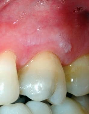 Oral frictional hyperkeratosis of the attached maxillary gingiva from inappropriate toothbrushing technique. Courtesy of Catherine M. Flaitz, DDS and Alfredo Aguirre, DDS.
Irritation from masticatory function may cause frictional keratosis when the alveolar mucosa and retromolar pad bear the stresses of eating. When lesions occur at these sites, they are referred to as alveolar ridge calluses. [3]
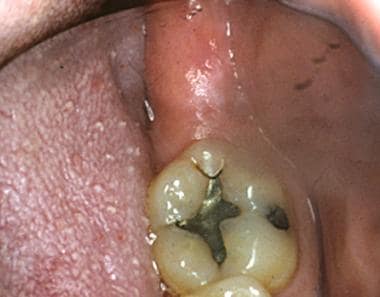 Oral frictional hyperkeratosis of the retromolar pad is also referred to as a ridge callus. This lesion is caused by masticatory irritation. Courtesy of Catherine M. Flaitz, DDS and Alfredo Aguirre, DDS.
Pregnancy may significantly increase the risk for cheek biting. [4] In rare cases, the overuse of topical anesthetics, overuse of antiseptic mouthrinses, or oromucosal delivery of medications (eg, cannabis) causes keratosis from chemical irritation. [5] The identification of such habits depends on obtaining a thorough history.
Epidemiology
United States data
Few large epidemiologic studies documenting the prevalence of various oral lesions, including oral frictional keratosis, have been published.
The most comprehensive survey on the prevalence of oral mucosal lesions is the Third National Health and Nutrition Examination Survey (NHANES III). Oral examinations were performed on 17,235 noninstitutionalized civilian adults. Cheek and lip biting had a point prevalence of 3.05% and ranked third in oral lesion prevalence, while frictional keratosis had a point prevalence of 2.67% and ranked fourth. [6]
In the same national survey, when 10,030 children aged 2-17 years were evaluated, the point prevalence for cheek and lip biting was 1.89% and 0.26% for frictional keratosis. [7] The reported prevalence of frictional keratosis in children and adolescents has ranged from 0.26% to 5.3% in various published series. [8]
In another extensive survey of 23,616 white American adults from Minnesota that evaluated a wide range of oral lesions, the number of cases of cheek-biting keratosis was 1.2 cases per 1000 individuals. [9] In this same study, frictional keratosis was not differentiated from leukoplakic lesions, so the prevalence of frictional keratosis alone cannot be determined.
Linea alba is a common mucosal variation that is rarely singled out as a specific entity in prevalence studies. In a limited study of young men, 13% had this mucosal alteration. [10]
International data
In a Danish study of 20,333 people aged 15 years and older, the prevalences of cheek and lip biting and frictional keratosis were slightly higher than those reported in the US studies. [11] The prevalence for cheek and lip biting was 5.1%, and the prevalence for frictional keratosis was 5.5%. Similarly, the prevalence for frictional keratosis from a small study sample [12] of Kenyan adults was 5.5%. In Slovenia, the prevalence was 2.7% for cheek and lip biting and 2.2% for frictional keratosis. [13] In a study of Turkish adolescents, linea alba was the second most common lesion, with a prevalence of 5.3%. [14]
When studies were limited to individuals seeking care in oral medicine clinics, a wider frequency of occurrence was noted. In a limited study of patients treated at a dental school in Spain, the rate was 11.5% for frictional keratosis, 10.7% for linea alba, and 6.8% for cheek biting. [15] In an India dental school study, frictional keratosis was the most common oral lesion detected, occurring in 5.8% of the patients. [16] When referred hospitalized and clinic patients were evaluated in an Australian oral medicine clinic, hyperkeratotic lesions, including tobacco-induced lesions, were documented in 11.6% of the hospitalized patients and 10.3% of the clinic patients. [17]
The largest study of 23,785 patients, attending a Mexican dental school clinic, found frictional keratosis to be the third most common oral mucosal finding, with a prevalence rate of 32 cases per 1000 patients, while cheek-biting lesions were ranked fifth, or 21.7 cases per 1000 patients. [18]
Race-, sex-, and age-related demographics
No racial predilection seems apparent for oral frictional keratosis.
In general, frictional keratosis has no known sex predilection, except for cheek biting and lip biting, which are twice as prevalent in women compared with men. [1]
Oral frictional keratosis affects persons from a wide range of ages, and contributing factors determine which age group is more commonly affected. In general, oral frictional keratosis lesions are more common in adults.
Prognosis
The prognosis for frictional keratosis is excellent. Resolution is usually accomplished when the frictional element is eliminated. Most lesions resolve in 1-3 weeks following the removal of the causative factor.
Morbidity/mortality
Frictional keratosis and its variants do not cause symptoms and are benign mucosal lesions that remain localized with no associated mortality or morbidity.
Complications
No significant complications are associated with frictional keratosis. This reactive lesion has no propensity for malignant transformation.
The risk of mutilating the buccal and labial mucosa following local anesthesia for dental treatment is increased, especially in children with lesions due to cheek biting. Both the child and the parent should be warned of the potential complication of unconsciously traumatizing the tissues because of this chronic habit. [19]
Patient Education
Encourage patients to stop any habit that may be implicated with this lesion. If the putative traumatic factors are eliminated and no resolution of the lesions ensues, advise the patient that a biopsy is indicated.
-
The white line observed on the cheek is level with the biting plane of the teeth. The wear on the occlusal surfaces of the molar teeth suggests that the patient had a habit of bruxism. Courtesy of Catherine M. Flaitz, DDS and Alfredo Aguirre, DDS.
-
Prominent linea alba with evidence of cheek biting. The white line shows a slightly scalloped appearance, which correlates with the buccal surfaces of the teeth against which the mucosa is rubbed. Courtesy of Catherine M. Flaitz, DDS and Alfredo Aguirre, DDS.
-
This wider area of roughened mucosa is typical of those produced by the habit of cheek biting or nibbling. Courtesy of Catherine M. Flaitz, DDS and Alfredo Aguirre, DDS.
-
This frictional keratotic line shows a roughened surface. A thicker patch of mucosa is at the anterior end (under the tongue blade edge). This area is exactly level with the occlusal plane and was being chewed constantly by the patient. Courtesy of Catherine M. Flaitz, DDS and Alfredo Aguirre, DDS.
-
Anterior rough surface area at the occlusal plane of the teeth. Courtesy of Catherine M. Flaitz, DDS and Alfredo Aguirre, DDS.
-
Oral frictional hyperkeratosis of the lateral border of the tongue from chronic biting habit. Courtesy of Catherine M. Flaitz, DDS and Alfredo Aguirre, DDS.
-
Oral frictional hyperkeratosis of the attached maxillary gingiva from inappropriate toothbrushing technique. Courtesy of Catherine M. Flaitz, DDS and Alfredo Aguirre, DDS.
-
Oral frictional hyperkeratosis of the retromolar pad is also referred to as a ridge callus. This lesion is caused by masticatory irritation. Courtesy of Catherine M. Flaitz, DDS and Alfredo Aguirre, DDS.
-
Low-power view of stratified squamous epithelium with marked hyperkeratinization, acanthosis, and a prominent granular cell layer. Courtesy of Catherine M. Flaitz, DDS and Alfredo Aguirre, DDS.
-
High-power view of the surface keratin layer and a prominent granular cell layer. Courtesy of Catherine M. Flaitz, DDS and Alfredo Aguirre, DDS.


