Continually Updated Clinical Reference
 
 
  All Sources     eMedicine     Medscape     Drug Reference     MEDLINE
 
eMedicine - Onchocerciasis (River Blindness) : Article by

Quick Find
Authors & Editors
Introduction
Clinical
Differentials
Workup
Treatment
Medication
Follow-up
Miscellaneous
Multimedia
References

Related Articles
Id Reaction (Autoeczematization)

Insect Bites

Leprosy

Lichen Planus

Lichen Sclerosus et Atrophicus

Morphea

Pityriasis Lichenoides

Pityriasis Rosea

Scabies

Syphilis

Tinea Capitis

Tinea Corporis

Tinea Cruris

Vitiligo

Yaws


Patient Education
Click here for patient education.


Author: Jason F Okulicz, MD, Assistant Professor of Medicine, Uniformed Services University of the Health Sciences; Fellow, Department of Infectious Disease, Wilford Hall United States Air Force Medical Center

Jason F Okulicz is a member of the following medical societies: Alpha Omega Alpha, American College of Physicians-American Society of Internal Medicine, Sigma Xi, and Southern Medical Association

Coauthor(s): Dirk M Elston, MD, Director, Department of Dermatology, Geisinger Medical Center; Robert A Schwartz, MD, MPH, Professor and Head of Dermatology, Professor of Medicine, Professor of Pediatrics, Professor of Pathology, Professor of Preventive Medicine and Community Health, UMDNJ-New Jersey Medical School

Editors: Donald Belsito, MD, Clinical Professor, Department of Internal Medicine, Division of Dermatology, University of Missouri at Kansas City; Private Practice, American Dermatology Associates, LLC; David F Butler, MD, Professor of Dermatology, Texas A&M University College of Medicine; Director, Division of Dermatology, Scott and White Clinic; Director Dermatology Residency Training Program, Scott and White Clinic; Jeffrey Meffert, MD, Assistant Clinical Professor of Dermatology, University of Texas Health Science Center-San Antonio; Catherine Quirk, MD, Clinical Assistant Professor, Department of Dermatology, Brown University; William D James, MD, Paul R Gross Professor of Dermatology, University of Pennsylvania School of Medicine; Vice-Chair, Program Director, Department of Dermatology, University of Pennsylvania Health System

Author and Editor Disclosure

Synonyms and related keywords: African river blindness, blinding filariasis, Robles disease, craw-craw, Onchocerca volvulus, O volvulus, Simulium species, Simulium damnosum, Simulium neavei, Simulium metallicum, Simulium ochraceum, Simulium exiguum, black fly, microfilariae, onchocercal dermatitis, hanging groin, leopard skin, sowda, onchocercal dermatitis, acute papular onchodermatitis, APOD, chronic papular onchodermatitis, CPOD, lichenified onchodermatitis, LOD,

Background

Onchocerciasis is a common, chronic, multisystemic disease caused by the nematode Onchocerca volvulus. The disease characteristically includes dermatologic, lymphatic, ophthalmologic, and systemic manifestations. Human transmission of the disease is caused by a bite from the intermediate host, the black fly (genus Simulium). Black flies breed along waterways, which can vary from small streams to broad rivers. Affected individuals usually live or work within a few kilometers of these sites. Onchocerciasis has long been associated with a high incidence of detrimental effects on socioeconomic development and public health in endemic areas.

Pathophysiology

In 1875, John O'Neill first observed O volvulus microfilariae in a case of "craw-craw," as onchocerciasis is known in West Africa. Almost 50 years later, Blacklock discovered the vector to be Simulium in Sierra Leone. The main vector in most of Africa is Simulium damnosum; in Ethiopia, Uganda, Tanzania, and the Democratic Republic of the Congo, Simulium neavei is common. In the Americas, the principal vectors are Simulium metallicum, Simulium ochraceum, and Simulium exiguum. Some vectors bite humans rather exclusively, whereas others are zoophilic to varying degrees. Animal reservoirs of O volvulus have not been found.

When a black fly takes a blood meal from an infected human, they also ingest onchocercal microfilariae in the skin. Surviving microfilariae in the black fly burst through the peritrophic membrane formed by the blood meal, invade the midgut, and advance to the thoracic muscles. The differentiation of these microfilariae into L1 larva begins in muscle within 28 hours after the blood meal. The first molt produces L2 larva within 96 hours, followed by the second molt, which produces L3 larva by day 7. The infective L3 larva migrates to the insect's head and mouth for future deposition into human skin during the next blood meal.

After L3 larvae are transmitted to human skin, those that survive molt within 1 week to form L4 larva. Their development into male and female forms is completed by 1-3 months. The adult worms reside in the deep dermis and fascial planes. Thick, fibrous, subcutaneous nodules called onchocercomas are formed as the result of the development of scar tissue around the adult worms. These onchocercomas average 3 cm in diameter, and they typically contain 2-3 female adults and 1-2 male adults. The nodules typically are surrounded by eosinophils and lymphocytes that are ready to attack the newly produced microfilariae.

Adult worms isolated in nodules are not directly harmful to the patient. Their progeny, which are released from the nodules, are responsible for most of the damage related to onchocerciasis. Nonetheless, to control the disease, the identification and removal of these onchocercomas is critical. Ivermectin is the most effective agent against microfilariae (see Medical Care). Currently, available agents that are effective against adult worms are toxic. For this reason, surgical removal of the adult worms is important.

Within 10-12 months after the initial infection, adult female worms start producing microfilariae, which have an average lifespan of 6 months to 2 years. The reproductive life of the adult averages 9-11 years. During this time, female worms may release 1300-1900 microfilariae per day. The maximal production of offspring occurs during the first 5 years of the worm's reproductive life, after which this activity declines in a linear fashion.

The microfilariae released from the nodules easily traverse the skin and connective tissue. The subepidermal lymphatics and the anterior chamber of the eye are the most common migration sites. The microfilariae can also be found in the blood, cerebrospinal fluid, urine, and internal organs. More than 100 million microfilariae may be present in severely affected individuals.

A symbiotic relationship has been demonstrated between Wolbachia bacteria and filarial nematodes, including O volvulus. Wolbachia species are essential for nematode fertility. Embryogenesis in the female worm is disrupted when Wolbachia numbers are depleted. In addition, murine experiments suggest that corneal inflammation secondary to onchocerciasis may be caused by endotoxins produced by Wolbachia. Results from small trials suggest that clearance of Wolbachia with antibiotic therapy may affect transmission and may reduce and prevent onchocerciasis-related blindness.

Frequency

United States

Onchocerciasis is rare in the United States. All reported cases result from the immigration of individuals from endemic areas.

International

Onchocerciasis is a major public health problem in many parts of the world. The disease is endemic in 37 countries in Africa, Latin America, and Yemen. In 1995, an estimated 123 million people were at risk of contracting the disease according to the World Health Organization Expert Committee on Onchocerciasis. Another 17-18 million people were estimated to be infected.

Approximately 95% of all infected people live in Africa. The disease is most severe along the major rivers in 30 countries across the northern and central areas of the continent. Nigeria, Ethiopia, Cameroon, Uganda, and the Democratic Republic of the Congo have the largest number of infected people.

In Latin America, onchocerciasis can be found in Brazil, Venezuela, Colombia, Ecuador, and Guatemala, as well as in the southern mountainous states of Chiapas and Oaxaca in Mexico.

Mortality/Morbidity

  • In 1995, an estimated 270,000 people were blinded and another 500,000 had severe visual impairment as a result of the disease.
  • A multicountry study showed that more than 30% of the population in endemic areas have onchocercal dermatitis.
  • In a survey of skin disease in 7 endemic sites in 5 African countries, 40-50% of adults reported troublesome itching.Blindness is not associated with excess mortality. However, increasing microfilarial load is associated with mortality in both males and females.

Race

To the author's knowledge, no current well-described studies have been performed to determine the frequency of onchocerciasis in specific races.

Sex

  • The disease generally affects more men than women, although sex-related differences may not be apparent until the patient reaches a certain age.
  • Sex-related differences are more pronounced in high-transmission areas, particularly the savanna.
  • The overall trend is partially attributed to increased exposures in men, which are related to the occupational risk in farmers, fishermen, and other workers.

Age

The prevalence of onchocerciasis is lowest in individuals aged 0-10 years. Afterward, the prevalence sharply increases, with a peak in individuals aged 20-30 years.


History

The most important task is determining if patients in endemic areas have been exposed to O volvulus via the black fly vector.

  • Obtain a detailed travel history if onchocerciasis is suspected. Travelers to endemic areas may have a particularly severe form of dermatitis.
  • Other manifestations of onchocerciasis include weight loss, musculoskeletal pain, inguinal hernias, and systemic embolization of microfilariae.
  • Many patients in endemic regions have associated the disease with secondary amenorrhea, lactation difficulties, spontaneous abortion, infertility, and sterility. However, these associations have never been proven.
  • Patients are asymptomatic in about 10% of cases.

Physical

Localized inflammatory responses to dead or dying microfilariae are almost entirely responsible for the clinical manifestations of the disease. In a severely infected person, 100,000 or more microfilariae die each day. The earliest symptoms are fever, arthralgia, and transient urticaria involving the trunk and face. Onchocercomas, the most characteristic skin lesions, are often present.

  • The following is a summary of the clinical classification system for onchocercal dermatitis, which Murdoch et al developed to standardize and facilitate the collection of data worldwide:
    • Acute papular onchodermatitis (APOD) is characterized by a solid, scattered, pruritic papular rash. The diameter of the papules is at least 1 mm. Vesicles or pustules at the apex may or may not be present. The obliteration of the skin creases due to edema also may or may not be present.
    • Chronic papular onchodermatitis (CPOD) involves a scattered, pruritic, hyperpigmented, and flat-topped papulomacular rash. The diameter of the papules is at least 3 mm, with or without excoriations.
    • Lichenified onchodermatitis (LOD) is characterized by raised, discrete, pruritic, and hyperpigmented papulonodular plaques associated with lymphadenopathy. The lesions may be confluent, with or without the presence of excoriations. APOD or CPOD may also be present.
    • Atrophy (ATR) involves wrinkled and dry skin. Firmly pressing the edge of a finger along the skin reveals additional fine wrinkles. In patients younger than 50 years, ATR is scored as a significant abnormality.
    • Depigmentation is characterized by areas of incomplete pigment loss, with associated islands or spots of normal pigment surrounding hair follicles. Leopard skin is similar, except that it is characterized by a complete loss of pigment, with islands or spots of normally pigmented skin around the follicles.
    • Palpable onchocercal nodule is a term used to describe onchocercomas that are palpable at bony prominences.
    • Lymphadenopathy is characterized by lymph nodes 1 cm or larger in diameter. They may or may not be tender.
    • Hanging groin (HG) involves the folds of inelastic, atrophic skin in the inguinal areas. The condition may be unilateral or bilateral, and it may involve enlarged lymph nodes.
    • Lymphedema (LYM) is characterized by edema of a limb or external genitalia.
  • Onchocercomas are fibrous, subcutaneous nodules containing adult worms. These nodules are generally located over bony prominences, and they are easily palpable. Deep nodules, commonly located around the pelvis, can be more difficult to detect. The number of palpable nodules is not correlated with the microfilarial load or the severity of disease. Identifying every nodule is important for proper management.
    • In Africa, the nodules are often observed along the iliac crests, ribs, greater trochanters, and ischial tuberosities. Juxta-articular areas, such as the knees, elbows, patella, and scalp, may also have nodules.
    • In the American forms, nodules are fewer and have a greater tendency to be located on the scalp. In patients with scalp nodules, the risk of ocular complications is generally higher than that of patients without scalp nodules.
    • Onchocercomas are less common in the Yemen form of the disease than in other forms.
  • Onchocercal dermatitis is the most common symptom of the disease.
    • Its initial manifestations, which can occur anywhere on the body, include itching, scratching, and alterations in skin pigmentation.
    • Pruritus may be intermittent and mild, continuous and severe, or absent.
    • A maculopapular rash may appear anywhere on the body at any time.
    • The papules may be small and densely packed or large and separated.
    • The maculopapular rash is often associated with severe pruritus. Excess scratching may lead to bleeding, ulceration, and secondary infection (a condition West Africans call craw-craw).
  • Sowda, a severe form of dermatitis first described in Yemen, is associated with an active delayed hypersensitivity response.
    • Many patients are travelers or temporary workers in nonendemic areas. These patients have dark, thickened, intensely pruritic skin with papules. The regional lymph nodes are soft, nontender, and enlarged.
    • Sowda is usually localized to a single lower extremity.
    • A less common, more generalized form can involve both lower extremities or other parts of the body. Patients have either focal swelling or a more diffuse LYM. Skin-snips do not usually contain microfilariae.
    • Sowda may also be found in patients in West Africa, Ethiopia, Sudan, Cameroon, Venezuela, and Ecuador.
  • Leopard skin is a characteristic finding in older patients.
    • Leopard skin involves depigmentation of the pretibial areas of the lower extremities. This pattern is initially seen as discrete depigmented macules, with sparing of the hair follicles.
    • Later, the macules may become confluent, involving a large area of the anterior portion below the knee. This pattern can sometimes be seen in the groin or lower abdomen as well.
  • HG or adenolymphocele is a severe degenerative condition in older individuals.
    • The inguinal and femoral lymph nodes become progressively enlarged and fibrotic, leading to lymphatic obstruction.
    • Concomitantly, progressive destruction of elastic fibers leaves the skin thinned and wrinkled.
    • The atrophied skin tends to hang in apronlike folds under the weight of the accumulating lymphedematous tissue.
    • This condition is more common in men than in women.
  • Ocular manifestations of onchocerciasis are late, serious reactions that occur in about 5% of affected persons.
    • Infection of the cornea produces punctate keratitis in the areas around dead microfilariae. This condition eventually clears when the inflammation settles.
    • Severe and prolonged infection over a number of years is likely to produce sclerosing keratitis, iridocyclitis, and uveitis. Permanent visual impairment, secondary glaucoma, or blindness is often the result.
    • Posterior segment changes may coexist with anterior segment lesions. The changes are caused by inflammation around microfilariae that invade the retina via the posterior ciliary vessels.
    • Choroidoretinal lesions are common around the optic disk or on the outer portion of the macula.
    • Active optic neuritis is a major cause of blindness in many areas with endemic disease.

Causes

In 1875, John O'Neill first observed O volvulus microfilariae in a case of craw-craw, as onchocerciasis is known in West Africa.

  • Almost 50 years later, Blacklock discovered that the vector in Sierra Leone was a Simulium organism. The main vector in most of Africa is S damnosum; in Ethiopia, Uganda, Tanzania, and the Democratic Republic of the Congo, S neavei is common.
  • The principal vectors in the Americas are S metallicum, S ochraceum, and S exiguum.
  • Some vectors bite humans rather exclusively, whereas others are zoophilic to varying degrees. Animal reservoirs of O volvulus have not been found.


Id Reaction (Autoeczematization)
Insect Bites
Leprosy
Lichen Planus
Lichen Sclerosus et Atrophicus
Morphea
Pityriasis Lichenoides
Pityriasis Rosea
Scabies
Syphilis
Tinea Capitis
Tinea Corporis
Tinea Cruris
Vitiligo
Yaws

Other Problems to be Considered

Leukoderma
Chronic dermatitis
Scleroderma


Lab Studies

  • The traditional standard diagnosis of onchocerciasis is based on the acquisition of 3- to 5-mg skin snips from an affected area.
    • These snips are immediately immersed in sodium chloride solution and placed under a microscope. The emerging microfilariae are then counted.
    • This method is specific, and it has been the most accurate. However, the use of skin snips is not sensitive for detecting early or mild infections, and this method is becoming increasingly unacceptable to people in endemic communities because of its invasiveness.
  • The Mazzotti test, now seldom used, involves the administration of 6 mg of diethylcarbamazine (DEC).
    • DEC inhibits neuromuscular transmission in nematodes.
    • Within 2 hours, a positive result produces pruritus and, sometimes, intense inflammation in the areas of dying microfilariae.
    • Other possible effects such as vomiting, conjunctivitis, albuminuria, hypotension, and sudden death (rare) limit its usefulness.
    • Oral DEC was formerly used in treatment of the disease.
  • The DEC patch test (ie, Mazzotti patch test) involves a topical application of DEC, which produces a local reaction to dying microfilariae at the patch site.
    • This noninvasive test is specific, but it is less sensitive than the skin-snip test.
    • In the future, this test may be more valuable in detecting the recrudescence of infection in onchocerciasis-free zones than in diagnosing the disease.
  • Enzyme-linked immunosorbent assays (ELISAs), which require only a finger-stick sample, are more sensitive and less invasive than skin-snip tests.
    • This test is used to recognize specific microfilarial antigens.
    • However, ELISA results cannot be used to distinguish past and current infections, a considerable problem in endemic areas.
  • The polymerase chain reaction (PCR) amplifies repetitive parasite DNA sequences in skin-snip specimens.
    • Compared with skin-snip tests, this method has greater sensitivity in patients with low-level infections. The return of microfilariae to the skin after drug treatment can also be identified earlier with this test.
    • The major disadvantages of PCR are its high cost and invasiveness.
    • A minimally invasive and painless alternative to the use of skin snips is the collection of skin scratches. The superficial layer of the epidermis is removed by carefully scraping the skin with the blunt edge of a disposable lancet.
    • PCR studies of skin scratches, instead of skin snips, have yielded similar results and may be used more often in the future.
  • Rapid-format antibody card tests are being developed to diagnose onchocerciasis.
    • In these tests, serum samples can be used to detect antibodies, such as immunoglobulin G4 (IgG4) antibodies to recombinant O volvulus antigen Ov16. Tests for additional antibodies are currently under development.
    • Initial studies show good sensitivity and specificity in small numbers of samples and controls using various methods.
    • Some tests can be modified to permit the use of blood samples obtained from less invasive finger sticks. This modification increases the utility of the test for field use.
    • Additional studies are needed to determine the value of these tests in the diagnosis of onchocerciasis.
  • Microfilariae can often be directly observed by means of slit lamp examination of the cornea and anterior chamber of the eye.
  • Recently, an oncho-C27 antigen detection dipstick assay using urine and tears has been developed. The test can be completed in as short as 3 hours, and the strips maintain reactivity when kept at room temperature for up to 8 months. Sensitivity and specificity were good in an initial study.

Histologic Findings

In early untreated cases, tissue biopsy samples may show a mild chronic inflammatory infiltration; eosinophils, lymphocytes, and histiocytes may surround the microfilariae. Microfilariae are often present without a surrounding cellular reaction. Later cases show hyperkeratosis, parakeratosis, tortuous dermal vessels, dilated lymphatics, and pigment incontinence. An increased number of dermal fibroblasts leads to perivascular fibrosis. In more advanced cases, hyalinized scar tissue replaces the collagen and elastic fibers in the dermis.

The microfilariae have pointed tails, elongated posterior nuclei, paired anterior nuclei, and large spaces between the tip or tail and the first nuclei. They lack sheaths.

Onchocercomas are made up of an outer vascular fibrous stroma embedded with groups of perivascular leukocytes. The inner layer is composed of hyaline connective tissue intermingled with coiled adult worms. A dense cellular infiltrate composed of eosinophils, lymphocytes, macrophages, and giant cells surrounds the worm. The coiled appearance of the worm, the presence of microfilaria in gravid females, and the presence of a gut help in identifying the worm.


Medical Care

The treatment of onchocerciasis was revolutionized with the introduction of ivermectin in 1987. Ivermectin therapy does not have the adverse reactions of DEC, and it eliminates the need for 6 weekly injections of suramin. The treatment is suitable for both clinical use and mass distribution in endemic areas.

  • Ivermectin is a compound derived from the bacterium Streptomyces avermitilis. The drug causes nematode paralysis by impairing neuromuscular function.
  • Ivermectin not only prevents ocular disease but also improves and eliminates the skin disease. A single dose of 150 mcg/kg clears the microfilariae from the skin for several months.
  • Ivermectin temporarily decreases the release of microfilariae, but it does not kill adult worms.
  • Adverse reactions are similar to the responses of the body to dying microfilariae, but the intensity and rate of development are increased. Adverse effects include fever, edema, pruritus, lymphadenitis, and body pains.
  • The frequency and duration of ivermectin therapy still is being debated. As many as 33% of patients in nonendemic areas are cured with only 1 dose of ivermectin, but most patients require additional therapy.
  • Small studies have shown that administration of doxycycline for 6 weeks in addition to ivermectin therapy led to Wolbachia depletion followed by interruption of embryogenesis and reduction in microfilarial loads lasting 18 months.
  • Suramin is used less often than ivermectin and given intravenously.

Surgical Care

Nodulectomy has been a traditional form of therapy in Mexico and Guatemala.

  • Moving from village to village, healthcare workers remove nodules from patients, especially nodules in the head.
  • This surgical approach may reduce the number of microfilariae that enter the eye, but no strong evidence supports its effectiveness in preventing blindness.
  • The removal of nodules may be a valuable adjunct in patients treated with ivermectin.


Ivermectin is the drug of choice in the treatment of onchocerciasis. The addition of 6 weeks of doxycycline to ivermectin has been shown to be effective in reducing the microfilarial load, which may affect transmission and may prevent or reduce onchocerciasis-related blindness. Suramin may be indicated for use only if ivermectin cannot adequately control the disease. Amocarzine has not been shown to be effective in treating onchocerciasis. Both suramin and amocarzine are capable of destroying adult worms. DEC therapy is no longer recommended.

Drug Category: Anthelmintics

Biochemical pathways in parasites are sufficiently different from those in the human host to allow selective interference by using chemotherapeutic agents in relatively small doses.

Drug NameIvermectin (Mectizan, Stromectol)
DescriptionOral, semisynthetic, broad-spectrum anthelmintic agent isolated from S avermitilis. Binds selectively with glutamate-gated chloride-ion channels in invertebrate nerve and muscle cells, causing cell death. Half-life is 16 h; metabolized in liver. Usual frequency is q12mo for mass treatment programs, but individual therapy may be given in intervals as short as 3 mo.
Adult Dose15-25 kg: 3 mg PO
26-44 kg: 6 mg PO
45-64 kg: 9 mg PO
65-84 kg: 12 mg PO
>85 kg: 150 mcg/kg PO
Pediatric Dose<15 kg: Not established
>15 kg: Administer as in adults
ContraindicationsDocumented hypersensitivity
InteractionsMay interact with other ligand-gated chloride channels (eg, GABA-gated channels)
PregnancyC - Safety for use during pregnancy has not been established.
PrecautionsPatients with sowda may be more likely to have severe adverse reactions, especially edema and aggravation of onchodermatitis; treat women who intend to breastfeed only when risk of delayed treatment outweighs possible risks to the newborn caused by excretion in milk; adverse reactions include arthralgia and/or synovitis, lymph node enlargement and tenderness, pruritus, edema, fever, and papular, pustular, and frank urticarial rash

Drug NameSuramin (Metaret)
DescriptionPolyanionic compound developed as an antiparasitic drug for treatment of CNS trypanosomiasis. IV antiparasitic agent effective against African trypanosomiasis and onchocerciasis. Most often, a 10% solution is used. Administration of a test dose of 100-200 mg is advised before initiating treatment.
Adult Dose66.7 mg/kg total dose slow IV injection divided in 6 weekly increments
Pediatric Dose<10 years: Not recommended
>10 years: Administer as in adults
ContraindicationsDocumented hypersensitivity; severe liver or renal disease; blindness; mild-to-moderate infections
InteractionsNone reported
PregnancyC - Safety for use during pregnancy has not been established.
PrecautionsImmediate adverse effects include nausea, vomiting, shock, and loss of consciousness; skin reactions and abdominal pain may occur; delayed effects include paresthesia, skin eruptions, fever, elevated LFTs results, photophobia, albuminuria, hematuria, and occasional adrenal insufficiency; monitor CBC; perform coagulation tests; evaluate for neuropathy during therapy

Drug Category: Antibiotics

When used concurrently with ivermectin therapy, tetracycline antibiotic therapy (eg, with doxycycline) depletes Wolbachia endosymbionts and may decrease transmission and reduce or prevent onchocerciasis-related blindness secondary to disruption of female nematode embryogenesis.

Drug NameDoxycycline (Doryx, Vibramycin)
DescriptionBroad-spectrum, synthetically derived bacteriostatic antibiotic in tetracycline class. Almost completely absorbed, concentrates in bile, and is excreted in urine and feces as a biologically active metabolite in high concentrations.
Inhibits protein synthesis and, thus, bacterial growth by binding to 30S and possibly 50S ribosomal subunits of susceptible bacteria. May block dissociation of peptidyl t-RNA from ribosomes, causing RNA-dependent protein synthesis to arrest.
Adult Dose100 mg PO qd/bid for 6 wk in conjunction with ivermectin therapy
Pediatric DoseNot established
ContraindicationsDocumented hypersensitivity; severe hepatic dysfunction
InteractionsBioavailability decreases with antacids containing aluminum, calcium, magnesium, iron, or bismuth subsalicylate; tetracyclines can increase hypoprothrombinemic effects of anticoagulants; tetracyclines can decrease effects of oral contraceptives, causing breakthrough bleeding and increased risk of pregnancy
PregnancyD - Unsafe in pregnancy
PrecautionsPhotosensitivity may occur with prolonged exposure to sunlight or tanning equipment; reduce dose in renal impairment; consider drug serum level determinations in prolonged therapy; tetracycline use during tooth development (last one-half of pregnancy through age 8 y) can cause permanent discoloration of teeth; Fanconilike syndrome may occur with outdated tetracyclines


Further Outpatient Care

  • Considerable debate exists regarding the proper dosing frequency of ivermectin in endemic areas. The drug is given from every 3 months to every year, depending on the degree of symptoms, cost constraints, and patient compliance.
  • In nonendemic areas, a reasonable approach is the administration of a single dose of ivermectin.
    • Depending on the patient's skin symptoms, the dose can be repeated every 3-6 months as needed.
    • Strict follow-up care to determine the need for therapy is important after several doses are administered.
    • The continuation of treatment throughout the entire 12- to 15-year life cycle of the worm has not been proven effective.
    • Nodulectomy remains an important adjunctive treatment.

Deterrence/Prevention

  • The Onchocerciasis Control Programme (OCP) began in 7 West African countries in 1974. The major strategy for interrupting transmission of onchocerciasis was vector control. Hand spraying of black fly breeding sites along rivers, combined with the aerial distribution of larvicide, has been successful in this region. To prevent reinvasion by black flies, parts of 4 other countries were also included in 1986. This program closed in 2002, with all subsequent control efforts transferred to the participating countries.
  • The introduction of ivermectin in 1987 allowed assistance to be extended to other areas. Merck & Co decided to provide the drug, at no cost, in whatever quantities were needed, for as long as it was needed. Community-based distribution programs were established in endemic areas to administer the drug 1-2 times per year, even to remote villages.
  • Encouraged by successes with the OCP and ivermectin, the World Bank launched the African Programme for Onchocerciasis (APOC) in the remaining areas of Africa in 1995. The goal of the program is to eliminate the disease as a public health issue in these areas by 2007. Unlike the OCP, the APOC uses the community-based distribution of ivermectin as its primary control strategy. The Onchocerciasis Elimination Program for the Americas (OEPA), a similar program, also aims to eliminate onchocerciasis by 2007 in the Americas.
  • These programs face many challenges in the future. Whether or not successful control of the disease can be accomplished without the use of vector control has yet to be determined. The organization and effectiveness of community control programs, as well as their funding, may need to be addressed.

Prognosis

  • The prognosis for onchocerciasis is good in patients who receive proper therapy before irreversible eye lesions develop.
  • Ivermectin is effective in reducing the skin manifestations of the disease; it thereby reduces morbidity and improves the patient's quality of life.


Medical/Legal Pitfalls

  • Failure to have a high degree of suspicion for onchocerciasis in patients from endemic areas can be a pitfall. The initial symptoms are generally nonspecific and include fever, arthralgia, pruritus, and transient urticaria.
  • Delayed treatment can result in irreversible ocular disease and unpleasant skin manifestations.


Media file 1:  Leopard-spot pattern of depigmentation on the shins. Courtesy of Brooke Army Medical Center teaching file. All images are in the public domain.
Click to see larger pictureClick to see detailView Full Size Image
Media type:  Photo

Media file 2:  
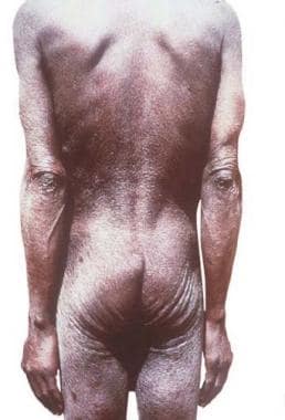
Dermatitis associated with microfilaria. Courtesy of Brooke Army Medical Center teaching file. All images are in the public domain.
Click to see larger pictureClick to see detailView Full Size Image
Media type:  Photo

Media file 3:  Hanging groin sign. Courtesy of Brooke Army Medical Center teaching file. All images are in the public domain.
Click to see larger pictureClick to see detailView Full Size Image
Media type:  Photo

Media file 4:  Hanging groin sign. Courtesy of Brooke Army Medical Center teaching file. All images are in the public domain.
Click to see larger pictureClick to see detailView Full Size Image
Media type:  Photo

Media file 5:  Onchocercoma. Courtesy of Brooke Army Medical Center teaching file. All images are in the public domain.
Click to see larger pictureClick to see detailView Full Size Image
Media type:  Photo

Media file 6:  Onchocercoma. Courtesy of Brooke Army Medical Center teaching file. All images are in the public domain.
Click to see larger pictureClick to see detailView Full Size Image
Media type:  Photo

Media file 7:  Photomicrograph of a skin biopsy specimen from a patient with onchocerciasis. A worm is shown in cross-section. Courtesy of Brooke Army Medical Center teaching file. All images are in the public domain.
Click to see larger pictureClick to see detailView Full Size Image
Media type:  Photo

Media file 8:  Photomicrograph from a gravid female worm (hematoxylin and eosin]). Courtesy of Brooke Army Medical Center. All images are in the public domain.
Click to see larger pictureClick to see detailView Full Size Image
Media type:  Photo


  • Awadzi K, Opoku NO, Attah SK, Addy ET, Duke BO, Nyame PK, et al. The safety and efficacy of amocarzine in African onchocerciasis and the influence of ivermectin on the clinical and parasitological response to treatment. Ann Trop Med Parasitol. Apr 1997;91(3):281-96. [Medline].
  • Ayong LS, Tume CB, Wembe FE, Simo G, Asonganyi T, Lando G, et al. Development and evaluation of an antigen detection dipstick assay for the diagnosis of human onchocerciasis. Trop Med Int Health. Mar 2005;10(3):228-33. [Medline].
  • Bandi C, Trees AJ, Brattig NW. Wolbachia in filarial nematodes: evolutionary aspects and implications for the pathogenesis and treatment of filarial diseases. Vet Parasitol. Jul 12 2001;98(1-3):215-38. [Medline].
  • Baraka OZ, Mahmoud BM, Ali MM, Ali MH, el Sheikh EA, Homeida MM, et al. Ivermectin treatment in severe asymmetric reactive onchodermatitis (sowda) in Sudan. Trans R Soc Trop Med Hyg. May-Jun 1995;89(3):312-5. [Medline].
  • Boatin BA, Toe L, Alley ES, Dembele N, Weiss N, Dadzie KY. Diagnostics in onchocerciasis: future challenges. Ann Trop Med Parasitol. Apr 1998;92 Suppl 1:S41-5. [Medline].
  • Borsboom GJ, Boatin BA, Nagelkerke NJ, Agoua H, Akpoboua KL, Alley EW, et al. Impact of ivermectin on onchocerciasis transmission: assessing the empirical evidence that repeated ivermectin mass treatments may lead to elimination/eradication in West-Africa. Filaria J. Mar 24 2003;2(1):8. [Medline].
  • Burnham G. Onchocerciasis. Lancet. May 2 1998;351(9112):1341-6. [Medline].
  • Burnham G. Ivermectin treatment of onchocercal skin lesions: observations from a placebo-controlled, double-blind trial in Malawi. Am J Trop Med Hyg. Mar 1995;52(3):270-6. [Medline].
  • Connor DH, Williams PH, Helwig EB, Winslow DJ. Dermal changes in onchocerciasis. Arch Pathol. Feb 1969;87(2):193-200. [Medline].
  • Debrah AY, Mand S, Marfo-Debrekyei Y, Larbi J, Adjei O, Hoerauf A. Assessment of microfilarial loads in the skin of onchocerciasis patients after treatment with different regimens of doxycycline plus ivermectin. Filaria J. 2006;5:1. [Medline].
  • Duke BO. Onchocerciasis. Br Med Bull. Jan 1972;28(1):66-71. [Medline].
  • Edgeworth JD, Abiose A, Jones BR. An immunohistochemical analysis of onchocercal nodules: evidence for an interaction between macrophage MRP8/MRP14 and adult Onchocerca volvulus. Clin Exp Immunol. Apr 1993;92(1):84-92. [Medline].
  • Enk CD. Onchocerciasis--river blindness. Clin Dermatol. May-Jun 2006;24(3):176-80.
  • Hagan M. Onchocercal dermatitis: clinical impact. Ann Trop Med Parasitol. Apr 1998;92 Suppl 1:S85-96. [Medline].
  • Hoerauf A, Volkmann L, Hamelmann C, Adjei O, Autenrieth IB, Fleischer B, et al. Endosymbiotic bacteria in worms as targets for a novel chemotherapy in filariasis. Lancet. Apr 8 2000;355(9211):1242-3. [Medline].
  • Hoerauf A, Mand S, Adjei O, Fleischer B, Buttner DW. Depletion of wolbachia endobacteria in Onchocerca volvulus by doxycycline and microfilaridermia after ivermectin treatment. Lancet. May 5 2001;357(9266):1415-6. [Medline].
  • Kale OO. Onchocerciasis: the burden of disease. Ann Trop Med Parasitol. Apr 1998;92 Suppl 1:S101-15. [Medline].
  • Lazarov A, Amihai B, Sion-Vardy N. Pruritus and chronic papular dermatitis in an Ethiopian man. Onchocerciasis (chronic papular onchodermatitis). Arch Dermatol. Mar 1997;133(3):382-3, 385-6. [Medline].
  • Little MP, Breitling LP, Basanez MG, Alley ES, Boatin BA. Association between microfilarial load and excess mortality in onchocerciasis: an epidemiological study. Lancet. May 8 2004;363(9420):1514-21. [Medline].
  • Martin-Tellaeche A, Ramirez-Hernandez J, Santos-Preciado JI, Mendez-Galvan J. Onchocerciasis: changes in transmission in Mexico. Ann Trop Med Parasitol. Apr 1998;92 Suppl 1:S117-9. [Medline].
  • Martinez RM, Saracho AO. Intramuscular onchocercoma. Case report. J Craniomaxillofac Surg. Aug-Sep 1992;20(6):261-3. [Medline].
  • Maso MJ, Kapila R, Schwartz RA, Wiltz H, Kaminski ZC, Lambert WC. Cutaneous onchocerciasis. Int J Dermatol. Nov 1987;26(9):593-6. [Medline].
  • Meyers WM, Neafie RC, Connor DH. Onchocerciasis: invasion of deep organs by Onchocerca volvulus. Am J Trop Med Hyg. Jul 1977;26(4):650-7. [Medline].
  • Murdoch ME, Hay RJ, Mackenzie CD, Williams JF, Ghalib HW, Cousens S, et al. A clinical classification and grading system of the cutaneous changes in onchocerciasis. Br J Dermatol. Sep 1993;129(3):260-9. [Medline].
  • Ogbuagu KF, Eneanya CI. A multi-centre study of the effect of Mectizan treatment on onchocercal skin disease: clinical findings. Ann Trop Med Parasitol. Apr 1998;92 Suppl 1:S139-45. [Medline].
  • Pacque M, Elmets C, Dukuly ZD, Munoz B, White AT, Taylor HR, et al. Improvement in severe onchocercal skin disease after a single dose of ivermectin. Am J Med. May 1991;90(5):590-4. [Medline].
  • Richards F, Hopkins D, Cupp E. Programmatic goals and approaches to onchocerciasis. Lancet. May 13 2000;355(9216):1663-4. [Medline].
  • Richards FO, Miri E, Meredith S, Guderian R, Sauerbrey M, Remme H, et al. Onchocerciasis. Bull World Health Organ. 1998;76 Suppl 2:147-9. [Medline].
  • Rodriguez-Perez MA, Dominguez-Vazquez A, Mendez-Galvan J, Sifuentes-Rincon AM, Larralde-Corona P, Barrera-Saldana HA, et al. Antibody detection tests for Onchocerca volvulus: comparison of the sensitivity of a cocktail of recombinant antigens used in the indirect enzyme-linked immunosorbent assay with a rapid-format antibody card test. Trans R Soc Trop Med Hyg. Sep-Oct 2003;97(5):539-41. [Medline].
  • Rozenman D, Kremer M, Zuckerman F. Onchocerciasis in Israel. Arch Dermatol. Apr 1984;120(4):505-7. [Medline].
  • Saint Andre A, Blackwell NM, Hall LR, Hoerauf A, Brattig NW, Volkmann L, et al. The role of endosymbiotic Wolbachia bacteria in the pathogenesis of river blindness. Science. Mar 8 2002;295(5561):1892-5. [Medline].
  • Somorin AO. Onchocerciasis. Int J Dermatol. Apr 1983;22(3):182-8. [Medline].
  • Stingl P. Onchocerciasis: clinical presentation and host parasite interactions in patients of southern Sudan. Int J Dermatol. Jan 1997;36(1):23-8. [Medline].
  • Stingl P, Ross M, Gibson DW, Ribas J, Connor DH. A diagnostic "patch test" for onchocerciasis using topical diethylcarbamazine. Trans R Soc Trop Med Hyg. 1984;78(2):254-8. [Medline].
  • Toe L, Adjami AG, Boatin BA, Back C, Alley ES, Dembele N, et al. Topical application of diethylcarbamazine to detect onchocerciasis recrudescence in west Africa. Trans R Soc Trop Med Hyg. Sep-Oct 2000;94(5):519-25. [Medline].
  • Toé L, Boatin BA, Adjami A, Back C, Merriweather A, Unnasch TR. Detection of Onchocerca volvulus infection by O-150 polymerase chain reaction analysis of skin scratches. J Infect Dis. Jul 1998;178(1):282-5. [Medline].
  • Vernick W, Turner SE, Burov E, Telang GH. Onchocerciasis presenting with lower extremity, hypopigmented macules. Cutis. May 2000;65(5):293-7. [Medline].
  • Weil GJ, Steel C, Liftis F, Li BW, Mearns G, Lobos E, et al. A rapid-format antibody card test for diagnosis of onchocerciasis. J Infect Dis. Dec 2000;182(6):1796-9. [Medline].

Onchocerciasis (River Blindness) excerpt

Article Last Updated: Jan 5, 2007