Practice Essentials
Exposure to aquatic life encompasses a variety of clinical situations. Dermatologists usually encounter patients with erythema, blisters, wheals, edema, scars, pigmentary changes, and paresthesias (see an example shown below). Whereas the circumstances under which they occur and the distribution of these injuries can be characteristic, most of these lesions are not specific.
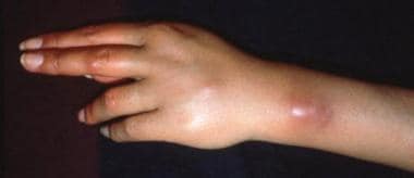 Mycobacterium marinum infection. Courtesy of the Department of Dermatology, UTMB at Galveston, Texas.
Cutaneous exposure to marine life occurs not only in the water but also when encountering living or dead marine animals on the beach. Commercial and recreational activities (eg, commercial or recreational fishing, beach combing, snorkeling, scuba diving, fish processing) have their specific concerns. Note that exposure to aquatic life can accompany various hobbies (eg, the keeping of saltwater fish tanks) or merchandising activities (eg, sellers of fish tank equipment of tropical fish). [1, 2] Exposure to freshwater life can also cause cutaneous injuries, sometimes manifesting with skin lesions similar to those caused by saltwater life. Ingestion of microorganisms can also cause cutaneous manifestations, some of which are of rapid onset.
Treatment of the severe acute sequelae of exposure to hazardous marine animals (eg, cardiorespiratory arrest, anaphylactic shock, bleeding) is in the realm of emergency medicine. This article focuses only on the cutaneous sequelae of exposure to aquatic life. Some first-aid measures are briefly mentioned. For further details, see the following articles:
See Deadly Sea Envenomations, a Critical Images slideshow, to help make an accurate diagnosis.
Signs and symptoms
Please see Presentation for a full discussion.
Examine location and characteristics of the affected areas. Certain contacts result in typical patterns and distribution (such as contact with tentacles resulting in characteristic linear eruptions on unprotected skin, seabather's eruption occurring on areas covered by clothing). Stingray injuries commonly affect the unprotected skin of the legs. Fingers are common areas for bites or injury when touching a life form that releases a toxin or inoculates microbes into the skin. Tips of sea urchin spines can break off and become embedded in the wound.
Carefully explore wounds. Look for penetrating injuries, especially over joints. Assess the wounded area for the presence of cellulitis, lymphangitis, pyogenic infections, cyanosis, and necrosis.
Some bacterial infections are typically observed on certain sites. Erysipeloid, caused by Erysipelothrix rhusiopathiae, typically affects the hands of anglers (see image below), although a diffuse cutaneous form has been described. Infection with this organism has been associated with septicemia and endocarditis. [3]
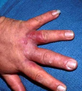 Erysipeloid. Courtesy of the Department of Dermatology, UTMB at Galveston, Texas.
Vibrio vulnificus infection usually manifests on the lower extremities as hemorrhagic bullae progressing to ulceration and necrosis. It may also cause life-threatening sepsis. [4]
M marinum infections may show sporotrichoid spread on the hand, wrist, and arm.
Venoms have rarely been described to cause injuries to other organs, such as acute renal failure. [5]
Delayed-type skin lesions such as erythema nodosum can occasionally be observed. [6]
Diagnostics
The following laboratory tests may be helpful:
-
Culture and sensitivity of infected wounds: Consider cultures for atypical Mycobacteria (M marinum) in the presence of granulomatous and ulcerating lesions or sporotrichoid spread. Other test results usually are noncontributory, unless systemic reactions are noted.
-
Intracutaneous tests with species-specific extracts: These can detect immediate and delayed-type reactions.
-
Serologic tests: These may reveal elevated immunoglobulins directed against specific antigens in patients with delayed cutaneous manifestations.
Radiography is indicated to detect foreign body or involvement of deeper structures (eg, joint, bone).
Consider taking a biopsy when diagnosis is in doubt or when tissue culture is indicated.
Histologic findings generally are nonspecific.
In seabather's eruption, superficial and deep perivascular and interstitial infiltrate consisting of lymphocytes, neutrophils, and eosinophils are described.
Biopsy of tissues infected with M marinum shows a mixed suppurative and granulomatous reaction with sparse-to-absent acid-fast bacilli.
Delayed skin reaction can be characterized by liquefaction degeneration of the basal layer.
Polarized light can reveal symmetric structures, corresponding to cross-sections of sea urchin spines.
Infections with V vulnificus show a nonspecific, yet characteristic, picture. Destruction extends into the dermis without an inflammatory cell infiltrate. Vasculitis may be present. Subepidermal noninflammatory bullae can be noted. Multiple organisms are observed.
Management
Also see Treatment and Medication.
Medical therapy in the dermatologist's office includes topical treatment of hypersensitivity reactions, systemic corticosteroid treatment of severe hypersensitivity reactions, and topical or systemic antibiotics for the prevention or treatment of infections.
Treatment with antibiotics either is initiated empirically because culture and sensitivity results are not yet available or is tailored according to the laboratory results.
Generally, the use of a first-generation cephalosporin is appropriate for empirical therapy. For mycobacterial infections, rifampin, isoniazid, or ethambutol can be used, often in combination therapy. Some authors recommend sulfonamide alone or in combination with trimethoprim; others used minocycline, doxycycline, tetracycline, or cefoxitin. Culturing and sensitivity testing of the organisms in these situations is strongly advisable to initiate specific therapy. Antibiotic therapy of some infections (especially those caused by Mycobacteria) can be lengthy. [7, 8]
Pathophysiology
The injuries can be grouped into several general categories. Overlap can also occur (such as when an abrasion also allows a toxin into injured skin or when microbes are inoculated into a puncture wound). Some of the injuries may be accompanied by bleeding and/or functional impairment of the affected area (eg, after extensive exposure to jellyfish tentacles, as shown below). Others, such as injuries followed by the exposure to venoms, can be very mild and self-limited but may also lead to fatal consequences.
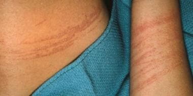 Jellyfish stings. Courtesy of the Department of Dermatology, UTMB at Galveston, Texas.
Mechanical injuries without infection
Wounds are caused by punctures (eg, sea urchin spine, stingray barbs), bites (eg, octopus or fish), cuts (eg, coral), suction (eg, octopus), abrasions, and lacerations (eg, shells). In rare instances, the human body can sustain a fatal injury to a vital organ, as documented by the case of "Crocodile Hunter" Steve Irwin. A strange case of a catfish sting causing fatal myocardial perforation was also documented. [9]
Mechanical injuries followed by infection
Wounds become secondarily infected by either debris or microscopic organisms found in the water. Improper wound care (eg, rinsing injuries with seawater possibly loaded with microorganisms) can be a source of wound infections. Inoculation of infectious agents can commonly occur with penetration injuries or lacerations caused by sea urchin spine, stingray, seal bite, [10] or other bites (eg, octopus, fish). Depending on the depth of the inoculation and on the microorganisms inoculated, severe infections of the underlying tissues and structures can also occur. In addition to possibly evolving into severe systemic infection, such processes may lead to deformities and loss of function.
Mechanical injuries accompanied by the inoculation of a venom or a substance with sensitizing properties (eg, from sea anemone, sponges, scorpionfish, stonefish, lionfish, or stingray)
The most commonly encountered phylum in this regard is the Cnidaria. These are animals that exhibit radial symmetry. Their body walls contain a jellylike substance. This phylum includes fire corals, hydroids, Portuguese man-of-war, jellyfish, sea anemones, and true corals. Almost all of these possess nematocysts, frequently on a tentacle. The nematocysts contain a toxin that is injected into the skin.
The sequelae of envenomations depend on the species involved, the nature and quantity of the toxin, and the size of the injured person. Cutaneous reactions can be immediate (eg, wheals, vesicles, bullae, angioedema) or delayed hypersensitivity reactions. Complications include pain, postinflammatory hyperpigmentation, scarring, and contractions. Systemic reactions range from mild to severe (eg, cardiac arrest, anaphylactic shock). Two species of box jellyfish around Queensland (Australia) are known to produce venom with hemolytic, dermatonecrotic, and cardiotoxic components.
The Irukandji syndrome is caused by a small amount of venom leading to severe muscle cramps, back pain, and systemic signs and symptoms, including psychological phenomena. The Irukandji jellyfish is very small (< 10 mm), but its tentacles can be 1 meter long. Whereas only a few fatal cases have been identified, the frequency and severity of jellyfish stings are significantly underestimated. [11] The length of the Irukandji season increased, but (possibly because of better beach management) the number of cases seems to decrease. [12] Underreporting of cases and very likely inconsistencies of classification can lead to difficulties in establishing effective and standard treatment. [13]
Cutaneous exposure to a dead animal or its parts (including tentacles drifted to beaches)
The lesions can be similar to those described in the above categories.
Invasion of the skin by organisms
Cercarial dermatitis (ie, clam digger's itch, swimmer's itch) is caused by Schistosoma organisms penetrating the unprotected skin.
Pyodermas and infections of the eyes, ears, or the urogenital tract
These can occur after bathing in contaminated water (eg, in areas where domestic or agricultural sewage mixes with water otherwise used for recreational activities), even in the absence of preceding mechanical injury. Existing wounds can become readily infected. [14]
Mixed or hard-to-classify manifestations
These include granulomatous processes that may actually be caused by a microorganism (eg, Mycobacterium marinum genome identified in some granulomas).
Toxins of dinoflagellates and algae can cause contact dermatitis and conjunctivitis. Ingestion of fish containing ciguatoxin may cause severe neurological manifestations (known as ciguatera), but dermatitis and pruritus are also described. Ingestion of pufferfish meat containing a potent neurotoxin (tetrodotoxin) is frequently fatal. [15]
Inhalation of coral vapor was described in a case series, resulting in respiratory and other systemic manifestations. [16]
Etiology
Bites and suction, where the injury is primarily mechanical, are caused by octopus, fish (eg, moray eel, barracuda, triggerfish), sharks, mammals (eg, sea lion, seal), and turtles.
Cuts, abrasions, lacerations, and punctures can be caused by any part of the animal that is sharp or pointed, including sea urchins, starfish, stingray, coral, barnacles, broken shells, cone shell, spines of fish fins (eg, surgeonfish, needlefish), and leeches. Inanimate objects can also cause these types of injuries.
Contact with toxic substances, including stings and other mechanical injuries resulting in the transfer of a venom, can occur after contact with fire coral, sea cucumber, bristleworm, sea anemone, sponges, stonefish, lionfish, scorpionfish, zebrafish, turkeyfish, catfish, jellyfish, Portuguese man-of-war (see image below), cone shell, starfish, and sea snakes (after bites).
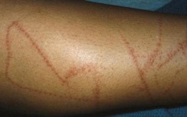 Envenomation caused by Portuguese-man-of-war. Courtesy of the Department of Dermatology, UTMB at Galveston, Texas.
Mechanical injuries can result in wounds inoculated with microorganisms during the injury or in the course of wound care.
Preexisting wounds can provide a readily available portal of entry for marine microorganisms (eg, V vulnificus) even in the absence of fresh trauma.
Irritant dermatitis attributed to algae has been described in anglers working with nets.
Delayed systemic sensitization has rarely been documented, manifesting as conjunctivitis, rhinitis, and asthma in anglers exposed to dragnets accidentally collecting red soft corals [17] and in a case of an angler using a marine worm as fishing bait. [18]
Epidemiology
Geographic area, season, and type of activity affect the prevalence of these injuries. With the increase of worldwide traveling, dermatologists may encounter lesions that would otherwise be unknown in their local area.
Predisposition to marine animal envenomation or injury is not limited by age, sex, or race.
Prognosis
Limited local reactions usually resolve without serious sequelae. Otherwise, prognosis depends on the extent of local and systemic reactions, such as cardiovascular collapse, or functional impairment of the limbs or other structures.
Mortality or morbidity depends on the nature and severity of the injury. Infection with Vibrio vulnificus in an immunocompromised host can lead to septicemia and death. Morbidity observed in dermatologic practice includes infection, pigmentary changes, scarring, severe deformities, and loss of function.
Children and elderly patients may be extremely susceptible to exposure to venoms. Immunocompromised patients are more vulnerable to systemic infections and life-threatening sequelae.
Patient Education
Prevention is important. Instruct patients to protect against re-exposure. Avoid touching marine animals, including beached organisms or animal parts (eg, broken jellyfish tentacles). Use protective equipment such as specially designed gloves when cleaning fish, shellfish, or invertebrates. Wear a wet suit while surfing, snorkeling, or diving. Avoid areas with known high risk of exposure such as shallow coral reefs where sea urchins can dwell.
Other methods of avoiding marine life are specific to given geographic areas (eg, walking with a shuffle in shallow water where stingrays may be encountered) and to a particular commercial or recreational activity (eg, fish cleaning).
-
Mycobacterium marinum infection. Courtesy of the Department of Dermatology, UTMB at Galveston, Texas.
-
Jellyfish stings. Courtesy of the Department of Dermatology, UTMB at Galveston, Texas.
-
Erysipeloid. Courtesy of the Department of Dermatology, UTMB at Galveston, Texas.
-
Envenomation caused by Portuguese-man-of-war. Courtesy of the Department of Dermatology, UTMB at Galveston, Texas.


