Background
The general term atheromatous emboli describes the embolization of any atheromatous material. Atheroemboli refers to the dislodgment of relatively large portions of atheromatous plaques containing RBCs and fibrin aggregates, which includes cholesterol crystals of sufficient size to occlude a major systemic artery and potentially result in major organ dysfunction. Cholesterol emboli (CE), on the other hand, result from ulceration of plaques and the subsequent release of cholesterol crystals. These emboli are smaller and usually more numerous, often producing multisystem disease.
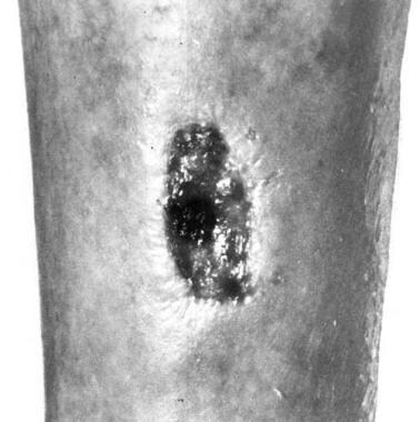 Chronic leg ulcer due to cutaneous cholesterol emboli on the leg of a 79-year-old woman.
The term cutaneous CE (CCE) is used when CE result in disease of the skin, as shown in the image below. The terms peripheral emboli, lower extremity atheromatous emboli syndrome, blue toe syndrome, purple toe syndrome, and trash foot refer to special cases of CE to the lower extremities in which cutaneous manifestations are usually present, the latter three occurring in association with anticoagulation or vascular surgery. [1]
In 1945, Flory was the first to suggest that CE may produce skin disease. [2] His hypothesis was later validated by Hoye and associates in 1959, who observed arteries occluded with cholesterol crystals in areas of gangrene on the feet and toes. [3]
It is more commonly associated with iatrogenic manipulation via invasive vascular procedures or therapies (anticoagulation or thrombolytics) and, most commonly, affects the kidneys, gastrointestinal system, and skin. [4] Skin findings can facilitate clinical diagnosis, since about 88% of patients with CCE have them, most commonly livedo reticularis and cyanotic changes of the toes.
CCE is a disease primarily of elderly white men with ulcerous atherosclerosis. Atherosclerotic foci release cholesterol crystals spontaneously or after anticoagulation or endovascular manipulation, inducing the obstruction of small arteries. Cholesterol embolization syndrome is a systemic disease due to distal showering of cholesterol crystals after angiography, major vessel surgery, or thrombolysis. Obstruction of cutaneous vasculature most often results in a clinical picture of livedo reticularis (LR). It is more common in patients with atherosclerotic disease, hypertension, a history of smoking, and elevated baseline plasma C-reactive protein levels.
Gangrene, cyanosis, ulcers, nodules, and purpura can also be observed. In cases of multisystem involvement, CCE may masquerade as many different diseases, but the clinical picture most often mimics a vasculitis. Skin or muscle specimens demonstrate the cholesterol crystals characteristic of this disease.
Treatment is based on the identification of the source of emboli through angiography and on the exclusion of that source from the circulation. Medical therapy has largely been unsuccessful. Gangrene necessitating amputation is the major complication of CCE, but complications may occur in practically any organ system. Without surgery, CCE is a recurrent process with a high mortality rate.
Pathophysiology
The most likely explanation for the cutaneous manifestation of CCE is trapping of cholesterol crystals in blood vessels leading to occlusion and tissue ischemia. Other contributing factors include underlying lowered arterial pressure from proximal atherosclerosis and the ability of emboli to activate the complement system.
The pattern of LR may be the first clinical sign of CCE and is thought to result from incomplete disturbance of circulation and desaturation of blood that initially occurs with subtotal occlusion of vessels. [5, 6, 7, 8, 9] As spasm and complete occlusion occur, the other signs of CCE become evident. In addition to the blockage of small vascular channels, lower arterial pressure from narrowing of larger proximal arteries may be necessary for the cutaneous manifestations of CCE because intact collateral supply should normally avert it. In one study, injections of a cholesterol suspension in the femoral arteries of dogs produced gangrene, but only in cases with associated thrombosis of the femoral artery. This indicates that embolism is a contributing factor in necrosis with a vascular supply already compromised by atherosclerosis or other occlusive disease. Neither thrombosis alone nor CE alone would produce necrosis.
Other evidence suggests that in addition to a purely mechanical effect, crystalline cholesterol may amplify infarctive tissue damage through the activation of plasma complement, which is capable of potently aggregating polymorphonuclear (PMN) leukocytes and provoking them to damage endothelial cells via toxic oxygen radical release. In both experimental and clinical infarction, evidence of plasma complement activation, often with depletion of complement components, is observed. Animals depleted of complement prior to experimental infarction experience smaller infarcts than controls. In one report, a man suspected of having CE with cutaneous lesions, including LR and digital infarcts, reportedly had plasma with PMN leukocyte–aggregating activity that contained a component of molecular weight and antigenicity consistent with C5a.
Cholesterol crystals and lipids from atheromata incubated with plasma or serum activate complement, as evidenced by immunoelectrophoresis that showed conversion of C3 to C5. On the other hand, serum or plasma depleted of complement or from a patient with congenital C5 deficiency resists activation. PMN leukocytes incubated with endothelial cells to which C5a or cholesterol-incubated plasma was added show evidence of endothelial damage via increased superoxide production, while the addition of plasma alone or cholesterol-incubated plasma without PMN leukocytes does not cause any damage beyond that which spontaneously occurs. This damage is partially inhibited by the addition of superoxide dismutase and catalase.
A related Medscape Reference article is Cutaneous Manifestations of Cholesterol Embolism.
Etiology
Ulcerated atherosclerosis is the primary risk factor for CCE and is especially prevalent in persons with aortic aneurysms; however, the size of the aneurysm does not correlate with the risk of emboli. Single cases of fibromuscular dysplasia of the external iliac arteries and aortic dissection leading to emboli have also been reported. In the latter 2 cases, abnormal turbulence near the diseased artery and disruption of an atheromatous plaque in the area of dissection were thought to be the cause of embolization. In all these patients, CCE can be spontaneous or precipitated by anticoagulant therapy, vascular procedures or surgery, or, rarely, trauma.
In one series of patients with CCE, 26 (36%) of 73 were taking anticoagulants and 31% had undergone vascular procedures. In a series of 15 cases of peripheral emboli, 13 were spontaneous and 2 followed infrarenal aortic operations. In a series of 13 patients with spontaneous CCE, 4 had aortic aneurysms, 2 had femoral aneurysms, and 7 had severe ulcerative atherosclerosis of the aortoiliac segments. In a larger review of 85 cases of peripheral atheroemboli, 38 (45%) were from proximal aneurysms, 37 (43%) were unexplained, 4 were from a nonaneurysmal source, 3 were from other sources, and 3 were iatrogenic.
While the abdominal aorta has traditionally been considered the source of embolization, some have observed embolization from more distal vessels of the arterial tree. In one study, patients with blue toe syndrome were found to have aortoiliac and peripheral (superficial femoral or popliteal artery) atherosclerosis. Surgical exploration of the peripheral lesions revealed ulcerated plaques or focal stenosis, both of which had adherent white thrombi that were interpreted as evidence of these lesions being the source of the emboli. Surgical correction of the peripheral lesions prevented recurrence at 8-24 months of follow-up. In another study on the source of peripheral emboli, 14 were from the aortic or iliac vessels and 28 were below the inguinal ligament. On the other hand, a smaller study showed 8 (57%) aortoiliac lesions versus 6 (43%) femoropopliteal lesions. Thoracic disease, which is more common in conditions such as syphilis or gout, has also been shown to lead to peripheral emboli.
Vascular manipulation, either for radiographic or surgical purposes, results in embolization through the mechanical disruption of atherosclerotic plaques by needles, wires, catheters, or clamps and is especially common after prolonged or difficult catheterizations. [10, 11] Less often, a stream of injected contrast material may dislodge material. Implicated vascular procedures and surgeries include angiography (most often aortography), heart catheterization, coronary artery bypass graft (CABG) surgery, and percutaneous transluminal coronary angioplasty. Procedures near vascular structures that may involve manipulation of such structures can also result in CE; this has been reported after transhiatal esophagectomy.
Anticoagulants are frequently a cause of CCE. Such occurrences are often designated blue or purple toe syndrome. [12] Anticoagulants are speculated to cause CE by preventing or removing adequate thrombosis over ulcerated atheromatous lesions. In one study, embolization was observed to occur 3 weeks to several months after initiation of therapy. Agents reported to cause CE include heparin, bishydroxycoumarin, warfarin sodium, streptokinase, [13, 14, 15] and intravenous tissue plasminogen activator. [16, 17, 18] In an attempt to define the role of anticoagulation in CE, a prospective study examined 60 patients with acute myocardial infarction who underwent CABG surgery. Twenty-nine received thrombolytic therapy for myocardial infarction, and 31 were treated conservatively. During the CABG surgeries, two muscle biopsy specimens and one skin biopsy specimen were taken from vein harvest sites. CE was observed in 4 of 29 and 3 of 31 specimens, respectively.
Embolization following blunt abdominal trauma from an automobile accident has been reported to precede CE. Vibration of the aortic wall may have dislodged the atheroma. Cholesterol crystal emboli can be induced by factor Xa inhibitor. [19]
Epidemiology
Frequency
United States
The incidence of CCE is rising in association with increased use of interventional vascular procedures, angioplasties, and anticoagulant/thrombolytic therapy. [4] The frequency of CCE is difficult to estimate because routine necropsy is often limited and does not include evaluation of the skin; however, because the abdominal aorta is usually the site of the most advanced intimal disease, one would anticipate that the lower extremities and associated skin and musculature would be one of the most frequently involved sites. In one study, skin and muscle biopsy specimens were obtained from the lower extremities of 100 consecutive autopsies and a 4% rate of CCE was observed. In a large review of 223 patients with all types of CE, 78 (35%) had skin involvement.
International
In the Netherlands, cutaneous involvement is observed in approximately 24% of cases of CE. [20]
Race
Of 31 cases of CCE in which race was mentioned, all patients were white.
Sex
In the same study, 64 (82%) of 78 cases of CCE were in men.
Age
Ages of those affected with CCE in the same study ranged from 26-90 years (mean, 63 y).
Prognosis
CCE is a repetitive process. Untreated patients often have recurrent episodes with significant morbidity and mortality; however, patients with only peripheral involvement, as opposed to both peripheral and visceral involvement, have a much better prognosis. In the most extensive review of CCE cases, the mortality rate was very high. Forty-nine (78%) of 68 patients died.
Patient Education
For patient education resources, see the Cholesterol Center, as well as High Cholesterol and Cholesterol FAQs.
-
Chronic leg ulcer due to cutaneous cholesterol emboli on the leg of a 79-year-old woman.
-
Skin biopsy specimen demonstrating ulceration and an occluded vessel at the right border of the specimen within the fat. Hematoxylin and eosin stain at 22X magnification.
-
Higher magnification of the same biopsy specimen, demonstrating cholesterol clefts within an occluded arteriole. Hematoxylin and eosin stain at 297X magnification.


