Background
In the United States, reports of severe envenomations by brown spiders began to appear in the late 1800s, and today, in endemic areas, brown spiders continue to be of significant clinical concern. See the current distribution map below.
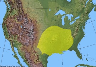 Complete distribution range of wild and domestic Loxosceles reclusa (brown recluse spider). Courtesy of Wikimedia Commons (By ReliefUSA_map.gif: Public domain, U.S. government derivative work: Bob the Wikipedian).
Of the 13 species of Loxosceles in the United States, at least five have been associated with necrotic arachnidism. Loxosceles reclusa, or the brown recluse spider, is the spider most commonly responsible for this injury.
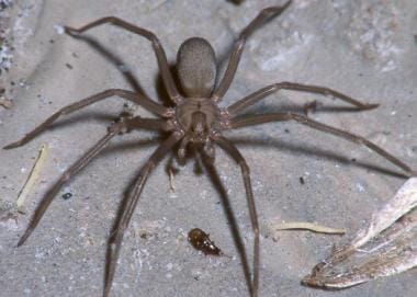 Typical appearance of a male brown recluse spider. Photo contributed by Michael Cardwell, Victorville, Calif.
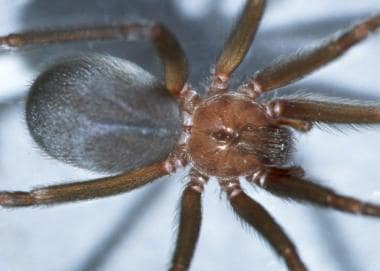 Spider envenomations, brown recluse. Close-up image of dorsal violin-shaped pattern. Photo contributed by Michael Cardwell, Victorville, Calif.
Dermonecrotic arachnidism refers to the local skin and tissue injury noted with this envenomation. Loxoscelism is the term used to describe the systemic clinical syndrome caused by envenomation from the brown spiders.
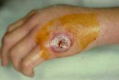 Dermonecrotic arachnidism represents a local cutaneous injury with tissue loss and necrosis.
See Arthropod Envenomation: From Benign Bites to Serious Stings and Venomous Spider Bites: Keys to Diagnosis and Treatment, Critical Images slideshows, for help identifying and treating various envenomations.
Pathophysiology
Brown recluse spider bites can cause significant cutaneous injury with tissue loss and necrosis. Less frequently, more severe reactions develop, including systemic hemolysis, coagulopathy, renal failure, and, rarely, death.
Brown recluse venom, like many of the other brown spider venoms, is cytotoxic and hemolytic. It contains at least 8 components, including enzymes such as hyaluronidase, deoxyribonuclease, ribonuclease, alkaline phosphatase, and lipase. Sphingomyelinase D is thought to be the protein component responsible for most of the tissue destruction and hemolysis caused by brown recluse spider envenomation. The intense inflammatory response mediated by arachidonic acid, prostaglandins, and chemotactic infiltration of neutrophils is amplified further by an intrinsic vascular cascade involving the mediator C-reactive protein and complement activation. Laboratory studies have shown a decrease in hemolysis from brown recluse venom in the presence of complement inhibitors. [1] These and other factors contribute to the local and systemic reactions of necrotic arachnidism.
Although numerous cases of cutaneous and viscerocutaneous reactions have been attributed to spiders of the genus Loxosceles, confirming the identity of the envenomating arachnid is difficult and rarely accomplished.
Etiology
Dermonecrotic arachnidism has been described in association with several species of Loxosceles spiders, but, in the United States, L reclusa venom is the most potent and the most commonly involved.
Epidemiology
US frequency
Although various species of Loxosceles are found throughout the world, L reclusa is found in the United States from the East to the West Coast, with predominance in the south. Recently, reports of persons with "spider bites" presenting to emergency departments have reached near urban legend proportions, prompting many physicians to question the diagnosis of a brown recluse bite in nonendemic areas. [2, 3, 4] The list of conditions that can present in a similar fashion to that of a brown recluse spider envenomation is extensive. A more likely explanation for this epidemic of spider bites is in fact community-acquired methicillin-resistant Staphylococcus aureus (MRSA) skin infections. [5]
The 2019 Annual Report of the American Association of Poison Control Centers recorded 790 individual exposures to brown recluse spiders, with 174 moderate outcomes, 24 major outcomes, and 0 dealths. [6]
Age
Systemic involvement, although uncommon, occurs more frequently in children than in adults. [7]
See the distribution map below.
Complete distribution range of wild and domestic Loxosceles reclusa (brown recluse spider). Courtesy of Wikimedia Commons (By ReliefUSA_map.gif: Public domain, U.S. government derivative work: Bob the Wikipedian).
Prognosis
Mortality/morbidity
Data regarding mortality rates are not reliable because diagnostic tests to detect brown recluse venom in tissue are not readily available.
Although deaths have been attributed to presumed brown recluse envenomation, severe outcomes are rare. [8] Typical cases involve only local soft tissue destruction. The 2014 Annual Report of the American Association of Poison Control Centers recorded 275 minor outcomes, 218 moderate outcomes, 11 major outcomes, and no deaths. [6]
In South America, the more potent venom of the species Loxosceles laeta is responsible for several deaths each year. [9]
Patient Education
For patient education information, see the First Aid and Injuries Center as well as Black Widow Spider Bite and Brown Recluse Spider Bite.
-
Classic finding of a vesicle with surrounding erythema at 24 hours following brown recluse envenomation. Photo by Thomas Arnold, MD.
-
Illustration of a brown recluse spider with the fiddle displayed prominently on its dorsum.
-
Spider envenomations, brown recluse. Envenomation site on inner thigh untreated at 1 week. Photo by Thomas Arnold, MD.
-
Typical appearance of a male brown recluse spider. Photo contributed by Michael Cardwell, Victorville, Calif.
-
Female brown recluse with size scale. Photo contributed by Michael Cardwell, Victorville, Calif.
-
Spider envenomations, brown recluse. Close-up image of dorsal violin-shaped pattern. Photo contributed by Michael Cardwell, Victorville, Calif.
-
Spider bite, brown recluse. Within an hour, the bite area swelled to the size of a quarter. The area turned blue and dark red by the evening of the first day, exceeding the boundaries of a circle drawn around the area of initial swelling by the patient's physician. Courtesy of Dale Losher.
-
Spider bite, brown recluse. The third day after the bite. The skin continues to die. Courtesy of Dale Losher.
-
Spider bite, brown recluse. Another view of the wound 3 days after the bite. Courtesy of Dale Losher.
-
Spider bite, brown recluse. Nine days after the bite. The patient endured 8 days with an open wound to drain the spider's toxins and needed multiple doses of intravenous antibiotics and pain medication. Courtesy of Dale Losher.
-
Spider bite, brown recluse. Eleven days after the bite. A 5-inch wide area of dead tissue was excised, necessitating skin grafting. Courtesy of Dale Losher.
-
Spider bite, brown recluse. Waiting to see skin graft results 38 days after the bite. Courtesy of Dale Losher.
-
Spider bite, brown recluse. Skin graft results 38 days after the bite. Courtesy of Dale Losher.
-
Spider bite, brown recluse. View of healed wound approximately 10 months after bite. Courtesy of Dale Losher.
-
Dermonecrotic arachnidism represents a local cutaneous injury with tissue loss and necrosis.
-
Brown recluse spider. Courtesy of US Centers for Disease Control and Prevention.
-
Brown recluse spider. Courtesy of US Centers for Disease Control and Prevention.
-
Complete distribution range of wild and domestic Loxosceles reclusa (brown recluse spider). Courtesy of Wikimedia Commons (By ReliefUSA_map.gif: Public domain, U.S. government derivative work: Bob the Wikipedian).


