Continually Updated Clinical Reference
 
 
  All Sources     eMedicine     Medscape     Drug Reference     MEDLINE
 
eMedicine - Ainhum : Article by

Quick Find
Authors & Editors
Introduction
Clinical
Differentials
Workup
Treatment
Follow-up
Miscellaneous
Multimedia
References

Related Articles
Leprosy

Lupus Erythematosus, Discoid

Morphea

Pityriasis Rubra Pilaris

Porokeratosis

Syphilis

Yaws


Patient Education
Click here for patient education.


Author: Samuel Selden, MD, Assistant Professor, Department of Dermatology, Eastern Virginia Medical School

Samuel T Selden is a member of the following medical societies: American Academy of Dermatology

Editors: Smeena Khan, MD, Private Practice, Adult and Pediatric Dermatology Associates; Richard P Vinson, MD, Assistant Clinical Professor, Department of Dermatology, Texas Tech University School of Medicine; Consulting Staff, Mountain View Dermatology, PA; Jeffrey J Miller, MD, Associate Professor, Department of Dermatology, Penn State University, Milton S Hershey Medical Center; Joel M Gelfand, MD, MSCE, Medical Director, Clinical Studies Unit, Assistant Professor, Department of Dermatology, Associate Scholar, Center for Clinical Epidemiology and Biostatistics, University of Pennsylvania; William D James, MD, Paul R Gross Professor of Dermatology, University of Pennsylvania School of Medicine; Vice-Chair, Program Director, Department of Dermatology, University of Pennsylvania Health System

Author and Editor Disclosure

Synonyms and related keywords: dactylolysis spontanea, bankokerend, sukhapakla, autoamputation of a digit, pseudoainhum, pseudo-ainhum, annular constriction of a digit

Background

Ainhum is the autoamputation of a digit, usually of the fifth toe bilaterally and as a result of a constricting scar in the form of a band or groove. Pseudoainhum is a similar condition that occurs as a secondary event resulting from certain hereditary and nonhereditary diseases that lead to annular constriction of digits.

Ainhum predominantly affects black patients in tropical regions. Although it has been reported in temperate areas, ainhum appears to be increasingly less common in the United States.

The origin of the term ainhum is unclear. In 1867, the term was used by da Silva Lima1 from Bahia, Brazil to report the first published case. The word ainhum means fissure in the language of the Nagos tribe of Brazil and may be related to ayun, the word for saw in the Lagos tribe of Nigeria. The synonym for ainhum is dactylolysis spontanea.

Pathophysiology

In true ainhum, dactylolysis of a toe (most commonly the fifth toe) most likely is triggered by trauma; however, the true cause remains unknown. The trauma may be related to walking barefoot in the tropics. A fibrotic band develops from a flexural groove and progressively constricts the full radius of the toe until spontaneous autoamputation occurs. A similar progression occurs in pseudoainhum because of a collagen band, rather than from fibrosis. Pseudoainhum may be acquired or congenital.

Frequency

United States

Approximately 130 cases have been reported in the United States, but only 30 cases have been reported since 1960. Pseudoainhum is a rare disorder.

International

Ainhum is a relatively common disease among black Africans. In Africa, the incidence range is 0.2-2%. The incidence of true ainhum outside of Africa appears to be low.

Mortality/Morbidity

Pain may be severe in ainhum and in pseudoainhum. Because ainhum occurs primarily in tropical areas, secondary infections and their complications may be a source of morbidity.

Race

Ainhum has been reported to affect all races but occurs predominately in blacks. No racial predilection exists for pseudoainhum.

Sex

In Nigeria, one study revealed an incidence of 2.48 cases per 1000 males and 1.08 cases per 1000 females; however, recent investigations suggest no sex preference.

Age

Full-blown ainhum is uncommon in persons younger than 30 years and older than 50 years. The reason ainhum appears to be age specific is unclear. Early lesions may be observed in childhood.


History

Cole2 has described 4 clinical stages of ainhum.

  • In the first stage, a small clavus or callus develops on the medial aspect of the plantar fold of a toe (usually the fifth) that progresses to a narrow groove or fissure. This groove deepens and slowly encircles the toe. Progression may be slow and can develop first in childhood. The deepening of the fissure is associated with pain in 78% of patients but is less intense than pain in the third stage.
  • The second stage is shorter because the toe becomes globular distal to the groove, which is associated with arterial narrowing and bone resorption.
  • In the third stage, the bone separates at the joint with hypermotility of the toe. Pain may be intense.
  • The fourth stage is characterized by a bloodless autoamputation of the toe at the site of the encircling band.

Physical

The clinical presentation depends on the stage to which the ainhum has progressed.

  • The initial sign of a painful fissure under a toe may not be defining, but the progressive constriction at the base of the toe with distal edema is diagnostic of ainhum.
  • The toe may become rotated, clawed, and dorsiflexed at the metatarsophalangeal joint.
  • Ultimately, before the toe is shed, it may be attached by an increasingly slender thread of fibrous tissue.
  • The clinical, histologic, and radiographic appearances in pseudoainhum are similar or identical to true ainhum.

Causes

In 1952, Wells and Robinson3 proposed 4 distinct sources of annular constrictions of the digits. The sources include (1) annular scarring from frostbite, burns, or trauma, (2) true ainhum, (3) constricting bands that simulate ainhum, and (4) congenital bands.

The exact etiology of true ainhum is unclear. Race and climate apparently are predisposing factors. Ainhum also may have a genetic component, since ainhum has been reported to occur within families. Infection and walking barefoot in childhood are linked to ainhum but probably are not major factors in its development. Abnormal scarring does not appear to be a cause; ainhum and keloid formation rarely occur in the same individual.

Pseudoainhum may be acquired or congenital.


Leprosy
Lupus Erythematosus, Discoid
Morphea
Pityriasis Rubra Pilaris
Porokeratosis
Syphilis
Yaws

Other Problems to be Considered

Congenital constricting bands of infants and young children
Trauma (burns, frostbite, ergot poisoning, tourniquet syndrome)
Connective tissue disease (scleroderma, morphea, Raynaud disease, discoid lupus erythematosus)
Infection (syphilis, Hansen disease)
Keratodermas (keratoderma hereditarium mutilans, mal de Meleda, focal acral hyperkeratosis, porokeratosis of Mibelli)
Pityriasis rubra pilaris
Pachyonychia congenita
Endocrine (diabetes mellitus)

Ainhum

Initial fissuring beneath toes may be mistaken for trauma or infection. Dermatophytosis complex may be a complication. In the tropics, where most true ainhum occurs, Hansen disease, syphilis, yaws, and tuberculosis must be excluded. Radiographically, osteolytic lesions may be observed both after trauma and in ainhum.

Following dactylolysis, ainhum may be confused with traumatic amputations, limb aplasia or hyperplasia, diabetic or vascular gangrene, or tourniquet syndrome (from human hair).

Pseudoainhum

Pseudoainhum most commonly is associated with scleroderma, Hansen disease, syringomyelia, and atypical keratoderma. Pseudoainhum refers to congenital annular bands or constrictions resulting from trauma or linked to other diseases.

Congenital bands usually constrict an extremity, but may encircle any portion of the body, and result from congenital collagen dysplasia and not from scarring. The bands frequently are associated with other congenital or developmental anomalies.


Imaging Studies

  • The radiographic manifestations of ainhum are diagnostic.
    • Initially, a radiolucent band can be observed constricting the base of the involved toe, with distal swelling.
    • Osteolysis develops in the distal and middle phalanges, with a characteristic tapering effect.
    • Ultimately, the bone narrows until it fractures and autoamputates.
  • The radiographic appearances in pseudoainhum are similar or identical to true ainhum.

Histologic Findings

Histologically, ainhum shows fissuring and epidermal hyperkeratosis and parakeratosis, which is followed by a fibrotic reaction under the deepening fissure. The fibrosis is predominately composed of collagen. As scar tissue contracts, it constricts and narrows neurovascular bundles. Histologic appearances in pseudoainhum are similar or identical to those observed in true ainhum.


Medical Care

No current treatment appears to halt the progression of ainhum.

  • In the early stages of ainhum, treatment with salicylic acid ointment applications and intralesional steroid injections (20-40 mg/mL triamcinolone acetonide suspension) have been reported to reduce pain.
  • Pseudoainhum may respond to treatment with etretinate.

Surgical Care

  • If discovered late, the definitive treatment for ainhum may be surgical amputation (if not autoamputation). The best treatment may be Z-plasty, which involves releasing the constricting base through a Z-shaped repair after surgical amputation.
  • In the early stages of ainhum, division of the fibrous band has been reported to reduce pain.
  • In pseudoainhum, congenital bands rarely need treatment other than surgery.


Complications

  • Secondary infections may complicate ainhum.
  • If more than just the 2 fifth toes are affected in ainhum or pseudoainhum, the individual's balance may be affected when walking.
  • Dermatophytosis complex may be a complication.

Prognosis

  • Outcome is related to the stage in ainhum when the disease is diagnosed.

Patient Education

  • Instructions in good foot care are critical. Since some cases of ainhum and pseudoainhum are familial, other family members may require examination.


Medical/Legal Pitfalls

  • Constricting, fissuring, and loss of digits can be mimicked by other diseases of the feet, and ainhum can remain unrecognized.
  • Ainhum may be difficult to diagnose in the United States because it is uncommon, and only 30 cases have been reported since 1960.


Media file 1:  
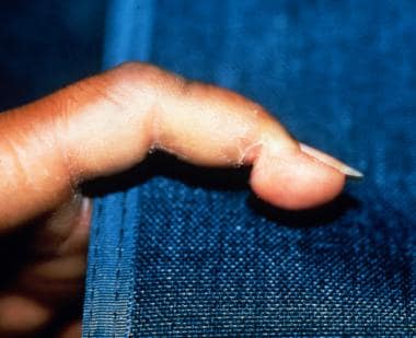
Ainhum of the finger. Courtesy of Hon Pak, MD, and reviewed by Ross Levy, MD.
Click to see larger pictureClick to see detailView Full Size Image
Media type:  Photo


  1. da Silva Lima JF. On ainhum. Arch Dermatol. 1880;6:367.
  2. Cole GJ. Ainhum: An account of fifty-four patients with special reference to etiology and treatment. J Bone Joint Surg Br. Feb 1965;47:43-51. [Medline].
  3. Wells TL, Robinson RC. Annular constrictions of the digits. AMA Arch Derm Syphilol. Nov 1952;66(5):569-72. [Medline].
  4. Allyn B, Leider M. Dactylolysis spontanea (ainhum). Report of a case treated by the surgical procedure known as Z-plasty. JAMA. May 25 1963;184:655-7. [Medline].
  5. Browne SG. Ainhum. Int J Dermatol. Jun 1976;15(5):348-50. [Medline].
  6. Cunliffe WJ. Ainhum and pseudo-ainhum. In: Rook A, Wilkinson DS, Ebling FJ. Textbook of Dermatology. Vol 2. Oxford: Blackwell Scientific; 1979:1638.
  7. Demis DJ. Ainhum, pseudoainhum, and tourniquet syndrome. In: Demis DJ, Dobson RL, McGuire J. Clinical Dermatology. 7th ed. Hagerstown, Md: Harper & Row; 1979:4-47.
  8. Dent DM, Fataar S, Rose AG. Ainhum and angiodysplasia. Lancet. Aug 22 1981;2(8243):396-7. [Medline].
  9. Fetterman LE, Hardy R, Lehrer H. The clinico-roentgenologic features of ainhum. Am J Roentgenol Radium Ther Nucl Med. Jul 1967;100(3):512-22. [Medline].
  10. Greene JT, Fincher RM. Case report: ainhum (spontaneous dactylolysis) in a 65-year-old American black man. Am J Med Sci. Feb 1992;303(2):118-20. [Medline].
  11. Hunt M, Glucksman EE. Ainhum presenting to the accident and emergency department. Arch Emerg Med. Dec 1993;10(4):324-7. [Medline].
  12. Kean BH, Tucker HA, Miller WC. Ainhum: a clinical summary of forty-five cases on the Isthmus of Panama. Trans R Soc Trop Med Hyg. 1946;39:331-4.
  13. Mendelson DS, Chan KF, Song IS. Spontaneous dactylolysis with pain in a 58-year-old American Black man. JAMA. Oct 2 1981;246(14):1591-2. [Medline].
  14. Olivieri I, Piccirillo A, Scarano E, Ricciuti F, Padula A, Molfese V. Dactylolysis spontanea or ainhum involving the big toe. J Rheumatol. Dec 2005;32(12):2437-9. [Medline].
  15. Ramesh V, Misra RS, Mahaur BS. Pseudoainhum in porokeratosis of Mibelli. Cutis. Feb 1992;49(2):129-30. [Medline].
  16. Rossiter JW, Anderson PC. Ainhum: treatment with intralesional steroids. Int J Dermatol. Jun 1976;15(5):379-82. [Medline].
  17. Schulz EJ. Genodermatoses. Dermatol Clin. Oct 1994;12(4):787-96. [Medline].
  18. Sharma RC, Sharma AK, Sharma NL. Pseudo-ainhum in discoid lupus erythematosus. J Dermatol. Apr 1998;25(4):275-6. [Medline].

Ainhum excerpt

Article Last Updated: Feb 15, 2007