Practice Essentials
Pemphigus vulgaris is an autoimmune, intraepithelial, blistering disease affecting the skin and mucous membranes. It is mediated by circulating autoantibodies directed against keratinocyte cell surfaces. A potentially life-threatening disease, it has a mortality rate of approximately 5-15%. [1]
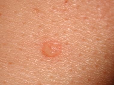
The primary lesion of pemphigus vulgaris is a flaccid blister filled with clear fluid that arises on healthy skin or on an erythematous base (see the image below).
See Clues in the Oral Cavity: Are You Missing the Diagnosis?, a Critical Images slideshow, to help identify the causes of abnormalities of the oral cavity.
Signs and symptoms
Mucous membranes
Mucous membranes of the oral cavity are involved in almost all patients with pemphigus vulgaris.
Patients may have ill-defined, irregularly shaped, gingival, buccal, or palatine erosions, which are painful and slow to heal.
Intact bullae are rare in the mouth.
Erosions may be seen on any part of the oral cavity, and they may spread to involve the larynx, with subsequent hoarseness.
In juvenile pemphigus vulgaris, stomatitis is the presenting complaint in more than 50% of cases.
Other mucosal surfaces may be involved, including the conjunctiva, [2] esophagus (causes odynophagia and/or dysphagia), [3] labia, vagina, cervix, vulva, [4] penis, urethra, nasal mucosa, and anus.
Skin
Primary lesion of pemphigus vulgaris is a flaccid blister filled with clear fluid that arises on healthy skin or on an erythematous base.
Blisters are fragile and may rupture, producing painful erosions (the most common skin presentation).
Nails
Acute or chronic paronychia, subungual hematomas, and nail dystrophies affecting one or several fingers or toes have been reported with pemphigus vulgaris. [5, 6]
Vegetating pemphigus vulgaris
Lesions in skin folds readily form vegetating granulations. In some patients, erosions tend to develop excessive granulation tissue and crusting; these individuals display more vegetating lesions.
See Clinical Presentation for more detail.
Diagnosis
Laboratory studies include the following:
-
Histopathology: Demonstrates an intraepidermal blister; the earliest changes consist of intercellular edema with loss of intercellular attachments in the basal layer
-
Indirect immunofluorescence (IDIF): If DIF results are positive; circulating intercellular antibodies are detected using IDIF in 80-90% of patients with pemphigus vulgaris [9]
See Workup for more detail.
Management
The aim of pharmacologic therapy for pemphigus vulgaris is to reduce inflammatory response and autoantibody production. Medications used in the disease’s treatment include the following:
-
Corticosteroids: Discourage the inflammatory process by inhibiting specific cytokine production
-
Immunosuppressants: Should be considered early in the course of disease as steroid-sparing agents
See Treatment and Medication for more detail.
Background
Pemphigus is derived from the Greek word pemphix meaning bubble or blister. Pemphigus describes a group of chronic bullous diseases, originally named by Wichman in 1791. The term pemphigus once included most bullous eruptions of the skin, but diagnostic tests have improved, and bullous diseases have been reclassified.
The term pemphigus refers to a group of autoimmune blistering diseases of the skin and mucous membranes characterized histologically by intraepidermal blister and immunopathologically by the finding of in vivo bound and circulating immunoglobulin G (IgG) antibody directed against the cell surface of keratinocytes. The 3 primary subsets of pemphigus include pemphigus vulgaris, pemphigus foliaceus, and paraneoplastic pemphigus. [10] Each type of pemphigus has distinct clinical and immunopathologic features. Pemphigus vulgaris accounts for approximately 70% of pemphigus cases.
Pathophysiology
Pemphigus vulgaris is an autoimmune, intraepithelial, blistering disease affecting the skin and mucous membranes and is mediated by circulating autoantibodies directed against keratinocyte cell surfaces. In 1964, autoantibodies against keratinocyte surfaces were described in patients with pemphigus. Clinical and experimental observations indicate that the circulating autoantibodies are pathogenic. An immunogenetic predisposition is well established.
Blisters in pemphigus vulgaris are associated with the binding of IgG autoantibodies to keratinocyte cell surface molecules. These intercellular or pemphigus vulgaris antibodies bind to keratinocyte desmosomes and to desmosome-free areas of the keratinocyte cell membrane. The binding of autoantibodies results in a loss of cell-to-cell adhesion, a process termed acantholysis. The antibody alone is capable of causing blistering without complement or inflammatory cells.
Pemphigus vulgaris antigen
Intercellular adhesion in the epidermis involves several keratinocyte cell surface molecules. Pemphigus antibody binds to keratinocyte cell surface the molecules desmoglein 1 and desmoglein 3. The binding of antibody to desmoglein may have a direct effect on desmosomal adherens or may trigger a cellular process that results in acantholysis. Antibodies specific for nondesmosomal antigens also have been described in the sera of patients with pemphigus vulgaris; however, the role of these antigens in the pathogenesis of pemphigus vulgaris is not known.
Antibodies
Patients with the mucocutaneous form of pemphigus vulgaris have pathogenic antidesmoglein 1 and antidesmoglein 3 autoantibodies. Patients with the mucosal form of pemphigus vulgaris have only antidesmoglein 3 autoantibodies. Patients with active disease have circulating and tissue-bound autoantibodies of both the immunoglobulin G1 (IgG1) and immunoglobulin G4 (IgG4) subclasses. [11, 12]
More than 80% of the patients with active disease produce autoantibodies to the desmosomal protein desmoglein. Disease activity correlates with antibody titers in most patients. [13] In patients with pemphigus vulgaris, the presence of antidesmoglein 1 autoantibodies, as determined by enzyme-linked immunosorbent assay (ELISA), is more closely correlated with the course of the disease compared with antidesmoglein 3 autoantibodies. Lack of in vivo antibody binding (reversion to a negative result on direct immunofluorescence) is the best indicator of remission and can help predict a lack of flaring when therapy is tapered.
Complement
Pemphigus antibody fixes components of complement to the surface of epidermal cells. Antibody binding may activate complement with the release of inflammatory mediators and recruitment of activated T cells. T cells are clearly required for the production of the autoantibodies, but their role in the pathogenesis of pemphigus vulgaris remains poorly understood. Interleukin 2 is the main activator of T lymphocytes, and increased soluble receptors have been detected in patients with active pemphigus vulgaris.
Epidemiology
United States
Pemphigus vulgaris is uncommon, and the exact incidence and prevalence depends on the population studied.
International
Pemphigus vulgaris has been reported to occur worldwide. Pemphigus vulgaris incidence varies from 0.5-3.2 cases per 100,000 population. Pemphigus vulgaris incidence is increased in patients of Ashkenazi Jewish descent and those of Mediterranean origin. Few familial cases have been reported. As with endemic pemphigus, there is some evidence to suggest clustering near industrial sites. [14]
Race
Pemphigus vulgaris affects persons of all races. The prevalence of pemphigus vulgaris is high in regions where the Jewish population is predominant. [15] For example, in Jerusalem, the prevalence of pemphigus vulgaris is estimated at 1.6 cases per 100,000 population; in Connecticut, the prevalence has been reported as 0.42 cases per 100,000 population. [16] The incidence in the United Kingdom is 0.68 case per 100 000 persons per year. The incidence of pemphigus vulgaris in Tunisia is estimated at 2.5 cases per million population per year (3.9 in women, 1.2 in men), while in France, the incidence is 1.3 cases per million population per year (no significant difference between men and women). [17] In Finland, where few people of Jewish or Mediterranean origin live, the prevalence is low, at 0.76 case per million population. [18]
Sex
The male-to-female ratio is approximately equal. In adolescence, girls are more likely to be affected than boys.
Age
The mean age of onset is approximately 50-60 years; however, the range is broad, and disease onset in older individuals and in children has been described. Patients are younger at presentation in India than in Western countries. [19]
Mortality/Morbidity
Pemphigus vulgaris is a potentially life-threatening autoimmune mucocutaneous disease with a mortality rate of approximately 5-15%. [1] Mortality in patients with pemphigus vulgaris is 3 times higher than the general population. Complications secondary to the use of high-dose corticosteroids contribute to the mortality rate. Morbidity and mortality are related to the extent of disease, the maximum dose of systemic steroids required to induce remission, and the presence of other diseases. Prognosis is worse in patients with extensive pemphigus vulgaris and in older patients.
Pemphigus vulgaris involves mucosa in 50-70% of patients. This may limit oral intake secondary to dysphagia. Blistering and erosions secondary to the rupture of blisters may be painful and may limit the patient's daily activities. Additionally, Patients with pemphigus vulgaris typically heal without scarring unless the disease is complicated by severe secondary infection.
Reversion of direct immunofluorescence (DIF) to negative can be useful to predict sustained remission after withdrawal of medication. Plucked hairs are an alternative to skin biopsy to provide a specimen for immunofluorescence, as the pilar sheath epithelium of the anagen hair typically demonstrates immunofluorescence comparable to skin. DIF on plucked hairs may be more acceptable to the patient than serial skin biopsies. [20, 21]
Prognosis
The severity and natural history of pemphigus vulgaris are variable, but before the advent of steroids, most patients with pemphigus vulgaris died. Treatment with systemic steroids has reduced the mortality rate dramatically. [22]
Untreated, pemphigus vulgaris is often fatal because of the susceptibility to infection and fluid and electrolyte disturbances.
Most deaths occur during the first few years of disease, and, if the patient survives 5 years, the prognosis is good. Early disease probably is easier to control than widespread disease, and mortality rates may be higher if therapy is delayed.
Morbidity and mortality are related to the extent of disease, the maximum dose of prednisolone required to induce remission, and the presence of other diseases. The outlook is worse in older patients and in patients with extensive disease.
Prognosis is usually better in childhood than in adulthood.
A few rare cases of pemphigus vulgaris transitioning to pemphigus foliaceus have been reported.
Patient Education
Minimize trauma to the skin because the patient's skin is fragile both from the disease and from the use of topical and systemic steroids.
The patient's understanding of the disease and education about pemphigus vulgaris is important because of the chronic nature of this disorder.
Educate patients regarding their medications. They should know about dose, adverse effects, and symptoms of toxicity so they can report adverse effects to the physician.
Educate patients about appropriate wound care.
-
Early, small blister filled with clear fluid arises on healthy skin.
-
Flaccid blister filled with clear fluid arises on healthy skin.
-
An erosion.
-
Erosions and healing areas on the back.
-
Healing areas on the chest and abdomen.
-
Direct immunofluorescence showing intercellular immunoglobulin G throughout the epidermis of a patient with pemphigus vulgaris.


