Background
Ecthyma gangrenosum (EG) is a well-recognized but uncommon cutaneous infection classically associated with Pseudomonas aeruginosa bacteremia. [1] EG usually occurs in patients who are critically ill and immunocompromised; it is almost always a sign of pseudomonal sepsis. Rarely, other bacteria, including Proteus species, Escherichia coli, and methicillin-resistant Staphylococcus epidermidis, have been implicated in similar lesions. [2, 3, 4] The characteristic lesions of EG are hemorrhagic vesicles or pustules that evolve into necrotic ulcers with a tender erythematous border. EG was first described in association with Pseudomonas septicemia by Barker in 1897 and was later given the name "ecthyma gangrenosum" by Hitschmann and Kreibich. Not all cases have been associated with sepsis. [5, 6, 7, 8, 9] See the image below.
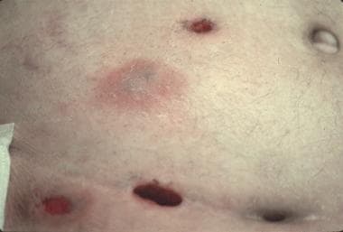 Violaceous plaques and necrotic ulcers on the abdomen of a renal transplant patient. Tissue cultures were positive for Pseudomonas aeruginosa.
Related Medscape articles include Pseudomonas aeruginosa Infections and Pseudomonas Infection.
Pathophysiology
Impaired humoral or cellular immunity leads to increased susceptibility to infections with P aeruginosa or other pathogens. In addition, breakdown of mechanical defensive barriers, such as the skin and mucosa, may allow infectious organisms to disseminate. The lesions of ecthyma gangrenosum (EG) result from perivascular bacterial invasion of arteries and veins in the dermis and subcutaneous tissues, producing a necrotizing vasculitis. [10] Perivascular involvement can occur by hematogenous seeding of the skin in bacteremic patients or by direct inoculation through the skin in non-bacteremic patients. Extravasation of blood, edema, and necrosis around the vessel interrupt the blood supply to these tissues, resulting in secondary ischemic necrosis of the epidermis and dermis, which manifests as nodular lesions that rapidly evolve through stages of central hemorrhage, ulceration, and necrosis.
Etiology
Ecthyma gangrenosum (EG) is typically and most commonly caused by P aeruginosa; however, EG-like lesions have been observed in patients with other bacterial and fungal infections. [11] Organisms that cause ecthyma and EG-like lesions include the following:
Gram-positive bacteria are as follows:
-
Staphylococcus aureus
-
Streptococcus pyogenes
Gram-negative bacteria are as follows:
-
Aeromonas hydrophila
-
Burkholderia cepacia [12]
-
Chromobacterium violaceum [13]
-
Citrobacter freundii
-
Corynebacterium diphtheriae
-
Escherichia coli
-
Klebsiella pneumoniae
-
Morganella morganii [14]
-
Neisseria gonorrhea
-
Pseudomonas aeruginosa
-
Pseudomonas stutzeri
-
Serratia marcescens
-
Vibrio vulnificus
-
Yersinia pestis
-
Xanthomonas maltophilia
Fungi are as follows:
-
Aspergillus fumigatus
-
Candida albicans [15]
-
Curvularia species [16]
-
Exserohilum species
-
Fusarium solani [17]
-
Mucor and Rhizopus species
-
Pseudallescheria boydii [16]
-
Scytalidium dimidiatum
Viral causes include herpes simplex virus. [18]
Epidemiology
Frequency
Ecthyma gangrenosum (EG) develops in 1.3-13% patients with P aeruginosa sepsis and, to a lesser extent, in patients who are not bacteremic.
Sex
No sexual predilection is evident in the overall prevalence of EG; however, a slight predominance of bacteremic EG in males (male-to-female ratio, 1.3-5:1) and nonbacteremic EG in females (female-to-male ratio, 2.3:1) has been observed.
Age
EG may affect patients of any age, although it is commonly reported in infants and elderly patients due to underdeveloped and/or compromised immune systems, including chronic granulomatous disease. [19]
Prognosis
A high mortality rate is reported with delayed diagnosis and therapy. Mortality rates of Pseudomonas sepsis in immunocompromised persons range from 18-96%, whereas the mortality rate of ecthyma gangrenosum (EG) in nonbacteremic patients is 15.4%. Coexisting conditions in patients prone to Pseudomonas sepsis may contribute to the morbidity and mortality rates.
Factors associated with a poor prognosis include the following:
-
Multiple lesions
-
Delayed diagnosis and institution of appropriate antibiotics
-
Persistent neutropenia
-
High bacteremic load
-
Repeated catheterization and instrumentation
-
Violaceous plaques and necrotic ulcers on the abdomen of a renal transplant patient. Tissue cultures were positive for Pseudomonas aeruginosa.


