Overview
Injury to the skin initiates a cascade of events that is collectively known as wound healing. Wound healing attempts to restore the barrier function as well the tensile strength to the injured skin. When healing deviates from its orderly pattern, abnormal or hypertrophic scarring can result. Although pigment and vascular alterations are often transient in nature, [1] textural changes caused by collagen disruption can be permanent, particularly in the case of keloid scarring. [2]
The wound-healing process is divided into 3 sequential yet overlapping stages known as inflammation, granulation, and remodeling. [3] The initial stage is characterized by a response to cutaneous injury involving inflammatory cells. [4] Neutrophils are the first cells present in an injury, and they serve to debride bacteria and necrotic tissue as well as recruit other mediators. [4] Subsequently, macrophages arrive and elaborate a variety of cytokines, including vascular endothelial growth factor (VEGF), thereby creating an environment that promotes granulation tissue formation. [3]
During the proliferative phase, fibroblasts arrive, proliferate, and deposit collagen, which initially consists of type III and later, type I. [5] Simultaneously, angiogenic factors released into the wound environment stimulate the formation of new capillaries. Keratinocytes also migrate across the wound, leading to reepithelialization. [5]
During remodeling, there is simultaneous collagen formation and degradation, while the presence of myofibroblasts contribute to increasing the tensile strength within the wound. [2] Granulation tissue deposition wanes as the cells responsible during this stage undergo apoptosis; failure for this to occur may result in a hypertrophic scar. [6] In the case of a hypertrophic scar, an overzealous healing response occurs, in which fibroblasts, small vessels, and collagen fibers are arranged in a nodular pattern. [2] Alternatively, collagen can be inadequately replaced and, as a result, can form a pitted appearance resembling the surface of a golf ball.
Although scars rarely pose a significant health risk, patients with exaggerated scarring can present with physical and psychosocial distress. [6] The physician should aim to minimize scar formation or at least anticipate its severity, considering the patient’s history of hypertrophic/keloid scarring, as well as body site (as certain regions sustain greater tension, leading to an increased risk of scarring). [7]
The purpose of this article is to review the strengths and limitations of current laser technology used to improve the appearance and symptomatology of hypertrophic scars, keloids, striae, atrophic scars, and acne scars.
Related Medscape articles include Keloid and Hypertrophic Scar and Scar Revision.
Background
During the early 1980s, Anderson and Parrish revolutionized dermatologic laser treatment by defining a concept known as selective photothermolysis. [8] This theory describes the use of laser energy to achieve localized photothermal injury of a targeted chromophore. From this idea, pulsed lasers were developed in which a short burst of photoenergy (photons) is delivered to a particular chromophore whose optimal wavelength of photoabsorption is distinct from its surrounding tissue. [8] This technique facilitates transfer of heat from photons, with the aim of keeping the converted thermal energy confined to a particular target, thereby diminishing widespread destructive or nonselective effects on normal surrounding tissue. [8] Despite the characterization of selective photothermolysis, to this day, the exact mechanism by which lasers improve the appearance and quality of scars remains largely unknown.
Early laser treatment of hypertrophic and keloid scars used ablative continuous-wave argon and carbon dioxide devices. [9] Early reports were encouraging, but subsequent studies could not confirm efficacy. [9]
Norris conducted a retrospective study of keloids treated with carbon dioxide laser and concluded this laser alone failed to suppress recurrence. [10] Another split-scar case series comparing carbon dioxide and argon laser treatment of keloids demonstrated temporary symptomatic improvement but showed sustained response in only one patient at 6-month follow-up. [11] Implementation of the continuous-wave neodymium:yttrium-aluminum-garnet (Nd:YAG) laser (1064 nm) also showed initial signs of efficacy. [9] This laser exerts a photobiologic effect on fibroblasts, causing suppressed collagen deposition. [12] The carbon dioxide, argon, and Nd:YAG lasers may be capable of attenuating collagen synthesis initially but fail to prevent recurrence, particularly in keloids. [11]
Following the aforementioned studies, further investigation demonstrated that the 585-nm pulsed-dye laser (PDL) was a viable treatment option for scars. Alster et al first used the PDL to treat scars created by argon laser–treated port-wine stains, with improvement in the clinical appearance of all 10 patients in the series. [13] The observed flattened topography was purported to occur owing to the laser’s ability to eradicate the enlarged, trapped blood vessels within the scar itself. [13] In 1994, Alster [14] reported clinical and textural improvement in long-standing erythematous and hypertrophic scars, with 57% improvement following one PDL treatment and 83% improvement after 2 treatments. For a better result, Ebrahimi et al recommend PDL within two weeks of surgery after sutures have been removed. [15]
Dierickx and colleagues [16] had similar findings the following year, in which they reported an average improvement of 77% after 1.8 laser treatments of erythematous or hypertrophic scars. Later, Alster and Williams [17] compared the clinical, textural, histologic, and symptomatic responses in a split scar study involving hypertrophic and keloidal median sternotomy scars. Significant improvement in texture, erythema, scar height, and pruritus was observed at 6 months after the PDL treatment. [17] In addition, they histologically demonstrated increased numbers of mast cells at the lasered sites. Subsequent studies also showed improvement in keloid scars following PDL treatment. In 1996, [18] Alster and McMeekin also reported improvement in erythematous and hypertrophic facial acne scars following treatment with the 585-nm PDL.
In 2003, Nouri and colleagues [19] showed that the 585-nm PDL can improve the quality and appearance of surgical scars when used as early as the day of suture removal. Scars were treated 3 times at monthly intervals and were significantly more improved compared with controls in overall Vancouver Scar Scale comparisons (ie, vascularity, pliability, height, and cosmetic appearance).
In a 1995 report, Goldman and Fitzpatrick [20] described a combination approach to scar management, in which they treated facial and nonfacial scars with the PDL and intralesional triamcinolone, concluding that PDL served as a useful adjuvant in erythematous, hypertrophic scars. Improvement in nonerythematous, minimally hypertrophic scars was also achieved following combination treatment involving pulsed dye technology and carbon dioxide laser vaporization. In 1998, Alster and Lewis [21] treated selected scars by performing carbon dioxide laser deepithelialization followed by PDL therapy. Significant and prolonged clinical and textural improvement was observed in all treatment areas.
No consensus exists regarding the mechanism by which the PDL achieves clinical improvement in scars. Purported mechanisms include (1) laser-induced microvasculature damage leading to tissue hypoxia with subsequent collagen degradation via release of collagenase [22, 23] ; (2) thermal damage to collagen fibers dissipated from adjacent vessels with dissociation of disulfide bonds and collagen realignment [22] ; and (3) increased regional mast cells, which may serve to stimulate collagen remodeling. [23]
Striae distensae, which are characterized by atrophic linear erythematous or fibrotic bands, due to excessive dermal stretching and elastolysis, have been notoriously challenging to treat. [23] Laser therapy of these aesthetically distressing lesions has been attempted secondary to the failure or inefficacy of topical treatments. Elmorsy et al found that fractional carbon dioxide laser resurfacing of striae distensae reduced dimensions of the lesions to a statistically significant degree. [24] McDaniel et al demonstrated improvement in surface topography and increased dermal elastin in striae treated with the 585-nm PDL at low fluence. [25] Alster and colleagues [21] also found that low-fluence PDL irradiation outperformed PDL fluences used for scars combined with pulsed carbon dioxide vaporization. Given that striae histologically demonstrate dysmorphic elastin and reduced collagen fibers, [23] the purported mechanism for improvement with the PDL likely involves influence on these dermal components. [23]
Types of Scars
Hypertrophic and keloid scars
Hypertrophic scars are pink, raised, firm, erythematous scars. They occur approximately within a month following surgery or trauma and result from overzealous collagen synthesis, mainly type III, coupled with limited collagen lysis during the remodeling phase of wound healing. [23] The result is the formation of thick collagen bundles consisting of fibroblasts and fibrocytes, arranged in nodules rather than in the normally smooth fashion. [26] Hypertrophic scars may be symptomatic, characterized by pruritus and/or dysesthesia, [23] and are more likely to arise in sites subjected to increased pressure or movement. [26] Despite obvious tissue proliferation, hypertrophic scars remain within the confines of the original integumental injury, in distinction to keloid scars, which extend beyond the original cutaneous injury. [23] Unlike keloids, which tend to persist indefinitely, hypertrophic scars may regress spontaneously. [26] This pathologic scarring is more common than keloid scaring. Seethe image below.
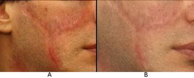 Erythematous and hypertrophic laceration scars before (A) and 3 months after (B) a second 585-nm pulsed dye laser treatment. Improvement in scar redness, symptomatology (decreased pruritus), and thickness were achieved. Courtesy of Tina S. Alster, MD.
Keloids are raised, reddish-purple, nodular scars that are firmer than hypertrophic scars, [23] and they may develop weeks or years after the initial insult, or even arise spontaneously. [26] Keloids develop during an extended proliferative phase of wound healing, [26] and they demonstrate thick, hyalinized bundles of collagen arranged haphazardly in whorls, in a mix of type I and type III, with increased hyaluronidase. [26] Unlike hypertrophic scars, keloids extend beyond wound margins and may even continue to grow over time. [26] Although they occur in all skin types, keloids are most common in patients with darker skin and, like hypertrophic scars, may be associated with pruritus and/or dysesthesia. [26] See the image below.
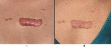 Keloid scar on the anterior chest before (A) and several months after (B) a second 585-nm pulsed dye laser treatment. While decreased erythema and scar bulk are noted, further pulsed dye laser treatments are necessary (at bimonthly intervals) to provide further scar improvement. Courtesy of Tina S. Alster, MD.
Striae distensae
Striae distensae, or stretch marks, are linear bands of atrophic or wrinkled skin. [26] They result from excessive dermal stretching, such as after rapid weight loss/gain, pregnancy, or pubertal growth spurts. [26] Dermal inflammation with mast cell degranulation, elastolysis, and dilated capillaries mark the initial presentation, [27] which results in an erythematous appearance of young striae distensae (stria rubra). [26] Later, striae appear hypopigmented and fibrotic owing to linear deposition of dermal bundles and thinning of the overlying epidermis (stria alba). [27] Their pathogenesis remains unclear, although estrogen and mast cell degranulation with elastolysis may be contributing factors. [26] See the image below.
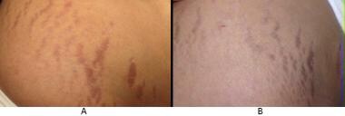 Erythematous (early) striae distensae before (A) and after (B) a single 585-nm pulsed dye laser treatment. Reduction in erythema and mild improvement of skin surface texture were observed. Courtesy of Tina S. Alster, MD.
Atrophic scars
Atrophic scars are dermal depressions that most commonly are the sequelae of an acute inflammatory process that has caused collagen destruction and dermal atrophy. [26] Examples of inciting events include cystic acne, varicella infection, surgery, and trauma. [26] On the skin, these pitted lesions form a surface resembling the dimples on a golf ball. This topographical irregularity tends to be difficult to treat, but therapy is generally aimed at resurfacing the distorted topography. [28]
Acne scars
Acne scars may be categorized as hypertrophic or atrophic, the latter of which can be further characterized as ice-pick, rolling, or boxcar scars. [28, 29] Ice-pick scars are usually narrow (< 2 mm), sharply demarcated tracts that can reach deep into the dermis or even the subcutaneous tissue. [29] They are typically wider at the epithelial surface and taper as they go deeper. [29] Rolling scars tend to be shallower, wider (4-5 mm), and produce an undulating appearance in otherwise normal-appearing skin. This rise and fall of the skin surface is due to abnormal fibrous attachment of the dermis to the subcutis. [29] Boxcar scars are wider at the base than ice-pick scars, but do not taper. These round- to oval-shaped skin dimples have sharp margins and can be either shallow (0.1-0.5 mm) or deep (>0.5 mm). [30] See the image below.
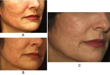 Atrophic acne scars before full-face carbon dioxide laser resurfacing (A). Six months after the procedure (B), mild improvement was observed in terms of scar severity and skin surface texture. One year later (C), further clinical improvement was apparent because of continued and prolonged collagen remodeling. Courtesy of Tina S. Alster, MD.
Patient Variables
When evaluating a potential candidate for laser surgery, the physician should consider certain factors that can complicate therapy. While not absolute contraindications to laser surgery, these factors can significantly influence treatment.
Darker skin phototypes
Racial background is an important consideration when assessing the likelihood of a scar developing into a keloid or hypertrophic scar. Both scar subtypes occur with greater frequency among individuals with darker skin types. [31] In fact, keloid prevalence among blacks and Latinos has been estimated to range from 4.5-16%. [32] Likewise, the physician should consider skin type when contemplating and discussing outcomes of laser treatment. For example, increased epidermal pigment in darker skin tones (phototypes III or greater) competes with hemoglobin to absorb energy delivered by vascular-specific lasers. [33] As a result, the amount of energy effectively delivered to the dermal scar tissue is reduced, yielding inferior results. [33]
Furthermore, laser destruction of epidermal melanin may result in inadvertent postoperative hypopigmentation. [33] When considering cutaneous laser resurfacing with the carbon dioxide or erbium:yttrium-aluminum-garnet (Er:YAG) laser, the patient and the treating physician must be prepared for the possibility of transient posttreatment hyperpigmentation. [34]
Concurrent inflammation or infection
Infectious and inflammatory processes must resolve before performing laser surgery. In cases of bacterial or viral infection (eg, herpes simplex, verrucae), the physician should consider that the infection can spread after laser irradiation. [34, 35]
In cases of concurrent inflammatory skin disorders (eg, cystic acne, psoriasis, dermatitis), the condition may worsen with laser treatment, and dermal inflammation may impede postoperative healing and clinical effects. [35]
Medication use
Patients seeking laser resurfacing for atrophic acne scars often have a history of severe acne, which may currently be or have previously been treated with isotretinoin. [35, 36] Recent use of this medication in combination with laser resurfacing can, ironically, yield hypertrophic scarring, due to isotretinoin’s effects, namely, impaired wound healing and inhibition of collagenase production. [36] Therefore, laser resurfacing should be delayed until at least 6 months after cessation of isotretinoin use. [35, 37]
However, Mysore et al disagree with this assessment, stating that recommendations against laser scar treatment with isotretinoin use are based on poor and limited evidence. Instead, they recommend a test procedure to ensure safety. [38]
Unrealistic expectations
Unfortunately, no currently available treatment yields 100% improvement. Patients must be informed that some degree of scarring can persist and multiple re-treatments may not completely eradicate the scar(s). [35] Nevertheless, patient compliance should be encouraged to optimize results and avoid complications. [35]
Treatment Options
Treatment of hypertrophic scars and keloids
The PDL is largely considered the first-line therapy amongst lasers for treating hypertrophic scars and keloids. [23] In these cases, PDL therapy is usually performed in an outpatient setting. If anesthesia is warranted, topical lidocaine cream (eg, 30% lidocaine powder in a water-miscible cream base) with or without occlusion for 30 minutes is sufficient. The eutectic mixture of lidocaine 2.5% and prilocaine 2.5% cream or liposomal lidocaine 4% cream with or without occlusion for 30-60 minutes prior to treatment are other reasonable alternatives. All creams and make-up should be removed with wet gauze prior to laser irradiation. Patients with scars in sensitive body locations (eg, lips, breast, perineum, fingers) may benefit from intralesional injections or nerve blocks. [35]
The surgical technique calls for a series of adjacent nonoverlapping laser pulses delivered across the entire scar breadth. [35] The entire scar should be treated at each session. The scar's size, thickness, location, and color, as well as the patient's skin type, determine the energy density that should be used. Less fibrotic scars in sensitive skin areas (eg, anterior chest and breast) require lower energy densities, while thicker or darker scars can be treated with higher fluences (see Pulsed dye laser treatment considerations and protocol in Summary).
In general, treatments should begin at lower fluences, allowing for flexibility of upward energy adjustment depending on the scar’s response to earlier treatments. [35] If the initial treatment session produces beneficial results, the energy density should remain constant on subsequent treatments. If minimal results were achieved, one should consider increasing treatment fluences in steps of 10%. If the patient reports postoperative vesiculation or crusting, consider a lower fluence with special attention to operative technique (ie, avoiding overlapping pulses).
Postoperative purpura following treatment with the PDL usually resolves within 7-10 days. [14] During the healing process, the patient should avoid extraneous manipulation of the treatment area. Showers are permitted, but care should be taken to lightly pat lasered areas dry. Gentle cleansing of the treatment area with water and a mild soap followed by application of a topical ointment can be used to keep the area clean. A nonstick bandage should cover the treated area. The treated area should be evaluated in approximately 6-8 weeks, at which time another laser treatment can be delivered.
The most common adverse effect is hyperpigmentation of lasered skin. Hyperpigmentation spontaneously fades with avoidance of or protection from sun exposure. If hyperpigmentation is present, consider postponing subsequent laser treatments to avoid interference from a competing chromophore (or target), such as melanin. Consider prescribing a hydroquinone-containing cream (applied qd-bid) to speed up the fading process.
Occasionally, patients develop allergic contact dermatitis secondary to topical antibiotic use or irritant dermatitis from an adhesive bandage. If a post-laser rash is present, determine if it is a normal purpuric response or nonpurpuric and unrelated to laser irradiation. If concurrent pruritus is reported, consider contact dermatitis. A mild topical corticosteroid cream should be applied until the dermatitis resolves. The offending agent should be immediately discontinued. [35]
Hypertrophic scars have an average of at least 50-80% improvement after 2 laser treatments. Keloid scars and more fibrotic hypertrophic scars usually require additional laser treatments to achieve desired results.
Of note, keloid scars have also been treated with chemotherapeutic agents such as 5-fluorouracil (5-FU). This has been noted to have a low rate of recurrence. Other options for treatment are concomitant radiation therapy, corticosteroid injection, and interferon. [39]
Treatment of striae distensae
The 585- or 595-nm PDL can be used in the treatment of striae distensae, which respond best to lower energy densities (3 J/cm2). [23] Adjacent nonoverlapping laser pulses are delivered such that each individual stria distensae is covered. Irradiated striae distensae do not typically exhibit the characteristic purpura observed with the treatment of hypertrophic scars and keloids. Because of lower fluences, striae usually appear mildly pink, which represents mild postoperative tissue hyperemia and edema. Vesiculation and crusting should not occur when proper fluences and operative technique are used. Typically, only 1-2 treatment sessions are necessary to obtain desired results. [35]
Ablative and nonablative carbon dioxide lasers have also been used in the treatment of striae. Yang and colleagues performed a study to compare these laser treatments in ethnic patients. Twenty-four South Korean patients with varying degrees of atrophic striae albae in the abdomen were enrolled in a randomized, blind, split study. The patients were treated with 1,550-nm fractional Er:glass laser and ablative fractional carbon dioxide laser resurfacing. Each half of the abdominal lesion was randomly selected and treated 3 times at intervals of 4 weeks using the same parameters. Although they do not differ statistically, both treatments with the nonablative fractional laser and ablative carbon dioxide fractional laser showed a significant clinical and histopathologic improvement of striae distensae over pretreatment sites. [40]
Postoperative management is similar to the protocol followed by patients treated for hypertrophic and keloid scars. Instruct patients to gently cleanse treatment areas with water and a mild soap. A topical ointment such as petrolatum should be applied daily and the treatment area covered with a nonstick bandage. Patients should be advised to avoid sun exposure to the treatment area during the course of treatment.
Treatment of atrophic scars
Recontouring of atrophic facial scars with carbon dioxide and Er:YAG laser vaporization has become a highly desired procedure by patients. [21, 25, 34, 41, 42] Through selective ablation of water-containing tissue, both laser systems offer predictable and reproducible vaporization of tissue, yielding greater precision than dermabrasion. [43, 44, 45, 46, 47, 48, 49] During laser resurfacing, the epidermis and a variable portion of the dermis are destroyed. [50, 51] In a 1996 study, Fitzpatrick and colleagues demonstrated that depth of skin vaporization and residual necrosis secondary to carbon dioxide laser resurfacing were directly proportional to pulse energy and number of laser passes delivered. [52]
The pulsed Er:YAG lasers are 10 times more selective for water than their carbon dioxide counterparts; therefore, they result in enhanced tissue vaporization and reduced residual thermal damage to the dermis. [49] Postoperative erythema is decreased; however, the limited photothermal effect on tissue is countered by an overall decrease in clinical improvement. [49] Thus, short-pulsed Er:YAG laser resurfacing delivers less collagen shrinkage compared with that observed with carbon dioxide laser treatment. [34] For milder atrophic scarring, the Er:YAG may be the preferred modality given the shorter postoperative recovery times.
Regardless of the system, the goals are 2-fold: (1) to soften the transition between the atrophic indentation and the intact (normal) skin surrounding it and (2) to stimulate collagen production within the atrophied area. [35] The entire cosmetic unit must be treated to minimize textural or color mismatch. If treating an isolated scar, one should consider spot resurfacing. In an effort to decrease treatment time when lasering large cutaneous areas, a scanning hand piece can be used. Once deepithelialization is achieved (typically requiring 1 pass with the carbon dioxide laser at 300 mJ and 2-3 passes with the Er:YAG laser at 5 J/cm2), the scar edges, or shoulders, can be further sculpted with additional vaporizing laser passes. Partially desiccated tissue should be removed with saline-soaked or water-soaked gauze after each laser pass to prevent charring. [52]
Typically, 300 mJ of energy and 60 watts of power with variable-sized and variable-shaped patterns are the parameters used with the computer pattern generator (CPG) scanning device (Coherent UltraPulse). [35] Scanning devices attached to other carbon dioxide laser systems (Sharplan FeatherTouch or Luxar NovaPulse) can be used at 5-20 watts per scan, depending on the system and severity of scarring. [35] Scan sizes ranging from 4-10 mm in diameter are delivered to the treatment area. Treatment usually requires 2-3 passes, and the physician should take care to remove all partially desiccated tissue between passes. Individual scar edges can be further sculpted using smaller-diameter spots or scans following treatment of the entire cosmetic unit.
The Er:YAG laser is used with a 5-mm spot size at 1-3 J (5-15 J/cm2) to deepithelialize and sculpt individual scars. A laser technique similar to the carbon dioxide system is used with Er:YAG; however, because Er:YAG vaporization does not typically produce a significant quantity of partially desiccated tissue, wiping between laser passes is not necessary except in hair-bearing areas (to reduce thermal conduction to skin through singed surface hairs). Bleeding typically is observed by the third laser pass as the result of dermal penetration and the inability of the Er:YAG laser to photocoagulate blood vessels. [34]
When using either the carbon dioxide or Er:YAG laser, treated skin appears erythematous and edematous immediately postoperatively, with further worsening for the next 48 hours due to sloughing of coagulated tissue. [52] Symptomatic palliation may be achieved with application of cool compresses, topical emollients, nocturnal head elevation, or semiocclusive dressings. [53] The first postoperative week is critical. It is important to closely monitor patients for appropriate healing responses and complications such as dermatitis and infection. For Er:YAG laser resurfacing, reepithelialization typically takes 4-7 days, whereas carbon dioxide laser resurfacing requires 7-10 days. [34]
A significant adverse effect that may occur with either carbon dioxide or Er:YAG is transient hyperpigmentation. Although hyperpigmentation is more common in patients with darker skin tones, it may occur in any skin type. Transient hyperpigmentation is observed early in the postoperative course, occurring approximately 1-2 months after treatment. [54, 55] While the process is usually self-limited, resolution may be hastened with bleaching creams (eg, hydroquinone, arbutin) or acid preparations (eg, glycolic, retinoic, azelaic, kojic, ascorbic). [34] Hypopigmentation is a relatively late sequela of treatment (typically observed ≥6 mo postoperatively) and appears to be permanent. [34]
Infection is another postoperative concern because reepithelializing skin is vulnerable to bacterial (eg, staphylococci, pseudomonas), viral (eg, herpes simplex), and fungal (eg, Candida organisms) infections. [34] Incidence can be reduced with appropriate use of prophylactic antibiotics and, more importantly, aggressive postoperative wound care. Suspected infection must be diagnosed and treated early. [54, 55]
The most severe complications of laser resurfacing include hypertrophic scarring and ectropion formation, which may result from overly aggressive intraoperative laser technique. [54] Hypertrophic burn scars can be effectively treated with 585 nm PDL irradiation as described earlier; ectropion typically requires surgical reconstruction.
Collagen remodeling with further scar improvement may occur for 12-18 months postoperatively, so consider postponing re-treatment of residual scars for at least 1 year to accurately gauge clinical improvement. The Er:YAG laser system, although effective in the treatment of atrophic scars, does not offer the same amount of collagen remodeling as the carbon dioxide laser system. The Er:YAG laser should be reserved for sculpting of individual scar edges and treatment of mild acne scars. [49, 56]
In a double-blind study of 20 patients with dermatologic surgery scars, 94% of patients preferred the results of fractionated Er:YAG laser treatment over the results of fully ablative Er:YAG laser treatment. [57]
Treatment of traumatic facial scars
Tripathi et al proposed a plan for laser treatment of traumatic facial scars. They recommend beginning with laser resurfacing of narrow facial scars parallel to relaxed skin tension lines (RSTLs), in a favorable location, and without contractures. Otherwise, their recommendation is to begin with surgical revision. In the case of laser resurfacing, erythematous scars should be treated with pulsed-dye, nonablative fractional, or erbium glass laser. Hypertrophic scars should undergo resurfacing with pulsed-dye, ablative, fractional carbon dioxide laser or Nd:YAG laser, and atrophic scars should be treated with nonablative, fractional Er:YAG laser or erbium glass laser. [58]
Treatment of acne scars
Lasers are relatively safe and effective options that remodel the skin to improve its appearance. In addition to lasers, numerous modalities can be used to treat acne scars, including excision, punch grafting, subcision, cryosurgery, dermal fillers, chemical peels, and silicone sheeting compression. [59] Ice-pick scars usually extend too deep into the dermis to be reached by conventional treatments and may require a punch excision for removal. [35, 59, 60] For rolling scars, therapy should be aimed at treating the irregular underlying anchoring between the dermis and subcutis. [35, 60] Therefore, laser revision is usually limited to shallow boxcar and superficial scars. [35, 59, 60]
Ablative laser resurfacing with either a carbon dioxide or Er:YAG laser may be beneficial. After the initial treatment, allow the skin to heal, which may take 6-8 weeks. The postoperative erythema may last for up to 12 weeks. The scars can be treated with additional laser sessions to achieve the desired dermal remodeling and skin appearance.
As stated, resurfacing with a carbon dioxide laser can carry many potential risks such as delayed posttreatment hypopigmentation and scarring, as well as prolonged healing after the procedure. [54] This ablative laser can be effective alone for scarring after acne, but the risks must be considered before a patient undergoes this procedure. To enhance the selectivity of the carbon dioxide laser, some have tried combining it with an Er laser, which is taken up more preferentially than the carbon dioxide beam (detailed below). The erbium laser's cutaneous destruction is much more localized because the energy dissipates quickly within the targeted tissues. This procedure is more selective and less damaging to the skin than carbon dioxide laser resurfacing.
The carbon dioxide laser can be used to first treat the scar, followed by irradiation with an Er laser to further remodel the ablated, carbon dioxide–treated tissue. This speeds the wound healing process and reduces the potential complications associated with only using a carbon dioxide laser. [54, 61]
In contrast to ablative resurfacing, nonablative lasers do not noticeably disrupt the epidermis, but deliver thermal energy and damage to the underlying dermis. [41] These lasers induce collagen remodeling and production, which is predominantly collagen type III. In time, the collagen expression changes to contain a greater proportion of type I collagen. [41] With these lasers, clinical improvement usually requires more than one treatment and results can continue to improve months after the laser treatments have been completed.
In 1996, Alster and McMeekin demonstrated that the 585-nm PDL could improve erythematous and hypertrophic acne scars. [18] In 22 patients, significant improvements in texture and redness were seen after 1 or 2 treatments (6-7 J/cm2; 7-mm spot size). Six weeks following only one laser treatment, the mean improvement was 67.5%. Eight of the patients received an additional laser treatment and saw an average improvement of 72.5% 6 weeks later.
Atrophic acne scarring has been treated with a 1064-nm Q-switched Nd:YAG laser. [62] Eleven patients with mild-to-moderate atrophic scarring were treated with 5 laser sessions at 3-week intervals. The laser was set at an average fluence of 3.4 J/cm2, 4- to 6-nanosecond pulse duration, and a 6-mm spot size. Skin roughness was significantly better (23.3%) 1 month after the fifth laser session. Further improvements continued with time. At the 6-month follow-up, patients demonstrated a statistically significant 39.2% improvement from baseline measurements. This sustained improvement is likely from an enduring dermal collagen remodeling after the laser treatments have concluded. The long-term results and safety profile (ie, mild-to-moderate erythema, pain, pinpoint petechiae) allow this laser to be a viable option for patients with mild-to-moderate atrophic acne scars.
The 1320 nm Nd:YAG laser with a built-in cryogen cooling spray has been used for acne scarring. In 2004, Sadick and Schecter [63] treated 8 patients with 6 monthly irradiations of 3 passes each and found a modest improvement. Ice-pick scars without fibrosis responded more favorably than those with fibrous tracts. Statistically significant improvements were noted in 7 of 8 patients 5 months and 1 year after their final treatments. When only 3 treatments were used, another study found that atrophic scars improved the most. In Asian patients, there may be only a mild response. Of 27 patients treated, 8 had no objective improvement and 9 were only mildly better than at baseline. This modality may produce better results if combined with another modality, such as surgery or an intense-pulsed light source (IPL).
A 2004 comparison by Tanzi and Alster [64] evaluated the efficacy of a 1450-nm diode laser versus a 1320-nm Nd:YAG for atrophic facial scars. Twenty patients with mild-to-moderate scarring each received 3 monthly treatments. Each half of the patient's face was randomized to 1 of the 2 lasers. After completing the treatments, the greatest clinical effect was seen at 6 months, which was consistent with the observed histologic increase in collagen production. Only modest improvements were seen with both lasers, but the 1450-nm diode laser resulted in greater improvements. Both lasers are safe, noninvasive options to improve the appearance of mild-to-moderate facial atrophic scars.
A recent laser technology, the fractional lasers, deliver high-intensity light fractionated through focused lenses to uniformly generate arrays of microscopic columns of thermal injury surrounded by uninjured tissue. [65] Tiny columns of injury are termed microscopic thermal-treatment zones. The operator can adjust the energy of the laser and the density of the microscopic thermal-treatment zones. Many fractional laser devices have been approved for the treatment of acne scars, periorbital rhytides, skin resurfacing, soft tissue coagulation, and melasma. [65]
The undamaged surrounding tissue allows for a reservoir of viable tissue, permitting rapid epidermal repair, decreasing patient downtime. [65] Both ablative and nonablative fractional lasers have been tried successfully for the treatment of scars.
In a 2009 study, the efficacy and safety of the nonablative, 1540-nm Er:glass fractional laser in the treatment of surgical and posttraumatic scars was evaluated. [66] A histological study was conducted on a postsurgical scar to follow the time course of healing post treatment and the impact of the fractional treatment on normalization of scar tissue, as compared with baseline histologic findings of the scar. Histologic findings demonstrated rapid reepithelialization of the epidermis within 72 hours of treatment. Remodeling of scar tissue with renewal and reorganization of collagen fibers in the dermis was noted 2 weeks post treatment. Relative to baseline, 73% of treated scars improved 50% or more and 43% improved 75% or more. [66]
Additionally, a new combination therapy is suggested that incorporates dot peeling (the focal application of higher trichloroacetic acid concentrations), subcision, and fractional laser irradiation. In a pilot study, the efficacy and safety of this method was investigated for the treatment of acne scars. Acne scar severity scores decreased by a mean of 55.3%. Eighty percent of the patients reported significant or marked improvement. [60]
Cho et al studied the efficacy and safety of single-session treatments of 1550-nm Er-doped fractional photothermolysis systems and 10,600-nm carbon dioxide fractional laser systems for acne scars through a randomized, split-face, evaluator-blinded study with 8 patients with acne scars. [67] Half of each subject's face was treated with a fractional photothermolysis system and the other half was treated with a carbon dioxide fractional laser system. At 3 months after treatment, the mean grade of improvement based on clinical assessment was 2 ± 0.5 for the fractional photothermolysis system and 2.5 ± 0.8 for the carbon dioxide fractional laser systems. [67]
A 2010 pilot clinical study demonstrated that lasers could also be used immediately after surgery to reduce the appearance of scars. The laser-assisted skin-healing (LASH) technique induces a temperature elevation in the skin, which modifies the wound healing process. Capon et al demonstrated that 810-nm diode laser treatment, performed immediately after surgery, can improve the appearance of a surgical scar. [68] The dose plays a significant role in scar improvement and must be well controlled. The authors additionally suggested that LASH could be used for hypertrophic scar revision.
Wada and colleagues performed an open study on 24 Japanese patients (17 female and 7 male, aged 15-44 y) with acne scars on the face treated with 5 sessions of low-energy, double-pass, 1450-nm diode laser at 4-week intervals. [69] The mean duration of the acne scars prior to receiving laser therapy was 4.8 years (range 1-9 y). Clinical evaluation by physicians and with photographs was conducted at baseline, 1 month after the final treatment, and at a 3-month follow-up visit. Topical therapies for acne vulgaris were permitted during the follow-up period. All patients completed the 5 treatment sessions. Seventy-five percent of the subjects showed at least 30% improvement of acne scars. At the 3-month follow-up evaluation, 92.9% of the subjects with greater than 30% improvement maintained the effectiveness. [69]
Summary
Current laser technology permits successful treatment of scars and striae distensae. It is imperative to appropriately classify the type of scars and striae distensae present to determine which laser is likely to be effective. The 585-nm PDL is best used to treat hypertrophic scars, keloids, and striae. The carbon dioxide and Er:YAG laser systems effectively resurface atrophic scars. Fractional lasers are becoming increasingly used for treating scars owing to their efficacy with reduced downtime compared with the ablative nonfractional carbon dioxide and Er:YAG lasers.
Table. Clinical Responses of Scars to Laser Therapy (Open Table in a new window)
Scar Type |
Laser Used |
Number of Treatments |
Hypertrophic |
585-nm pulsed dye |
2-4 |
Keloid |
585-nm pulsed dye |
2-6 |
Striae |
585-nm pulsed dye |
1-2 |
Atrophic |
High-energy, pulsed carbon dioxide or Er:YAG |
1-2 |
Pulsed dye laser treatment considerations and protocol
Preoperative consideration
Skin types I-III are best suited for treatment.
Hypertrophic scars are more amenable to treatment.
Treat all body locations when possible.
Patients should not take anticoagulants.
Intraoperative considerations
In most circumstances, use topical anesthesia or no anesthesia.
Energy densities may be as follows:
-
4.5-5.5 J/cm2 (10-mm spot)
-
6.0-7.0 J/cm2 (7-mm spot)
-
6.5-7.5 J/cm2 (5-mm spot
Deliver adjacent, nonoverlapping spots.
Postoperative considerations
Patients should use a topical antibiotic ointment.
Advise the patient to use sunscreen and avoid sun exposure.
Evaluate for re-treatment at 6-8 weeks.
Consider bleaching for hyperpigmentation.
Carbon dioxide or Er:YAG laser resurfacing of atrophic scars
Preoperative considerations
Skin types I-II are best suited for treatment.
This laser is used for nonpitted atrophic scars.
Facial scars respond best.
Previously treated scars are more difficult to treat.
Patients should discontinue isotretinoin at least 6 months prior to treatment.
Patients should be free of concurrent infection and inflammatory disease.
Intraoperative considerations
Intravenous anesthesia is used in most cases.
Use energies that exceed critical vaporization threshold (>5 J/cm2).
Remove partially desiccated skin between passes.
Dampen hair-bearing areas.
Avoid flammable substances.
Postoperative considerations
Patients should use healing ointments and semiocclusive dressings.
Patients should use ice or cooling masks as needed.
Close follow-up is necessary.
Patients should frequently clean skin.
Consider prophylactic antibiotics and pain or sleep medications.
Observe for and treat adverse effects and complications as early as possible.
-
Erythematous and hypertrophic laceration scars before (A) and 3 months after (B) a second 585-nm pulsed dye laser treatment. Improvement in scar redness, symptomatology (decreased pruritus), and thickness were achieved. Courtesy of Tina S. Alster, MD.
-
Keloid scar on the anterior chest before (A) and several months after (B) a second 585-nm pulsed dye laser treatment. While decreased erythema and scar bulk are noted, further pulsed dye laser treatments are necessary (at bimonthly intervals) to provide further scar improvement. Courtesy of Tina S. Alster, MD.
-
Erythematous (early) striae distensae before (A) and after (B) a single 585-nm pulsed dye laser treatment. Reduction in erythema and mild improvement of skin surface texture were observed. Courtesy of Tina S. Alster, MD.
-
Atrophic acne scars before full-face carbon dioxide laser resurfacing (A). Six months after the procedure (B), mild improvement was observed in terms of scar severity and skin surface texture. One year later (C), further clinical improvement was apparent because of continued and prolonged collagen remodeling. Courtesy of Tina S. Alster, MD.
Tables
Scar Type |
Laser Used |
Number of Treatments |
Hypertrophic |
585-nm pulsed dye |
2-4 |
Keloid |
585-nm pulsed dye |
2-6 |
Striae |
585-nm pulsed dye |
1-2 |
Atrophic |
High-energy, pulsed carbon dioxide or Er:YAG |
1-2 |


