Practice Essentials
Varicose veins and telangiectasia (spider veins) are the visible surface manifestations of an underlying problem with reverse venous flow, which is also termed venous insufficiency syndrome. Mild forms of venous insufficiency are merely uncomfortable, annoying, or cosmetically disfiguring, but severe venous disease can produce serious systemic consequences and can lead to loss of life or limb. See the image below.
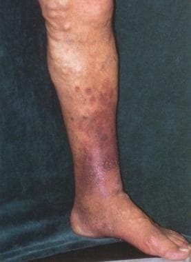 Patient with large tortuous varicose veins, high-volume venous reflux, and early stasis changes of the medial ankle.
See Superficial Venous Insufficiency: Varicose Veins and Venous Ulcers, a Critical Images slideshow, to help identify the common risk factors and features of this condition and its management options.
Signs and symptoms
Common chronic symptoms of varicose veins that should be elicited include the following:
-
Leg heaviness
-
Exercise intolerance
-
Pain or tenderness along the course of a vein
-
Pruritus
-
Burning sensations
-
Restless legs
-
Night cramps
-
Edema
-
Skin changes
-
Paresthesias
Common symptoms of telangiectasia include the following:
-
Burning
-
Swelling
-
Throbbing
-
Cramping
-
Leg fatigue
Aspects of symptoms include the following:
-
Subjective symptoms usually are more severe early in the progression of disease, less severe in the middle phases, and worse again with advancing age
-
Symptoms do not correlate with the size or extent of visible varices or with the volume of reflux
-
Not all symptomatic patients are aware of their symptoms, because the onset may be extremely gradual; after treatment, patients are often surprised to realize how much chronic discomfort they had accepted as normal
-
Pain associated with larger varicose veins is usually a dull ache that is worse after prolonged standing
-
Pain caused by venous insufficiency is often improved by walking or by elevating the legs, in contrast to the pain of arterial insufficiency, which is worse with ambulation and elevation
-
Pain and other symptoms may worsen with the menstrual cycle, with pregnancy, and in response to exogenous hormonal therapy (eg, oral contraceptives)
-
A small number of women regularly experience pain associated with their varicose veins after sexual intercourse
Inspection may reveal the following findings:
-
Ulceration
-
Telangiectasias
-
Atrophie blanche
-
Interdigital mycosis
-
Acrocyanosis
-
Eczematous lesions
-
Microulcers
-
Stasis dermatitis
-
Flat angiomata
-
Prominent varicose veins
-
Scars from a prior surgical operation
-
Evidence of previous sclerosant injections
Findings on palpation may include the following:
-
A firm, thickened, thrombosed superficial vein in an area of leg pain or tenderness
-
Deep boggy or spongy pockets in the calf muscle and deep palpable bony notches, especially over the anterior tibia, caused by erosion from chronic varices
-
Fascial defects in the calf along the course of an abnormal vein at sites where superficial tributaries emerge through openings in the superficial fascia
See Clinical Presentation for more detail.
Diagnosis
The following are the most useful modalities available for venous imaging:
-
Contrast venography
-
MRI
-
Color-flow duplex ultrasonography
Duplex ultrasonography is the standard imaging modality for diagnosis of varicose insufficiency syndromes and for treatment planning and preoperative mapping
For complex cases, the following physiologic tests of venous function may reveal more information:
-
Venous refilling time (VRT) – Results correlate with severity of venous insufficiency and reflux
-
Maximum venous outflow (MVO) – Detects obstruction to venous outflow from the lower leg, regardless of cause
-
Calf muscle pump ejection fraction (MPEF) – Detects failure of the calf muscle pump to expel blood from the lower leg.
See Workup for more detail.
Management
The following are the modern techniques used to ablate varicosities:
-
Sclerotherapy – The most widely used medical procedure for varicose veins and spider veins [1]
-
Laser and intense-pulsed-light therapy
-
Radiofrequency (RF) or laser ablation
-
Ambulatory phlebectomy
Common surgical approaches to large-vein varicose disease include the following:
-
Ligation of the saphenofemoral junction with vein stripping
-
Phlebectomy performed through microincisions
-
Endovenous RF thermal ablation
-
Endovenous laser thermal ablation
The principal surgical approach to small-vein disease is by microincisional phlebectomy followed by sclerotherapy.
See Treatment and Medication for more detail.
Background
Varicose veins and telangiectasia (spider veins) are the visible surface manifestations of an underlying problem with reverse venous flow, which is also termed venous insufficiency syndrome. Venous insufficiency syndromes describe venous blood deviating from a normal flow path and flow in a retrograde direction so that fluid accumulates, causing a "congested" leg.
Mild forms of venous insufficiency are merely uncomfortable, annoying, or cosmetically disfiguring, but severe venous disease can produce serious systemic consequences and can lead to loss of life or limb.
Most patients with venous insufficiency have subjective symptoms that may include pain, soreness, burning, aching, throbbing, cramping, muscle fatigue, and restless legs. Over time, chronic venous insufficiency leads to cutaneous and soft tissue breakdown that can be debilitating.
Chronic venous insufficiency eventually produces chronic skin and soft tissue changes that begin with mild swelling and then progress to include discoloration, inflammatory dermatitis, recurrent or chronic cellulitis, cutaneous infarction, ulceration, and even malignant degeneration. See the image below.
Patient with large tortuous varicose veins, high-volume venous reflux, and early stasis changes of the medial ankle.
Chronic nonhealing leg ulcers, bleeding from varicose veins, and recurrent phlebitis are serious problems that are caused by venous insufficiency and can be relieved by the correction of venous insufficiency. See the image below.
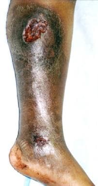 Typical chronic medial leg ulceration associated with long-standing venous insufficiency. The ulcer had been present for 12 years and was refractory to every treatment approach until treatment of the refluxing superficial varices was performed. Treatment consists of endovenous ablation, foam sclerotherapy, or ambulatory phlebectomy.
Pathophysiology
Varicose veins and spider veins are normal veins that have dilated under the influence of increased venous pressure.
In healthy veins, one-way valves direct the flow of venous blood upward and inward. Blood is collected in superficial venous capillaries, flows into larger superficial veins, and eventually passes through valves into the deep veins and then centrally to the heart and lungs. Superficial veins are suprafascial, while deep veins are within the muscle fascia. Perforating veins allow blood to pass from the superficial veins into the deep system.
Within muscle compartments, muscular contraction compresses deep veins and causes a pumping action that can produce transient deep venous pressures as high as 5 atmospheres. Deep veins can withstand this pressure because of their construction and because their confining fascia prevents them from becoming excessively distended. In contrast to deep veins, the venous pressure in superficial veins normally is very low. Exposure to high pressures causes superficial veins of any size to become dilated and tortuous.
Perfectly normal veins dilate and become tortuous in response to continued high pressure, as is observed in patients with dialysis shunts or with spontaneous arteriovenous malformations. In a subset of patients with hereditary vein wall weakness, even normal venous pressures produce varicose changes and venous insufficiency.
Elevated venous pressure most often is the result of venous insufficiency due to valve incompetence in the deep or superficial veins. Varicose veins are the undesirable pathways by which venous blood refluxes back into the congested extremity. Ablation of the varicose pathways invariably improves overall venous circulation.
Chronically increased venous pressure can also be caused by outflow obstruction, either from intravascular thrombosis or from extrinsic compression. In patients with outflow obstruction, varicosities must not be ablated because they are an important bypass pathway allowing blood to flow around the obstruction. Specific diagnostic tests can distinguish between patients who will benefit from ablation of dilated superficial veins and those who will be harmed by the same procedure.
Deep vein thrombosis initially produces an obstruction to outflow, but in most cases the thrombosed vessel eventually recanalizes and becomes a valveless channel delivering high pressures from above downward.
Most commonly, superficial venous valve failure results from excessive dilatation of a vein from high pressure of reverse flow within the superficial venous system. Valve failure can also result from direct trauma or from thrombotic valve injury. When exposed to high pressure for a long enough period, superficial veins dilate so much that their delicate valve leaflets no longer meet.
In the most common scenario, a single venous valve fails and creates a high-pressure leak between the deep and superficial systems. High pressure within the superficial system causes local dilatation, which leads to sequential failure (through over-stretching) of other nearby valves in the superficial veins. After a series of valves have failed, the involved veins are no longer capable of directing blood upward and inward. Without functioning valves, venous blood flows in the direction of the pressure gradient: outward and downward into an already congested leg.
As increasing numbers of valves fail under the strain, high pressure is communicated into a widening network of dilated superficial veins in a recruitment phenomenon. Over time, large numbers of incompetent superficial veins acquire the typical dilated and tortuous appearance of varicosities.
Varicose veins of pregnancy most often are caused by hormonal changes that render the vein wall and the valves themselves more pliable. The sudden appearance of new dilated varicosities during pregnancy still warrants a full evaluation because of the possibility that these may be new bypass pathways related to acute deep vein thrombosis.
The sequelae of venous insufficiency are related to the venous pressure and to the volume of venous blood that is carried in a retrograde direction through incompetent veins. Unfortunately, the presence and size of visible varicosities are not reliable indicators of the volume or pressure of venous reflux. A vein that is confined within fascial planes or is buried beneath subcutaneous tissue can carry massive amounts of high-pressure reflux without being visible at all. Conversely, even a small increase in pressure can eventually produce massive dilatation of an otherwise normal superficial vein that carries very little flow.
Etiology
Intrinsic pathological conditions and extrinsic environmental factors combine to produce a wide spectrum of varicose disease.
Most varicose disease is due to elevated superficial venous pressures, but some people have an inborn weakness of vein walls and can develop varicosities even in the absence of elevated venous pressures. Some patients with varicose veins of the legs also have abnormally distensible veins in the forearm and hand veins.
Heredity is important in determining susceptibility to primary valvular failure, but the specific genetic factors responsible for varicosities have not yet been elucidated. Reflux at the saphenofemoral junction (where the superficial greater saphenous vein joins the deep common femoral vein) is twice as likely when a parent had a similar condition. Monozygotic twins are concordant with regard to varicose veins in 75% of cases. The prevalence of varicose veins is 43% in female relatives of patients with varicose veins but is only 19% in male relatives.
Prolonged standing leads to increased hydrostatic pressures that can cause chronic venous distention and secondary valvular incompetence anywhere within the superficial venous system. If proximal junctional valves become incompetent, high pressure passes from the deep veins into the superficial veins and the condition rapidly progresses to become irreversible. Women are particularly susceptible to this type of varicose problem because vein walls and valves periodically become more distensible under the influence of cyclic increases in progesterone.
Pregnancy is a common cause of varicosities. During pregnancy, circulating hormonal factors increase the distensibility of vein walls and soften valve leaflets. At the same time, the veins must accommodate a greatly expanded circulating blood volume. Late in pregnancy, the enlarged uterus compresses the inferior vena cava, causing further venous hypertension and secondary distension of leg veins. Depending on the relative contributions of these mechanisms, varicose veins of pregnancy may or may not spontaneously regress after delivery. Treatment of existing varicose veins prior to pregnancy has been shown to prevent the progression of disease and reduce the recruitment of other veins during pregnancy.
Age is an independent risk factor for varicosities. With advancing age, the elastic lamina of the vein becomes atrophic and the smooth muscle layer begins to degenerate, leaving a weakened vein that is more susceptible to dilatation.
Wherever a venous outflow obstruction exists, varicose veins may arise as a bypass pathway. Such veins are an important pathway for venous return and must not be ablated.
Epidemiology
United States
Approximately 23% of adults in the United States have varicose veins. This figure rises to 80% for men and 85% for women if reticular veins and spider telangiectasias are included. [2]
International
The prevalence of venous disease is higher in Westernized and industrialized countries, most likely due to alterations in lifestyle and activity.
Sex
Because of hormonal factors, varicosities and telangiectasia are more common in women than in men at any age. [3]
Age
Most varicose and spider veins in adults have their genesis in childhood. Serial examinations of children aged 10-12 years and again 4 and 8 years later showed that symptoms are experienced (and venous test results are abnormal) before any abnormal veins are visible at the surface of the skin.
When abnormal veins do become visible, reticular veins usually appear first and are followed after several years by incompetent perforators. Smaller telangiectatic webs and large varicose veins usually become visible only in adulthood, many years after the true onset of disease.
Although varicosities continue to worsen and to recruit new areas of involvement throughout life, only a small number of new cases appear after the childbearing years.
Prognosis
Patients with significant venous reflux are at high risk for progression to chronic venous ulcers that can be very difficult to treat effectively. With appropriate treatment, the vast majority of patients have a good outcome.
Death can occur because of bleeding from friable varicose veins, [4] but the mortality associated with varicose veins is almost entirely due to the association of this condition with venous thromboembolism. When treating a patient with varicose veins, the possibility of associated deep venous thrombosis (DVT) must always be considered because the mortality rate of unrecognized and untreated thromboembolism is 30-60%. A 2018 study from Taiwan compared 212,984 subjects with varicose veins and 212,984 subjects without varicose veins, all approximately the same average age (55 y) and of similar sex-based proportions (70% women). [5] The observation spanned 14 years, and researchers found 10,630 cases of DVT in the varicose vein subjects versus 1,980 cases in the non–varicose vein subjects. The results suggest that varicose vein patients may have an approximately 5 times greater risk of developing DVT compared with those without varicose veins.
Patients with varicose veins are at increased risk of deep vein thrombosis because venous stasis and injury often cause superficial phlebitis that can pass through perforating vessels to involve the deep venous system.
Varicose veins may arise after an unrecognized episode of deep vein thrombosis that causes damage to venous valves. Such patients have some underlying risk factor for thromboembolism and are at especially high risk for recurrence.
Varicose veins may sometimes serve as an important pathway for venous return in a patient with acute blockage of the deep venous system from any cause. This most often occurs after an episode of deep vein thrombosis, but it may also be a response to tumor growth or to impaired portal flow through a cirrhotic liver.
Patient Education
For patient education resources, see the patient education articles Varicose Veins, Blood Clot in the Legs, and Phlebitis.
-
Patient with large tortuous varicose veins, high-volume venous reflux, and early stasis changes of the medial ankle.
-
Typical chronic medial leg ulceration associated with long-standing venous insufficiency. The ulcer had been present for 12 years and was refractory to every treatment approach until treatment of the refluxing superficial varices was performed. Treatment consists of endovenous ablation, foam sclerotherapy, or ambulatory phlebectomy.