Background
Tinea capitis is a disease caused by superficial fungal infection of the skin of the scalp, eyebrows, and eyelashes, with a propensity for attacking hair shafts and follicles (see the image below). The disease is considered to be a form of superficial mycosis or dermatophytosis. Several synonyms are used, including ringworm of the scalp and tinea tonsurans. In the United States and other regions of the world, the incidence of tinea capitis is increasing. [1]
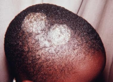 Gray-patch ringworm (microsporosis) is an ectothrix infection or prepubertal tinea capitis seen here in an African American male child. Gray patch refers to the scaling with lack of inflammation, as noted in this patient. Hairs in the involved areas assume a characteristic dull, grayish, discolored appearance. Infected hairs are broken and shorter. Papular lesions around hair shafts spread and form typical patches of ring forms, as shown. Culture from the lesional hair grew Microsporum canis.
See 15 Rashes You Need to Know: Common Dermatologic Diagnoses, a Critical Images slideshow, for help identifying and treating various rashes.
Also, see the 15 Back-to-School Illnesses You Should Know slideshow to help identify conditions that may occur in young patients after they return to the classroom.
Dermatophytosis includes several distinct clinical entities, depending on the anatomic site and etiologic agents involved. Clinically, the conditions include tinea capitis, tinea favosa (favus resulting from infection by Trichophyton schoenleinii), tinea corporis (ringworm of glabrous skin), tinea imbricata (ringworm resulting from infection by Trichophyton concentricum), tinea cruris (ringworm of the groin), tinea unguium or onychomycosis (ringworm of the nail), tinea pedis (ringworm of the feet), tinea barbae (ringworm of the beard), and tinea manuum (ringworm of the hand).
Clinical presentation of tinea capitis varies from a scaly noninflamed dermatosis resembling seborrheic dermatitis to an inflammatory disease with scaly erythematous lesions and hair loss or alopecia that may progress to severely inflamed deep abscesses termed kerion, with the potential for scarring and permanent alopecia. The type of disease elicited depends on interaction between the host and the etiologic agents.
The term tinea originally indicated larvae of insects that fed on clothes and books. Subsequently, it meant parasitic infestation of the skin. By the mid 16th century, the term was used to describe diseases of the hairy scalp. The term ringworm referred to skin diseases that assumed a ring form, including tinea. The causative agents of tinea infections of the beard and scalp were described first by Remak and Schönlein, then by Gruby, during the 1830s. Approximately 50 years later, in Sabouraud's dissertation, the endothrix type of tinea capitis infection was demonstrated, and it was known that multiple species of fungi cause the disease. Simple culture methods were described and treatment using x-ray epilation was reported in 1904. Effective treatment of tinea capitis by griseofulvin became available in the 1950s.
Pathophysiology
Tinea capitis is caused by fungi of species of genera Trichophyton and Microsporum. Tinea capitis is the most common pediatric dermatophyte infection worldwide. The age predilection is believed to result from the presence of Pityrosporum orbiculare (Pityrosporum ovale), which is part of normal flora, and from the fungistatic properties of fatty acids of short and medium chains in postpubertal sebum.
Causative agents of tinea capitis include keratinophilic fungi termed dermatophytes. These molds usually are present in nonliving cornified layers of skin and its appendages and sometimes are capable of invading the outermost layer of skin, stratum corneum, or other keratinized skin appendages derived from epidermis, such as hair and nails.
Dermatophytes are among the most common infectious agents of humans, causing a variety of clinical conditions that are collectively termed dermatophytosis. From the site of inoculation, the fungal hyphae grow centrifugally in the stratum corneum. The fungus continues downward growth into the hair, invading keratin as it is formed. The zone of involvement extends upwards at the rate at which hair grows, and it is visible above the skin surface by days 12-14. Infected hairs are brittle, and by the third week, broken hairs are evident.
Three types of in vivo hair invasion are recognized.
Ectothrix invasion is characterized by the development of arthroconidia on the exterior of the hair shaft. The cuticle of the hair is destroyed, and infected hairs usually fluoresce a bright greenish-yellow color under a Wood lamp ultraviolet light. Common agents include Microsporum canis, Microsporum gypseum, Trichophyton equinum, and Trichophyton verrucosum. [2]
Endothrix hair invasion is characterized by the development of arthroconidia within the hair shaft only. The cuticle of the hair remains intact and infected hairs do not fluoresce under a Wood lamp ultraviolet light. All endothrix-producing agents are anthropophilic (eg, Trichophyton tonsurans, Trichophyton violaceum). [3]
Favus, usually caused by T schoenleinii, produces favuslike crusts or scutula and corresponding hair loss.
Etiology
Infection of the scalp by dermatophytes usually is the result of person-to-person transmission. The organism remains viable on combs, brushes, couches, and sheets for long periods. Certain species of dermatophytes are endemic only in particular parts of the world. Zoophilic fungal infections of the scalp are rare.
In the United States, T tonsurans has replaced M audouinii and M canis as the most common cause of tinea capitis. T tonsurans also is the most common cause of the disease in Canada, Mexico, and Central America.
Historically, M audouinii was the classic causative agent in Europe and America and M ferrugineum was most common in Asia. Currently, M audouinii and M canis remain prevalent in most parts of Europe, although T violaceum also is common in Romania, Italy, Portugal, Spain, and the former USSR, as well as in Yugoslavia. In Africa, T violaceum, T schoenleinii, and M canis commonly are isolated. [4] T violaceum and M canis are prevalent agents in Asia. [5] T schoenleinii is common in Iran and Turkey, while M canis is common in Israel. Epidermophyton floccosum and T concentricum do not invade scalp hair. Trichophyton rubrum, which is the most common dermatophyte isolated worldwide, is not a common cause of tinea capitis.
Dermatophytic fungi causing tinea capitis can be divided into anthropophilic and zoophilic organisms. Anthropophilic fungi grow preferentially on humans, and the most common type forms large conidia of approximately 3-4 µm in diameter within the hair shaft. Zoophilic fungi are acquired through direct contact with infected animals. Smaller conidia of approximately 1-3 µm in diameter typically are present, extending around the exterior of the hair shaft.
Dermatophytosis customarily is divided into endothrix (inside the hair shaft) and ectothrix (extending outside the hair shaft) infection based on the location of proliferation of pathogenic fungi and destruction of the hair structure.
Common causes of endothrix infection include T tonsurans, characterized by chains of large spores and T schoenleinii, characterized by hyphae with air spaces. Infected hairs break off sharply at the follicular orifice, leaving a conidia-filled stub or black dot. Suppuration and kerion formation (see the image below) commonly are associated with T tonsurans infection.
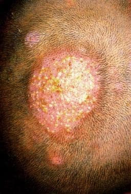 Typical lesions of kerion celsi on the vertex scalp of a young Chinese boy. Note numerous bright yellow purulent areas on skin surface, surrounded by adjacent edematous, erythematous, alopecic areas. Culture from the lesion grew Trichophyton mentagrophytes. Courtesy of Skin Diseases in Chinese by Yau-Chin Lu, MD. Permission granted by Medicine Today Publishing Co, Taipei, Taiwan, 1981.
In ectothrix infection, fragmentation of the mycelium into spores occurs just beneath the cuticle. In contrast to endothrix infection, destruction of the cuticle occurs. This type of infection is caused by T verrucosum, T mentagrophytes, and all Microsporum species.
Epidemiology
Frequency
United States
Occurrence of the disease is no longer registered by public health agencies; therefore, true incidence is unknown. The reported peak incidence occurs in school-aged African American male children, at rates of 12.9%. [6] In Northern California, the reported incidence among the pediatric population is 0.34%. [7]
Tinea capitis is predominantly a disease of preadolescent children. Typical age of onset is between 5 and 10 years. [6] Tinea capitis accounts for up to 92.5% of dermatophytoses in children younger than 10 years. The disease is rare in adults, although occasionally, it may be found in elderly patients. Tinea capitis occurrence is widespread in some urban areas in the United States.
International
Tinea capitis is widespread in some urban areas, particularly in children of Afro-Caribbean extraction, in North America, Central America, and South America. It is common in parts of Africa and India. [8, 9, 10, 11] In Ethiopia, the incidence of tinea capitis is 8.7% among children aged 4-14 years. [12] In Southeast Asia, the rate of infection has been reported to have decreased dramatically from 14% (average of male and female children) to 1.2% in the last 50 years because of improved general sanitary conditions and personal hygiene. In northern Europe, the disease is sporadic.
In the United Kingdom and North America, T tonsurans accounts for greater than 90% of cases of infection . [13] In the nonurban communities, sporadic infections acquired from puppies and kittens are due to M canis, which accounts for less than 10% of cases in the United Kingdom. Occasional infection from other animal hosts (eg, T verrucosum from cattle) occurs in rural areas.
Sex
The incidence of tinea capitis may vary by sex, depending on the causative fungal organism. Microsporum audouinii –related tinea capitis has been reported to be up to 5 times more common in boys than in girls. After puberty, however, the reverse is true, possibly because of women having greater exposure to infected children and possibly because of hormonal factors. In infection by M canis, the ratio varies, but the infection rate usually is higher in boys. Girls and boys are affected equally by Trichophyton infections of the scalp, but in adults, women are infected more frequently than are men.
Age
Tinea capitis occurs primarily in children and occasionally in other age groups. It is seen most commonly in children aged between 5 and 10 years. [6] Mean age of onset is in patients aged 6.9-8.1 years. [7]
Prognosis
Tinea capitis carries a positive prognosis, with the vast majority of those treated obtaining resolution of the infection. Those who have maintained untreated or resistant-to-treatment tinea capitis are at risk for abscess development, referred to as a kerion. [14]
Continuous shedding of fungal spores may last several months despite active treatment; therefore, keeping patients with tinea capitis out of school is impractical. The causes of treatment failure include reinfection, relative insensitivity of the organism, suboptimal absorption of the medication, and lack of compliance with the long courses of treatment. T tonsurans and Microsporum species are typical offending agents in persistent positive cases. If fungi can still be isolated from the lesional skin at the completion of treatment, but clinical signs have improved, the recommendation is to continue the original regimen for another month.
Classification and severity of tinea capitis depend on the site of formation of their arthroconidia.
Ectothrix infection is defined as fragmentation of the mycelium into conidia around the hair shaft or just beneath the cuticle of the hair, with destruction of the cuticle. Inflammatory tinea related to exposure to a kitten or puppy usually is a fluorescent small spore ectothrix. Some mild ringworm or prepubertal tinea capitis infections are of the ectothrix type, also termed the gray-patch type (microsporosis; see the image below). Some ectothrix infections involute during the normal course of disease without treatment. Depending on the extent of associated inflammation, lesions may heal with scarring.
Gray-patch ringworm (microsporosis) is an ectothrix infection or prepubertal tinea capitis seen here in an African American male child. Gray patch refers to the scaling with lack of inflammation, as noted in this patient. Hairs in the involved areas assume a characteristic dull, grayish, discolored appearance. Infected hairs are broken and shorter. Papular lesions around hair shafts spread and form typical patches of ring forms, as shown. Culture from the lesional hair grew Microsporum canis.
Endothrix infections are noted in which arthrospores are present within the hair shaft in both anagen and telogen phases, contributing to the chronicity of the infections. Endothrix infections tend to progress, become chronic, and may last into adult life. Lesions can be eradicated by systemic antifungal treatment. Since the organisms usually remain superficial, little potential for mortality exists. Disseminated systemic disease has been reported in patients who are severely immunocompromised.
Patient Education
Patient education is paramount in eradicating tinea capitis. The current recommendations of the Committee on Infectious Diseases of the American Academy of Pediatrics state that "Children receiving treatment for tinea capitis may attend school. Haircuts, shaving of the head, wearing a cap during treatment are not necessary."
-
Gray-patch ringworm (microsporosis) is an ectothrix infection or prepubertal tinea capitis seen here in an African American male child. Gray patch refers to the scaling with lack of inflammation, as noted in this patient. Hairs in the involved areas assume a characteristic dull, grayish, discolored appearance. Infected hairs are broken and shorter. Papular lesions around hair shafts spread and form typical patches of ring forms, as shown. Culture from the lesional hair grew Microsporum canis.
-
Typical lesions of kerion celsi on the vertex scalp of a young Chinese boy. Note numerous bright yellow purulent areas on skin surface, surrounded by adjacent edematous, erythematous, alopecic areas. Culture from the lesion grew Trichophyton mentagrophytes. Courtesy of Skin Diseases in Chinese by Yau-Chin Lu, MD. Permission granted by Medicine Today Publishing Co, Taipei, Taiwan, 1981.
-
Discrete patches of hair loss or alopecia caused by Trichophyton violaceum infection of the vertex scalp of a young Taiwanese boy. Courtesy of Skin Diseases in Chinese by Yau-Chin Lu, MD. Permission granted by Medicine Today Publishing Co, Taipei, Taiwan, 1981.
-
Photomicrograph depicting an endoectothrix invasion of a hair shaft by Microsporum audouinii. Intrapilary hyphae and spores around the hair shaft are seen (hematoxylin and eosin stain with Periodic acid-Schiff counterstain, magnification X 250).
-
Fungal hyphae and yeast cells of Trichophyton rubrum seen on the stratum corneum of tinea capitis. Periodic acid-Schiff stain, magnification 250X.
-
Pronounced inflammatory tissue reaction with follicular pustule formation surrounding a hair follicle seen in a patient with clinical form of infection, termed kerion celsi. No fungal hyphae or spores were identified in the lesion in either tissue sections or culture. Fluorescein-labeled Trichophyton mentagrophytes antiserum cross-reacted with antigens of dermatophyte in the infected hairs within the pustule (hematoxylin and eosin stain, magnification X 75).
-
Wood lamp examination of a gray-patch area on the scalp. In Microsporum canis infection, scalp hairs emit a diagnostic brilliant green fluorescence. Trichophyton tonsurans does not fluoresce with Wood lamp.
-
Tinea capitis, presenting as alopecia with scale, in an African American child.


