Background
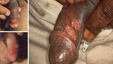
Syphilis is an infectious venereal disease caused by the spirochete Treponema pallidum. Syphilis is transmissible by sexual contact with infectious lesions, from mother to fetus in utero, via blood product transfusion, and occasionally through breaks in the skin that come into contact with infectious lesions. If untreated, it progresses through 4 stages: primary, secondary, latent, and tertiary. The image below depicts the characteristic chancre observed in primary syphilis.
 Syphilis. These photographs depict the characteristic chancre observed in primary syphilis. Used with permission from Wisdom (Left) A. Color Atlas of Sexually Transmitted Diseases. Year Book Medical Publishers Inc; 1989. (Right) Centers for Disease Control and Prevention
See 20 Signs of Sexually Transmitted Infections, a Critical Images slideshow, to help make an accurate diagnosis.
Also see the Clues in the Oral Cavity: Are You Missing the Diagnosis? slideshow to help identify the causes of abnormalities of the oral cavity.
Syphilis has a myriad of presentations and can mimic many other infections and immune-mediated processes in advanced stages. Hence, it has earned the nickname “the great impostor.” The complex and variable manifestations of the disease prompted Sir William Osler to remark, “The physician who knows syphilis knows medicine.”
Many famous persons throughout history are thought to have suffered from syphilis, including Bram Stoker, Henry VIII, and Vincent Van Gogh. Since the discovery of penicillin in the mid-20th century, the spread of this once very common disease has been largely controlled, but efforts to eradicate the disease entirely have been unsuccessful.
Pathophysiology
Three genera of spirochetes cause human infection:
-
Borrelia, which causes Lyme disease and relapsing fever
-
Leptospira, which causes leptospirosis
The particular spirochete responsible for syphilis is Treponema pallidum.
T pallidum is a fragile spiral bacterium 6-15 micrometers long by 0.25 micrometers in diameter. Its small size makes it invisible on light microscopy; therefore, it must be identified by its distinctive undulating movements on darkfield microscopy. It can survive only briefly outside of the body; thus, transmission almost always requires direct contact with the infectious lesion.
Syphilis is usually classified into 4 stages: primary, secondary, latent, and tertiary. It can be either acquired or congenital. That is, it can be transmitted either by intimate contact with infectious lesions (most common) or via blood transfusion (if blood has been collected during early syphilis), and it can also be transmitted transplacentally from an infected mother to her fetus.
Acquired syphilis
In acquired syphilis, T pallidum rapidly penetrates intact mucous membranes or microscopic dermal abrasions and, within a few hours, enters the lymphatics and blood to produce systemic infection. Incubation time from exposure to development of primary lesions, which occur at the primary site of inoculation, averages 3 weeks but can range from 10-90 days. Studies in rabbits show that spirochetes can be found in the lymphatic system as early as 30 minutes after primary inoculation, suggesting that syphilis is a systemic disease from the outset.
The central nervous system (CNS) is invaded early in the infection; during the secondary stage, examinations demonstrate that more than 30% of patients have abnormal findings in the cerebrospinal fluid (CSF). During the first 5-10 years after the onset of untreated primary infection, the disease principally involves the meninges and blood vessels, resulting in meningovascular neurosyphilis. Later, the parenchyma of the brain and spinal cord are damaged, resulting in parenchymatous neurosyphilis. Go to Neurosyphilis for complete information on this topic.
Regardless of the stage of disease and location of lesions, histopathologic hallmarks of syphilis include endarteritis (which in some instances may be obliterative in nature) and a plasma cell–rich infiltrate. Endarteritis is caused by the binding of spirochetes to endothelial cells, mediated by host fibronectin molecules bound to the surface of the spirochetes. The resultant endarteritis can heal with scarring in some instances.
The syphilitic infiltrate reflects a delayed-type hypersensitivity response to T pallidum, and in certain individuals with tertiary syphilis, this response by sensitized T lymphocytes and macrophages results in gummatous ulcerations and necrosis. Antigens of T pallidum induce host production of treponemal antibodies and nonspecific reagin antibodies. Immunity to syphilis is incomplete.
For example, host humoral and cellular immune responses may prevent the formation of a primary lesion on subsequent infections with T pallidum, but they are insufficient to clear the organism. This may be because the outer sheath of the spirochete is lacking immunogenic molecules, or it may be because of down-regulation of helper T cells of the TH1 class. [1, 2]
Primary syphilis is characterized by the development of a painless chancre at the site of transmission after an incubation period of 3-6 weeks. The lesion has a punched-out base and rolled edges and is highly infectious.
Histologically, the chancre is characterized by mononuclear leukocytic infiltration, macrophages, and lymphocytes. The inflammatory reaction causes an obliterative endarteritis. In this stage, the spirochete can be isolated from the surface of the ulceration or the overlying exudate of the chancre. Whether treated or not, healing occurs within 3-12 weeks, with considerable residual fibrosis.
Secondary syphilis develops about 4-10 weeks after the appearance of the primary lesion. During this stage, the spirochetes multiply and spread throughout the body. Secondary syphilis lesions are quite variable in their manifestations. Systemic manifestations include malaise, fever, myalgias, arthralgias, lymphadenopathy, and rash.
Widespread mucocutaneous lesions are observed over the entire body and may involve the palms, soles, and oral mucosae. Most often, the lesions are macular, discrete, reddish brown, and 5 mm or smaller in diameter; however, they can be pustular, annular, or scaling. Vesicular rash is typically absent. All such lesions contain treponemes. Of these, wet mucous patches are the most contagious. Histologically, the inflammatory reaction is similar to but less intense than that of the primary chancre.
Other skin findings of secondary syphilis are condylomata lata and patchy alopecia. Condylomata lata are painless, highly infectious gray-white lesions that develop in warm, moist sites. The alopecia is characterized by patchy hair loss of the scalp and facial hair, including the eyebrows. Patients with this finding have been referred to as having a “moth-eaten” appearance. During secondary infection, the immune reaction is at its peak and antibody titers are high.
Latent syphilis is a stage at which the features of secondary syphilis have resolved, though patients remain seroreactive. Some patients experience recurrence of the infectious skin lesions of secondary syphilis during this period. About one third of untreated latent syphilis patients go on to develop tertiary syphilis, whereas the rest remain asymptomatic.
Currently, tertiary syphilis disease is rare. When it does occur, it mainly affects the cardiovascular system (80-85%) and the CNS (5-10%), developing over months to years and involving slow inflammatory damage to tissues. The 3 general categories of tertiary syphilis are gummatous syphilis (also called late benign), cardiovascular syphilis, and neurosyphilis.
Gummatous syphilis is characterized by granulomatous lesions, called gummas, which are characterized by a center of necrotic tissue with a rubbery texture. Gummas principally form in the liver, bones, and testes but may affect any organ. Histological examination shows palisaded macrophages and fibroblasts, as well as plasma cells surrounding the margins. Gummas may break down and form ulcers, eventually becoming fibrotic. Treponemes are rarely visualized or recovered from these lesions.
Cardiovascular syphilis occurs at least 10 years after primary infection. The most common manifestation is aneurysm formation in the ascending aorta, caused by chronic inflammatory destruction of the vasa vasorum, the penetrating vessels that nourish the walls of large arteries. Aortic valve insufficiency may result.
Neurosyphilis has several forms. If the spirochete invades the CNS, syphilitic meningitis results. Syphilitic meningitis is an early manifestation, usually occurring within 6 months of the primary infection. CSF shows high protein, low glucose, high lymphocyte count, and positive syphilis serology.
Meningovascular syphilis occurs as a result of damage to the blood vessels of the meninges, brain, and spinal cord, leading to infarctions causing a wide spectrum of neurologic impairments.
Parenchymal neurosyphilis includes tabes dorsalis and general paresis. Tabes dorsalis develops as the posterior columns and dorsal roots of the spinal cord are damaged. Posterior column impairment results in impaired vibration and proprioceptive sensation, leading to a wide-based gait.
Disruption of the dorsal roots leads to loss of pain and temperature sensation and areflexia. Damage to the cortical regions of the brain leads to general paresis, formerly called “general paresis of the insane,” which mimics other forms of dementia. Impairment of memory and speech, personality changes, irritability, and psychotic symptoms develop and may advance to progressive dementia.
The Argyll-Robertson pupil, a pupil that does not react to light but does constrict during accommodation, may be seen in tabes dorsalis and general paresis. The precise location of the lesion causing this phenomenon is unknown.
Congenital syphilis
Congenital syphilis, discussed briefly here, is a veritable potpourri of antiquated medical terminology. The treponemes readily cross the placental barrier and infect the fetus, causing a high rate of spontaneous abortion and stillbirth. Within the first 2 years of life, symptoms are similar to severe adult secondary syphilis with widespread condylomata lata and rash. “Snuffles” describes the mucopurulent rhinitis caused by involvement of the nasal mucosae.
Later manifestations of congenital syphilis include bone and teeth deformities, such as “saddle nose” (due to destruction of the nasal septum), “saber shins” (due to inflammation and bowing of the tibia), “Clutton’s joints” (due to inflammation of the knee joints), “Hutchinson’s teeth” (in which the upper incisors are widely spaced and notched), and “mulberry molars” (in which the molars have too many cusps).
Tabes dorsalis and general paresis may develop as in adults, with 8th cranial nerve deafness and optic nerve atrophy as well as a variety of other ophthalmologic involvement leading to blindness being additional features.
From 2012-2014, the number of congenital syphilis cases in the United States increased from 334 to 458. This appears to be associated with an increase in the rate of primary and secondary syphilis among women. [3]
Go to Pediatric Syphilis for complete information on this topic.
Etiology
The cause of syphilis is infection with the spirochete T pallidum.T pallidum is solely a human pathogen and does not naturally occur in other species. T pallidum has, however, been cloned in Escherichia coli and has been used experimentally in rabbits.
Transmission of T pallidum occurs via penetration of the spirochetes through mucosal membranes and abrasions on epithelial surfaces. It is primarily spread through sexual contact but can be spread by exposure to blood products and transferred in utero. T pallidum is a labile organism that cannot survive drying or exposure to disinfectants; thus, fomite transmission (eg, from toilet seats) is virtually impossible.
Unprotected sex is the major risk factor for the acquisition of syphilis, especially among men who have sex with men (MSM), who accounted for 83.7% of all syphilis cases in the United States. [4]
Epidemiology
United States statistics
Since reporting began in 1941, the incidence of primary and secondary syphilis in the United States has varied. The incidence dropped from 66.4 cases per 100,000 in 1947 to 3.9 cases per 100,000 in 1956 following the introduction of penicillin.
During the mid-1980s, however, this trend reversed. Increases in the use of intravenous (IV) drugs and crack cocaine, the exchange of sex for drugs, indiscriminate or anonymous sex, and the number of people with multiple sexual partners contributed to the turnaround. From 1986-1990, the rate of syphilis nearly doubled, reaching a peak of 53.8 cases per 100,000 population in 1990.
After 1990, the incidence decreased again; there were 53,000 reported cases (11,387 primary and secondary cases) in 1996, compared with 113,000 cases (33,962 primary and secondary cases) reported in 1992. In 2000, the number of syphilis cases reported was at an all-time low, with rates falling to 2.1 cases per 100,000 population. Increased awareness, aggressive screening, and emphasis on primary prevention contributed to the decrease.
Since 2000, however, the number of syphilis cases in the United States has been on the rise. From 2005-2013, the number of primary and secondary syphilis cases reported each year in the United States nearly doubled, from 8,724 to 16,663; the annual rate increased from 2.9 to 5.3 cases per 100,000 population. [5] Most of this increase has been noted in men, particularly among MSM, who accounted for 87.3% of all primary and secondary syphilis cases in 2013. Rates have increased in all racial groups in the past decade, but black and Hispanic men have an overall higher rate than other racial groups. The overall highest rate was in the western United States, not in the South, for the first time in at least 50 years. [6]
International statistics
Internationally, the prevalence of syphilis varies by region. Syphilis remains prevalent in many developing countries and in some areas of North America, Asia, and Europe, especially Eastern Europe. The highest rates are in South and Southeast Asia, followed closely by sub-Saharan Africa. The third highest rates are in the regions of Latin America and the Caribbean. [7] In some regions of Siberia, as of 1999, prevalence was 1300 cases per 100,000 population. [8]
Age distribution of syphilis
Syphilis is most common during the years of peak sexual activity. Most new cases occur in men and women aged 20-29 years. In 2013, the rate of primary and secondary syphilis was highest in people aged 25-29 years (27 per 100,000). [4]
The incidence of congenital syphilis has increased to 11.6 cases per 100,000 live births in 2014, the highest congenital syphilis rate reported since 2001. The number of congenital syphilis cases declined in the United States during 2008-2012, from 446 to 334 cases (10.5 to 8.4 cases per 100,000 live births) but is increasing; from 2012-2014, the number of reported congenital syphilis cases in the United States increased from 334 to 458. [3]
Sex distribution of syphilis
Men are affected more frequently with primary or secondary syphilis than women. This difference has varied over time. Male-to-female ratios of primary and secondary syphilis increased from 1.6:1 in 1965 to nearly 3:1 in 1985. After, the ratio decreased, reaching a nadir in 1994-95. The past decade has seen a sharp rise in syphilis cases among men, driven mostly by the MSM community. Males with primary and secondary syphilis outnumber females 10 to 1. Among women, the reported primary and secondary syphilis rate increased from 0.9 to 1.5 per 100,000 population per year during 2005- 2008 and decreased to 0.9 in 2013. [4]
Prevalence of syphilis by race or ethnicity
In the United States, syphilis is more prevalent among persons of minority race and ethnicity. Non-Hispanic blacks are at higher risk for syphilis than all other racial groups. In 2013, the primary and secondary syphilis rate among black men was 5.2 times that among white men (27.9 vs 5.4 cases per 100,000 population); the rate among black women was 13.3 times that among white women (4 vs 0.3). The rate among Hispanic men was 2.1 times that among white men (11.6 vs 5.4), and the rate among Hispanic women was 2.7 times that among white women (0.8 vs 0.3). These disparities were similar to disparities observed in 2005 and represent an increase in syphilis rates in all racial groups. [4]
HIV and syphilis co-infection
Syphilis acquisition increases the risk of HIV acquisition by 2- to 5-fold and makes transmission of HIV more efficient via various methods. First, primary syphilis infection causes a genital ulcer, which disrupts the mucous membrane, making it more vulnerable to penetration by the HIV virus. Second, genital ulcers bleed easily during sex, increasing the risk of viral transmission. Third, genital ulcers attract CD4 cells to the ulcer surface, increasing targets for the HIV virus to infect. Fourth, the risk behaviors associated with acquiring syphilis also increase the likelihood of acquiring HIV. [9]
The rate of HIV and syphilis co-infection is high. More than 50% of MSM with syphilis are also infected with HIV, and this number increases with each recurrence. [10, 11]
Prognosis
The morbidity of syphilis ranges from the relatively minor symptoms of the primary stages of infection to the more significant constitutional systemic symptoms of secondary syphilis and the significant neurological and cardiovascular consequences of tertiary disease. Since latent syphilis can persist for years or decades, the manifestations of tertiary syphilis often occur much later in life, causing significant morbidity and even death if not identified and treated.
These figures have continued to increase since the emergence of the AIDS epidemic, since genital ulcer diseases (including syphilis) are cofactors for the sexual transmission of HIV. Additionally, untreated patients who are HIV seropositive have an increased risk for rapid progression to neurosyphilis. In addition, patients with HIV are at greater risk for development or relapse of early symptomatic neurosyphilis for up to 2 years after treatment with intramuscular or intravenous penicillin.
The morbidity and mortality of untreated syphilis must be estimated from the limited data available regarding its natural course. These data are largely from one retrospective study of autopsies and two prospective studies, most notably the famous Tuskegee Study of Untreated Syphilis in the Negro Male, which fell under serious ethical scrutiny in later years for exploiting a vulnerable patient population and not offering treatment for the disease when it became available after the study was underway.
These data indicate that approximately one third of patients left untreated will develop late complications, with 10% of the total developing cardiovascular syphilis; 6%, neurosyphilis; and 16%, gummatous syphilis. Mortality rates in general are greater among those affected, and late complications appear to occur more commonly in men than in women. [12, 13]
For patients diagnosed with either primary or secondary syphilis (without auditory/neurologic/ocular involvement), the prognosis is good following appropriate treatment. T pallidum remains highly responsive to the penicillins, and cure is likely. Among patients diagnosed with tertiary syphilis, the prognosis is less sanguine. Twenty percent of untreated patients with tertiary syphilis die of the disease, making syphilis one of the few sexually transmitted diseases (SDTs) capable of killing its host. However, with adequate treatment, 90% of patients with neurosyphilis have a clinical response.
Overall prognosis for tertiary syphilis depends on the extent of scarring and tissue damage, as treatment arrests further damage and inflammation but cannot reverse previous tissue damage. For example, the prognosis or advanced symptomatic disease in cardiovascular syphilis is poor. In contrast, syphilitic gummas typically resolve promptly with high-dose penicillin.
Congenital syphilis is the most serious outcome of syphilis in women. It has been shown that a higher proportion of infants are affected if the mother has untreated secondary syphilis, compared to untreated early latent syphilis. Since T pallidum does not invade the placental tissue or the fetus until the fifth month of gestation, syphilis causes late abortion, stillbirth, or death soon after delivery in more than 40% of untreated maternal infections. [14, 15] Neonatal mortality usually results from pulmonary hemorrhage, bacterial superinfection, or fulminant hepatitis.
For patients who are pregnant and have early syphilis, it is likely that the mother will deliver a child not infected by syphilis (assuming the mother was treated appropriately).
Patient Education
Patients who abuse IV drugs should be advised to avoid sharing needles and to use clean needles. Needle exchange programs are in place in some areas; however, the establishment and existence of these programs remain controversial in many communities.
It is also important to stress to patients the importance of compliance with their entire antibiotic course and follow-up visits.
As with all STDs, patient education must stress the importance of safer sexual practices and the need for prompt medical evaluation of chancres and other symptoms of STDs. All patients diagnosed with an STD should be screened for HIV infection. All MSM who test positive for syphilis should be considered at risk for HIV if not already infected and should be targeted for intensive HIV prevention interventions with pre-exposure prophylaxis (PrEP).
Patients with syphilis should be counseled to notify their partners of infection and to inform them of the need to be treated. Information regarding management of sex partners infected with syphilis can be found at the CDC Web site.
For patient education information, see the Sexually Transmitted Diseases Center and Pregnancy and Reproduction Center, as well as Sexually Transmitted Diseases, Syphilis, Birth Control Overview, and Birth Control FAQs.
-
Syphilis. These photographs depict the characteristic chancre observed in primary syphilis. Used with permission from Wisdom (Left) A. Color Atlas of Sexually Transmitted Diseases. Year Book Medical Publishers Inc; 1989. (Right) Centers for Disease Control and Prevention
-
Syphilis. These photographs show the disseminated rash observed in secondary syphilis. Used with permission from Wisdom A. Color Atlas of Sexually Transmitted Diseases. Year Book Medical Publishers Inc; 1989.
-
Syphilis. These photographs show close-up images of gummas observed in tertiary syphilis. Used with permission from Wisdom A. Color Atlas of Sexually Transmitted Diseases. Year Book Medical Publishers Inc; 1989.
-
Syphilis. This photograph depicts primary syphilis "kissing" lesions. Used with permission from Wisdom A. Color Atlas of Sexually Transmitted Diseases. Year Book Medical Publishers Inc; 1989.
-
Syphilis. Palmar lesions observed in secondary syphilis. Used with permission from Wisdom (Left) A. Color Atlas of Sexually Transmitted Diseases. Year Book Medical Publishers Inc; 1989. (Right) Centers for Disease Control and Prevention
-
These photographs illustrate examples of condylomata lata. The lesions resemble genital warts (condylomata acuminata). Fluids exuding from these lesions are highly infectious. Used with permission from Wisdom A. Color Atlas of Sexually Transmitted Diseases. Year Book Medical Publishers Inc; 1989.
-
Syphilis. These photographs illustrate typical facies of congenital syphilis. Used with permission from Wisdom A. Color Atlas of Sexually Transmitted Diseases. Year Book Medical Publishers Inc; 1989.
-
Syphilis. This photograph shows an example of Hutchinson teeth in congenital syphilis. Note notching. Used with permission from Wisdom A. Color Atlas of Sexually Transmitted Diseases. Year Book Medical Publishers Inc; 1989.
-
Syphilis. This photograph illustrates chorioretinitis of congenital syphilis. Used with permission from Wisdom A. Color Atlas of Sexually Transmitted Diseases. Year Book Medical Publishers Inc; 1989.
-
Syphilitic chancre
-
Secondary syphilis - Exanthem
-
Secondary syphilis - Exanthem
-
Lues hematoxylin and eosin stain. Histopathological examination shows a lichenoid infiltrate that is stereotypical of the secondary stage of syphilis. Note that vacuolar alteration of the superjacent epithelium can be seen much like a noninfectious form of lichenoid dermatitis. The subjunctional infiltrate is rich in histiocytes and plasmacytes. At times, an overtly granulomatous lichenoid infiltrate can be seen.
-
Lues TP stain. Immunoperoxidase staining for T pallidum highlights many slender coiled organisms residing in the perijunctional zone. Occasionally, organisms can also be found in the upper dermis or around adnexal structures.


