Background
Seborrheic keratoses are the most common benign tumor in older individuals. Seborrheic keratoses have a variety of clinical appearances, as seen in the images below, and they develop from the proliferation of epidermal cells. No specific etiologic factors have been identified.
 Closer view of multiple seborrheic keratoses in an autosomally dominant mode of inheritance.
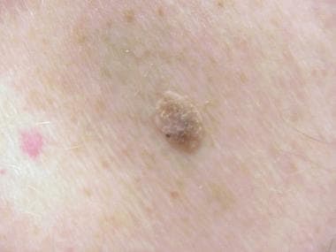
 Seborrheic keratosis showing lackluster surface and appearance of being stuck on the skin surface.
See Mole or Melanoma? Test Yourself With These Suspicious Lesions, a Critical Images slideshow, to help identify various skin lesions.
Pathophysiology
Seborrheic keratoses are thought to result from a clonal expansion of a mutated epidermal keratinocyte. [1] Seborrheic keratoses exhibit histologic evidence of proliferation. Increased cell replication has been demonstrated in seborrheic keratoses with bromodeoxyuridine incorporation studies and immunohistochemistry for proliferation-associated antigens.
Reticulated seborrheic keratoses are usually found on sun-exposed skin, and the reticulated type of seborrheic keratoses may develop from solar lentigines.
Epidermal growth factors and their receptors have been studied in the development of seborrheic keratoses. [2, 3, 4] No difference was observed in the expression of immunoreactive growth hormone receptors in keratinocytes from normal epidermis and keratinocytes from seborrheic keratoses. The expression of BCL2, an apoptosis-suppressing oncogene, is low in seborrheic keratosis in contrast to the high values in basal cell and squamous cell carcinoma. [5] No increase is observed in the sonic hedgehog signal transducers patched (ptc) and smoothened (smo) messenger RNA (mRNA) in seborrheic keratosis over normal skin. [6]
A high frequency of mutations in the gene encoding the tyrosine kinase receptor FGFR3 (fibroblast growth factor receptor 3) has been found in certain types of seborrheic keratoses. This was the first clue into the genetic basis for the pathogenesis of seborrheic keratoses. FGFR3 belongs to a class of transmembrane tyrosine kinase receptors involved in signal transduction to regulate cell growth, differentiation, and migration, as well as wound healing and angiogenesis. Upon ligand binding, FGFR3 dimerizes, which, in turn, induces phosphorylation of the kinase domain. Activating mutations in FGFR3 have been found in approximately 40% of hyperkeratotic seborrheic keratoses, 40% of acanthotic seborrheic keratoses, and 85% of adenoid seborrheic keratoses. [7, 8, 9]
More than 80% of seborrheic keratoses have at least one mutation, and 45% have more than one mutation in an oncogene such as FGFR3, PIK3CA, KRAS, and EGFR. [10] The most frequently mutated genes in seborrheic keratoses are FGFR3 (found in 71% or sporadic seborrheic keratosis) and the p110 catalytic subunit of phosphatidylinositol 3 kinase (PI3K) (found in 50% of sporadic seborrheic keratoses). [11] Seborrheic keratoses have a higher proliferative rate than normal keratinocytes, and apoptosis is suppressed in seborrheic keratoses compared with healthy skin. [12, 13] Both of these proteins effect the activity of Akt kinase. The activation of Akt kinase enhances the survival of cells by blocking the p53 pathway and the FOXO-mediated proapoptotic cascade. [14] Thus, the signaling kinase Akt is critical in preventing seborrheic keratosis cells from undergoing programmed cell death. When Akt is inhibited, seborrheic keratosis cells quickly die through apoptosis. [15]
Seborrheic keratoses have a varying degree of pigmentation. In pigmented seborrheic keratoses, the proliferating keratinocytes trigger the activation of neighboring melanocytes by secreting melanocyte-stimulating cytokines. Endothelin-1 has dual stimulatory effects on DNA synthesis and melanization of human melanocytes and has been implicated as playing a part in the hyperpigmentation observed in seborrheic keratoses. [16] Immunohistochemically, the keratinocytes of seborrheic keratoses express low molecular weight keratin but often exhibit a partial lack of the high molecular weight forms of keratin.
Etiology
Seborrheic keratoses are thought to result from a clonal expansion of a mutated epidermal keratinocyte. [1] (see Pathophysiology). Some cases are inherited through an autosomal dominant mode of inheritance. Sunlight seems to play a role in the development of some seborrheic keratoses. [17] Evidence indicates that at least some seborrheic keratoses have a clonal nature. Activating mutations in the gene encoding the tyrosine kinase receptor FGFR3 have been found in 85% of adenoid seborrheic keratoses. This is discussed in Pathophysiology section.
Epidemiology
Frequency
United States
Seborrheic keratoses are the most common benign tumor in older individuals. The frequency appears to increase with age. In 1963, Tindall and Smith examined a population of individuals older than 64 years in North Carolina and found that 88% of the people had at least one seborrheic keratosis. [18] In this study, 38% of the white women, 54% of the white men, and 61% of the black men and women were found to have 10 or more seborrheic keratoses. In 1965, Young examined 222 residents of the Orthodox Jewish Home for the Aged in New York and found that 29.3% of the men and 37.9% of the women had seborrheic keratosis.
International
In 2000, Memon et al found in a British population younger than 40 years that 8.3% of the males and 16.7% of the females had at least one seborrheic keratosis. [19] In an Australian population, 23.5% of individuals aged 15-30 years were found to have at least one seborrheic keratosis, with no significant differences between the sexes. In another Australian study of 100 people composed of hospital staff and nondermatologic day patients, 12% of people aged 15-25 years (n = 34), 79% of people aged 26-50 years (n = 24), 100% of people aged 51-75 years (n = 25), and 100% of people older than 75 years (n = 17) had seborrheic keratoses. The median number of seborrheic keratoses per person was 6 in the group aged 15-25 years, 5 in the group aged 26-50 years, 23 in the group aged 51-75 years, and 69 in those older than 75 years.
Race
Seborrheic keratoses are less common in populations with dark skin compared to those having white skin; however, black individuals develop a variant of seborrheic keratoses termed dermatosis papulosa nigra. These lesions affect the face, especially the upper cheeks and lateral orbital areas. They are small, pedunculated, and heavily pigmented with a minimal keratotic element. The onset of these lesions generally is earlier than that of ordinary seborrheic keratoses.
Sex
No sex difference is apparent in the frequency of occurrence of seborrheic keratoses.
Age
Seborrheic keratoses are the most common benign tumor in older individuals. They appear to increase with age. Seborrheic keratoses have also been found to occur in younger individuals.
Prognosis
Seborrheic keratoses are benign, but secondary tumors and Bowen disease (squamous cell carcinoma in situ) or malignant melanoma may occasionally arise within the lesion. Seborrheic keratoses can also catch on clothing and become irritated. They can itch, grow, and bleed. Scratching seborrheic keratoses or trying to pick them off the skin can result in a secondary infection.
People sometimes have many seborrheic keratoses, and they may obscure the detection of a dysplastic nevus or malignant melanoma.
The lesions generally do not resolve and usually grow larger and thicker with time.
Patient Education
Patients often have heard that they need to have a changing mole examined, and the appearance of seborrheic keratoses prompts them to seek medical care. This provides an excellent opportunity for a complete skin examination to search for skin cancer and a discussion on using sunscreens for both the patient and their family.
Patients can generally be reassured that the lesions are benign and do not need to be removed unless they are changing or become irritated.
Continued follow-up is important. Patients who see a doctor and who are assured that these lesions are benign may not pay attention to newly appearing lesions that continue to develop over time. One of the newly appearing lesions may not be a seborrheic keratosis but, in fact, a malignant tumor.
For patient education resources, visit the Skin Conditions and Beauty Center and Cancer Center. Also, see the patient education article Skin Cancer.
-
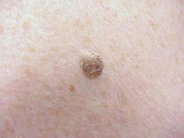
Sharply circumscribed elevated seborrheic keratoses.
-
Closer view of multiple seborrheic keratoses in an autosomally dominant mode of inheritance.
-
Seborrheic keratoses projecting above the level of the epidermis. Cysts represent sections of hyperkeratotic follicles.
-
Seborrheic keratosis showing lackluster surface and appearance of being stuck on the skin surface.
-
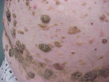
This is an autosomal dominant form of multiple seborrheic keratoses. This man's daughter is developing a similar distribution and quantity of seborrheic keratoses.
-
The back of this same patient as in the image above with multiple seborrheic keratoses. His face had a similar number of seborrheic keratoses.
-
Acanthotic type of seborrheic keratosis.
-
Higher-power view of the cells in an acanthotic seborrheic keratosis.
-
Hyperkeratotic type of seborrheic keratosis.
-
Reticulated (or adenoid) type of seborrheic keratosis.
-
This is a reticulated (or adenoid) seborrheic keratosis with abundant pigment.
-
Seborrheic keratosis with inflammation in the dermis.
-
This seborrheic keratosis was a pedunculated lesion in an axillary fold. Clinically, it had some resemblance to a skin tag.