Practice Essentials
Seborrheic dermatitis is a papulosquamous disorder patterned on the sebum-rich areas of the scalp, face, and trunk (see the image below). In addition to sebum, this dermatitis is linked to Malassezia, [1] immunologic abnormalities, and activation of complement. Its severity ranges from mild dandruff to exfoliative erythroderma.
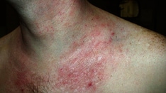
 Seborrheic dermatitis may affect any hair-bearing area, and the chest is frequently involved. Courtesy of Wilford Hall Medical Center Dermatology Teaching slides.
Signs and symptoms
History findings in seborrheic dermatitis may include the following:
-
Intermittent, active phases manifesting with burning, scaling, and itching, alternating with inactive periods; activity is increased in winter and early spring, with remissions commonly occurring in summer.
-
In active phases, potential secondary infection in intertriginous areas
-
Candidal overgrowth
-
Generalized seborrheic erythroderma (rare)
Physical findings may include the following:
-
Scalp appearance ranging from mild, patchy scaling to widespread, thick, adherent crusts; plaques are rare; lesions may spread from the scalp onto the forehead, the posterior part of the neck, and the postauricular skin
-
Seborrheic skin lesions manifesting as scaling over red, inflamed skin; hypopigmentation (in dark-skinned individuals); oozing and crusting; blepharitis (occurring independently)
-
Lesion distribution following the oily and hair-bearing areas of the head and the neck; extension to submental skin can occur
-
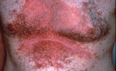
Either of two distinct truncal patterns: (1) annular or geographic petaloid scaling or (2) pityriasiform variety (rare)
Malassezia organisms are probably not the cause of seborrheic dermatitis but a cofactor linked to a T-cell depression, increased sebum levels, and an activation of the alternative complement pathway. Various medications may also flare or induce seborrheic dermatitis. [2, 3]
See Clinical Presentation for more detail.
Diagnosis
The diagnosis of seborrheic dermatitis is usually made on clinical grounds, based on a history of waxing and waning severity and by the distribution of involvement upon examination.
A skin biopsy may be needed in persons with exfoliative erythroderma, and a fungal culture can be used to rule out tinea capitis, though tinea capitis is rare in adults. Dermatopathologic findings of seborrheic dermatitis are nonspecific and typically include the following:
-
Hyperkeratosis
-
Acanthosis
-
Accentuated rete ridges
-
Focal spongiosis
-
Parakeratosis
See Workup for more detail.
Management
Early treatment of flares is encouraged. Behavior modification techniques in reducing excoriations are especially helpful with scalp involvement.
Pharmacologic agents that may be used include the following:
-
Topical corticosteroids (discouraged except for short-term use and at risk for tachyphylaxis when used as monotherapy)
-
For acute flares, class IV or lower corticosteroid creams, lotions, or solutions
-
For severe or unresponsive lesions, systemic fluconazole [16]
Treatment of dandruff may involve the following:
-
More frequent shampooing or longer lathering
-
Discontinuance of hair spray or hair pomades
-
Use of shampoos containing salicylic acid, tar, selenium, sulfur, or zinc [17, 18] ; selenium sulfide (2.5%), ketoconazole, and ciclopirox shampoos may help by reducing Malassezia yeast scalp reservoirs [19, 20, 21] ; an alternative to a shampoo with zinc is a conditioner rinse with zinc, 0.01% fluocinolone, and acetonide topical oil
-
Overnight application of tar, bath oil, or Baker’s P&S solution; Derma-Smoothe F/S oil is especially helpful for widespread plaques
See Treatment and Medication for more detail.
Background
Seborrheic dermatitis is a papulosquamous disorder patterned on the sebum-rich areas of the scalp, face, and trunk. When localized to the scalp, it is commonly referred to as dandruff. In addition to sebum, this dermatitis is linked to Malassezia, [1] immunologic abnormalities, and activation of complement. It is commonly aggravated by changes in humidity, changes in seasons, trauma (eg, scratching), or emotional stress. The severity varies from mild dandruff to exfoliative erythroderma. Seborrheic dermatitis may worsen in Parkinson disease and in AIDS. [22, 23] Increased perspiration in Parkinson disease may be the link to seborrheic dermatitis.
Pathophysiology
Seborrheic dermatitis is associated with normal levels of Malassezia but an abnormal immune response. Helper T cells, phytohemagglutinin and concanavalin stimulation, and antibody titers are depressed compared with those of control subjects. Zani et al report that even with antifungal treatments, there was no reduction in levels of Malassezia. [24] The contribution of Malassezia species to seborrheic dermatitis may come from its lipase activity—releasing inflammatory free fatty acids—and from its ability to activate the alternative complement pathway. [25]
Etiology of Seborrheic Dermatitis
The exact pathophysiology remains unclear. Malassezia organisms are probably not the cause but are a cofactor linked to a T-cell depression, increased sebum levels, and an activation of the alternative complement pathway. Persons prone to this dermatitis also may have a skin-barrier dysfunction. [26, 27]
Because seborrheic dermatitis is uncommon in preadolescent children, and tinea capitis is uncommon after adolescence, dandruff in a child is more likely to represent a fungal infection. A fungal culture should be completed for confirmation.
Various medications may flare or induce seborrheic dermatitis. These medications include auranofin, aurothioglucose, buspirone, chlorpromazine, cimetidine, ethionamide, fluorouracil, gold, griseofulvin, haloperidol, interferon alfa, lithium, methoxsalen, methyldopa, phenothiazines, psoralens, stanozolol, thiothixene, and trioxsalen. [2, 3]
Epidemiology
The prevalence rate of seborrheic dermatitis is 3-5%, with a worldwide distribution. In the United States alone, $300 million is spent annually on dandruff over-the-counter treatments. [28] Dandruff, the mildest form of this dermatitis, is probably far more common and is present in an estimated 15-20% of the population.
Race
Seborrheic dermatitis occurs in persons of all races.
Sex
Seborrheic dermatitis is slightly worse in males than in females.
Age
The usual onset occurs with puberty. It peaks at age 40 years and is less severe, but present, among older people. In infants, it occurs as cradle cap or, uncommonly, as a flexural eruption or erythroderma. [29]
-
Seborrheic dermatitis affecting the scalp line and the eyebrows with red skin and scaling. Courtesy of Wilford Hall Medical Center Dermatology slide files.
-
Seborrheic dermatitis may affect any hair-bearing area, and the chest is frequently involved. Courtesy of Wilford Hall Medical Center Dermatology Teaching slides.
-
African Americans and persons from other darker-skinned races are susceptible to annular seborrheic dermatitis, also called petaloid seborrheic dermatitis or seborrhea petaloides. Sarcoidosis, secondary syphilis, and even discoid lupus may be in the differential in such cases. Courtesy of Jeffrey J. Meffert, MD.