Practice Essentials
Rosacea is a common condition characterized by symptoms of facial flushing and a spectrum of clinical signs, including erythema, telangiectasia, coarseness of skin, and an inflammatory papulopustular eruption resembling acne. [1, 2] See the images below.
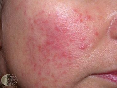 Mild papules and erythema. Courtesy of DermNet New Zealand (https://www.dermnetnz.org/imagedetail/2367).
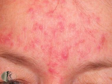 Moderate papules and early pustules. Courtesy of DermNet New Zealand (https://www.dermnetnz.org/imagedetail/2369).
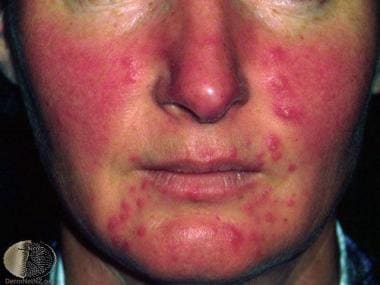 Severe erythema, papules, and pustules. Courtesy of DermNet New Zealand (https://www.dermnetnz.org/imagedetail/2371).
Classification and diagnosis
Based on specific clinical signs and symptoms, an expert committee assembled by the National Rosacea Society explicitly defined and classified rosacea into the following subtypes:
-
Erythematotelangiectatic type
-
Papulopustular
-
Phymatous
-
Ocular
Although didactically successful, the subtype designations were widely used individually and construed as distinct disorders, ignoring the frequent simultaneous occurrence of more than one subtype and potential progression of one subtype to another.
In 2016, the global rosacea consensus panel recommended a new classification: at least one diagnostic or two major phenotypes are required for the diagnosis of rosacea. [3]
Diagnostic phenotypes
A diagnosis of rosacea may be considered in the presence of one of the following diagnostic cutaneous signs:
-
Fixed centrofacial erythema in a characteristic pattern that may periodically intensify
-
Phymatous changes: Patulous follicles, skin thickening or fibrosis, glandular hyperplasia, and bulbous appearance of the nose (rhinophyma is the most common form)
Major phenotypes
Without a diagnostic phenotype, the presence of two or more of the following major features may be considered diagnostic:
-
Papules and pustules
-
Flushing: Frequent and typically prolonged
-
Telangiectasia: Predominantly centrofacial in phenotypes I-IV, rarely seen in darker phenotypes
-
Ocular manifestations
Secondary phenotypes
The following secondary signs and symptoms may appear with one or more diagnostic or major phenotypes:
-
Burning and stinging
-
Edema: Facial edema
-
Dry appearance: Central facial skin may be rough and scaly
Ocular rosacea
Major features of ocular rosacea are as follows:
-
Lid margin telangiectasia
-
Interpalpebral conjunctival injection
-
Spade-shaped infiltrates in the cornea
-
Scleritis and sclerokeratitis
Secondary features of ocular rosacea are as follows:
-
"Honey crust" and collarette accumulation at the base of the lashes
-
Irregularity of the lid margin
-
Evaporative tear dysfunction (rapid tear breakup time)
Although ocular manifestations may precede the cutaneous signs by years, in many cases they develop concurrently with dermatologic manifestations.
The diagnosis of rosacea is made clinically, based on the 2016 global rosacea consensus that one diagnostic or two major phenotypes are required for the diagnosis of rosacea. A skin biopsy is sometimes performed to exclude other cutaneous diseases, such as lupus or sarcoidosis.
See Clinical Presentation for more detail.
Histological findings
Histologic findings include the following:
-
Nonpustular lesions show a nonspecific perivascular and perifollicular lymphohistiocytic infiltrate, accompanied by occasional multinucleated cells, plasma cells, neutrophils, and eosinophils
-
Papulopustular lesions demonstrate more pronounced granulomatous inflammation and sometimes perifollicular abscesses
-
Demodex folliculorum may be abundant in nearby follicles
See Workup for more detail.
Management
Laser treatment
Vascular lasers, the mainstay of rosacea therapy, use wavelengths that allow selective absorption by oxyhemoglobin, leading to vessel reduction and causing minimal scarring or damage to surrounding tissue.
Surgery
Permanent telangiectasia may be treated by electrosurgery or the 585-nm pulsed dye laser. However, facial erythema is not improved, and new telangiectasias develop with the passage of time.
Cosmetic improvement of rhinophyma may be produced by mechanical dermabrasion, carbon dioxide laser peel, and surgical shave techniques.
Deterrence
Before the initiation of therapy, the triggering factors that exacerbate the patient's rosacea should be identified and avoided if possible. Common triggering factors include the following [4, 5] :
-
Hot or cold temperatures
-
Wind
-
Hot drinks
-
Caffeine
-
Exercise
-
Spicy food
-
Alcohol
-
Emotions
-
Topical products that irritate the skin and decrease the barrier
-
Medications that cause flushing
In addition, the use of daily broad-spectrum sunscreen is recommended for all patients with rosacea. [6]
See Treatment and Medication for more detail.
Background
Rosacea is a common condition characterized by symptoms of facial flushing and a spectrum of clinical signs, including erythema, telangiectasia, coarseness of skin, and an inflammatory papulopustular eruption resembling acne. [1, 2]
An expert committee assembled by the National Rosacea Society explicitly defined and classified rosacea into four different subtypes (erythematotelangiectatic type, papulopustular, phymatous, and ocular) based on specific clinical signs and symptoms. Although didactically successful, the subtype designations were widely used individually and construed as distinct disorders, ignoring the frequent simultaneous occurrence of more than one subtype and potential progression of one subtype to another. In 2016, the global rosacea consensus panel recommended a new classification: at least one diagnostic or two major phenotypes are required for the diagnosis of rosacea. [3] Currently, the therapeutics of rosacea empirically target the signs and symptoms of the disease because investigators do not understand the details of its pathophysiology. The classification systems aide clinicians in treatment by highlighting the preponderance of one or more of the clustering signs of presentation and, thus, help to specify which therapeutic approach to initiate.
The diagnosis of rosacea is made clinically, based on the 2016 global rosacea consensus that one diagnostic or two major phenotypes are required for the diagnosis of rosacea. Skin biopsy may be necessary to exclude other disease states that mimic the clinical presentation of rosacea. For example, the clinician must exclude polycythemia vera, connective-tissue diseases (eg, lupus erythematous, dermatomyositis, mixed connective-tissue disease), photosensitivity, carcinoid syndrome, mastocytosis, long-term application of topical steroids, contact dermatitis, and photosensitivity before making the diagnosis of rosacea.
Rosacea is defined by persistent erythema of the central portion of the face lasting for at least 3 months. Supporting criteria include flushing, papules, pustules, and telangiectasias on the convex surfaces. Secondary characteristics are burning and stinging, edema, plaques, a dry appearance, ocular manifestations, and phymatous changes. The prevalence of these findings designates the subclassification of the presentation and, additionally, the therapeutic options. [7, 8, 9]
Erythematotelangiectatic type
Central facial flushing, often accompanied by burning or stinging, is the predominant sign in erythematotelangiectatic rosacea (ETR). The redness usually spares the periocular skin. These patients typically have skin with a fine texture that lacks a sebaceous quality characteristic of other subtypes. The erythematous areas of the face at times appear rough with scale likely due to chronic, low-grade dermatitis. Frequent triggers to flushing include acutely felt emotional stress, hot drinks, alcohol, spicy foods, exercise, cold or hot weather, and hot baths and showers. These patients also report that the burning or stinging is exacerbated when topical agents are applied.
Papulopustular rosacea
Papulopustular rosacea (PPR) is the classic presentation of rosacea. Patients are typically women of middle age who predominately present with a red central portion of their face that contains small erythematous papules surmounted by pinpoint pustules. One may elicit a history of flushing. Telangiectasias are likely present but may be difficult to distinguish from the erythematous background in which they exist.
Phymatous rosacea
Phymatous rosacea is defined as marked skin thickenings and irregular surface nodularities of the nose, chin, forehead, one or both ears, and/or the eyelids. Four distinct histologic variants can occur with rhinophyma (associated changes of the nose) that include glandular, fibrous, fibroangiomatous, and actinic. The mainstays of treatment are isotretinoin topical application and surgical correction. This varies from other rosacea subtypes.
Ocular rosacea
Ocular manifestations may precede the cutaneous signs by years. Yet, frequently they develop concurrently with dermatologic manifestations. The ocular manifestations include blepharitis, conjunctivitis, inflammation of the lids and meibomian glands, interpalpebral conjunctival hyperemia, and conjunctival telangiectasias. Patients may describe eye stinging or burning, dryness, irritation with light, or foreign body sensation. Ocular rosacea, similar to phymatous rosacea, has a distinct therapeutic management. Therefore, dermatologists must ask their patients specifically about ocular symptoms and perform a thorough physical examination to rule out this type of rosacea.
Pathophysiology
The etiology of rosacea is unknown. However, several factors, such as vasculature, climatic exposures, dermal matrix degeneration, chemicals and ingested agents, pilosebaceous unit abnormalities, microbial organisms, ferritin expression, reactive oxygen species (ROS), increased neoangiogenesis, and dysfunction of antimicrobial peptides (AMPs), likely play a role in its development. [10] Furthermore, the distinct subtype of rosacea is likely determined by a patient's unique sensitivity to these triggers.
Vasculature
Increased blood flow to the blood vessels of the face and increased numbers of blood vessels that are closer to the surface of the face are thought to be responsible for the redness and flushing associated with rosacea. Furthermore, vasodilatation, the normal response to hyperthermia, is thought to be more pronounced or exaggerated in those individuals with rosacea.
Climatic exposures
Some evidence suggests that harsh climatic exposures damage cutaneous blood vessels and dermal connective tissue. This also includes exposure to solar irradiation, which may explain why rosacea predominately affects the facial convexities and has a tendency to flare in the spring. However, other studies suggest the contrary, in that most patients' symptoms do not worsen in the sunlight and do not flare with an acute exposure to ultraviolet (UV) light.
Dermal matrix degeneration
Rosacea involves associated damage to the endothelium and degeneration of the dermal matrix. However, it is not known whether the initial damage is in the dermal matrix and this leads to poor tissue support of cutaneous vessels, causing pooling of serum, inflammatory mediators, and metabolic waste, or whether the initial abnormality exists in the cutaneous vasculature and this leads to leaky vessels and delayed clearance of serum proteins, inflammatory mediators, and metabolic waste, thus resulting in matrix degeneration.
Chemicals and ingested agents
Spicy foods, alcohol, and hot beverages were traditionally thought to trigger flushing in patients with rosacea. However, most evidence does not support dietary factors playing a central role in the pathogenesis. Moreover, certain medications, such as amiodarone, topical steroids, nasal steroids, and high doses of vitamins B-6 and B-12, may cause flares for patients with rosacea.
Perivascular versus perifollicular inflammation
An inflammatory infiltrate may exist in a perivascular and/or a perifollicular location; however, evidence is conflicting regarding which location predominates. To answer this question, more studies need to be designed to categorize subtypes of rosacea because the answer varies depending on the subclassification.
Microbial organisms
Demodex species (mites that normally inhabit human hair follicles) may play a role in the pathogenesis of rosacea. Some studies suggest that Demodex prefers the skin regions that are affected in rosacea, such as the nose and cheeks. [11] Research also supports that an immune response of helper-inducer T-cell infiltrates occurs, surrounding the Demodex antigens in patients with rosacea. Yet, conflicting evidence indicates that Demodex does not induce an inflammatory response in patients with rosacea. Moreover, Demodex is found in large numbers of healthy individuals without rosacea. More studies need to be performed to determine whether Demodex truly is pathogenic.
Additionally, inconclusive evidence suggests that Helicobacter pylori is associated with the etiology of rosacea. However, many of the studies have not controlled for confounding variables that influence H pylori prevalence, such as sex, age, socioeconomic status, and medications. Furthermore, these studies were not statistically powered to account for the ubiquitous nature of H pylori infection.
Ferritin expression
Iron catalyzes the conversion of hydrogen peroxide to free radicals, which leads to tissue injury by damaging cellular membranes, proteins, and DNA. At the cellular level, iron that is not metabolized is stored as ferritin. In a 2009 study, skin biopsy specimens from patients with rosacea were immunohistochemically analyzed, and the number of ferritin-positive cells was significantly higher in affected individuals compared with control subjects. Additionally, higher ferritin positivity correlated with more advanced subtypes of rosacea. Thus, increased release of free iron from proteolysis of ferritin can result in oxidative damage to the skin, which may contribute to the pathogenesis of rosacea. [12]
Reactive oxygen species
Early in the inflammatory process, reactive oxygen species (ROS) are released by neutrophils, which are postulated to have a central role in the inflammation associated with rosacea. Free radicals, such as superoxide anions and hydroxyl radials, in addition to other reactive molecules, such as molecular oxygen, singlet oxygen, and hydrogen peroxide, comprise many of the ROS that lead to oxidative tissue damage. Several mechanisms explain how ROS result in skin inflammation, most notably the deactivation of natural defenses caused by excessive oxidant stress from ROS; chemical and oxidative modification of proteins and lipids by ROS; alteration of the lipid balance in rosacea patients, which, in normal proportions would suppress the creation of ROS; production of cytokines and other inflammatory mediators by keratinocytes, fibroblasts, and endothelial cells damaged by ROS; and the generation of ROS by cathelicidins, which are found in greater amounts in the facial skin of affected individuals. [13]
Neoangiogenesis and vascular endothelial growth factor (VEGF) overexpression
Studies performed using video capillaroscopy on erythematotelangiectatic rosacea lesions showed increased neoangiogenesis and blood vessel enlargement. Multiple immunohistochemistry studies showed increased VEGF expression in vascular endothelium in lesional versus nonlesional skin of rosacea patients. Cuevas et al [14] used topical dobesilate, an inhibitor of angiogenic growth factor, for the treatment of erythematotelagiectatic rosacea and reported an improvement in erythema and telangiectasia after 2 weeks. [10]
Antimicrobial peptides
AMPs are small molecular weight proteins that are a part of the innate immune response and have demonstrated broad-spectrum antimicrobial activity against bacteria, viruses, and fungi. They are rapidly released upon injury and/or infection of the skin, and they have been implicated in the pathogenesis of many inflammatory skin diseases. Cathelicidins and β-defensins are 2 well-known types of AMPs, of which the former has been shown to be expressed in abnormally high levels in patients with rosacea.
Specifically, the LL-37 peptide form of cathelicidin, in addition to proteolytically processed forms of LL-37, have been found in significantly different amounts in rosacea patients compared with healthy individuals. LL-37 is expressed by polymorphonuclear leukocytes and lymphocytes. LL-37 interacts with endothelial cells and stimulates angiogenesis both in vitro and in vivo. It also modulates the expression of VEGF. [10] Injection of LL-37 and these novel peptides derived from LL-37 into mice induced inflammation, erythema, and telangiectasia; therefore, researchers hypothesized that an excess of cathelicidins coupled with abnormal processing caused disease. [15]
Etiology
A rosacealike syndrome (including perioral dermatitis) can result from the indiscriminate use of potent corticosteroids on the face. A number of aggravating factors may be recognized. Excess wind and UV light (weathering) exposure may accelerate the disease process. See Pathophysiology for more information.
Epidemiology
Accurate incidence data are not available; however, rosacea is a common skin condition that disproportionately affects persons of fair-skinned European and Celtic origin. In the United States, more than 16 million people are affected by rosacea, and, worldwide, as high as 18% of the population is affected. [16] A study in Sweden revealed an incidence of 1 case in 10 middle-class workers. The caseating granulomatous variant (acne agminata) may occur more commonly in people of Asian or African origin.
Rosacea also occurs in people with dark skin but is less frequently diagnosed in such populations. It is unknown whether factors such as masking of facial redness by abundant skin pigment, protective effects of melanin against UV light, and genetic predisposition to rosacea contribute to the lower rate of diagnosis in people with skin color. [16]
Prognosis
A spectrum of clinical features is seen, and progression may be step-wise. The condition ranges from minor cosmetic disability to severe disfiguring disease. In most patients who receive treatment, a stable state is reached with variable residual symptomatology. The disease takes a chronic relapsing or progressive course for some patients.
Patient Education
Patients should be advised to avoid known exacerbating factors, such as hot or cold temperatures, wind, hot drinks, caffeine, exercise, spicy food, alcohol, strong emotions, topical products that irritate the skin and decrease the barrier, and medications that cause flushing. Patients should be encouraged to use a noncomedogenic, high-factor sunscreen when exposed to sunlight and wind.
For patient education resources, see the Skin Conditions and Beauty Center.
-
Histopathology of rosacea. Perifollicular chronic inflammation and vascular ectasia. Courtesy of Dirk Elston, MD.
-
Mild papules and erythema. Courtesy of DermNet New Zealand (https://www.dermnetnz.org/imagedetail/2367).
-
Moderate papules and early pustules. Courtesy of DermNet New Zealand (https://www.dermnetnz.org/imagedetail/2369).
-
Severe erythema, papules, and pustules. Courtesy of DermNet New Zealand (https://www.dermnetnz.org/imagedetail/2371).


