Background
Cutaneous hyperpigmentation is a frequent dermatologic complaint, which most commonly affects people with skin of color (Fitzpatrick types III-VI) around the world, and often these conditions have an enormous negative psychosocial impact. [1] Until recently, there has been no consensus regarding the terminology used to describe a variety of possibly interrelated acquired macular dermal hyperpigmentation disorders of unknown etiology, including ashy dermatosis (AD), lichen planus pigmentosus (LPP)/lichen dyschromicum perstans, erythema dyschromicum perstans (EDP), and idiopathic eruptive macular pigmentation. [2] Although many people have also included Riehl melanosis within the aforementioned categories of acquired pigmentary disorders, in 2018 the international pigmentary consensus meetings determined that Riehl melanosis is better classified as a separate entity because the clinical presentation slightly differs and it is hypothesized to arise as a result of contact dermatitis. [3]
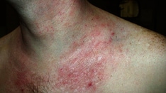
In the spring of 1917, during World War I, Riehl first identified 17 patients with striking dark-brown to grayish-brown facial pigmentation, which was most pronounced on the lateral aspects of the face and neck and primarily concentrated on the forehead, ears, temple, and zygomatic regions. [4, 5] Hyperpigmentation was also noted on the thorax, but it was less pronounced in this region and primarily consisted of small follicular-based pigmented macules. Subtle hyperpigmentation was also identified on the hands, forearms, and intertriginous regions. [5] The patients ranged in age and sex, but all were White from Vienna and without evidence of an underlying illness. In addition to hyperpigmentation, erythematous macules and papules were also identified. [5, 6, 7] Histologically, the lesions were marked by a dense inflammatory cell infiltrate in the superficial dermis admixed with melanophages. [5, 6]
Although Riehl was unable to identify the cause of the eruption, he speculated that the etiology of hyperpigmentation was a nutritional alteration attributed to wartime conditions. [4, 5, 6, 7] Concomitant with the end of the war, no further cases were identified, thereby supporting his hypothesis. [5] Subsequently in World War II, a similar eruption surfaced in approximately 165 people in France, again this was associated with the scarce food supplies and disappeared with the end of the war; however, this latter situation differed from what had been previously seen in World War I in that the majority of cases were reported in women. [5, 6]
Later, Hoffmann and Habermann described a condition referred to as melanodermatitis toxica and hypothesized that it may represent a type of contact dermatitis associated with the use of certain oils and hydrocarbons. Although these authors emphasized the clinical similarities between Riehl melanosis and melanodermatitis toxica, Riehl could not accept that the melanosis he described was due to a local chemical irritant and thought the conditions were separate entities. The role of nutrition as a possible cause of this unusual melanosis was further addressed in a paper by Findlay, who described several cases of Riehl melanosis in the Bantu people in South Africa; however, no further reports in the literature linked Riehl melanosis to nutritional deficiencies. [8]
Subsequent to the great wars, the majority of cases of Riehl melanosis described in the literature differ from the cases originally described by Riehl. In 1950, Minami and Noma described a pigmented dermatitis in Asian women unrelated to the war and named the condition melanosis faciei feminae. [6] The etiology of this latter pigmentation was unknown for many years, until 2 studies from Argentina in the late 1940s and 1950s described facial pigmentation similar to Riehl melanosis that was subsequently attributed to the use of cosmetics. In the first study, patch testing identified aniline dye (orange II) present in facial powder as the cause of the pigmented contact dermatitis, whereas the second study emphasized that photosensitizing may play an addition role in the pigmentation. [5, 9]
In 1970, Osmundsen subsequently reported 7 patients who had a similar bizarre hyperpigmentation that occurred as a result of contact dermatitis to an optical whitener, Tinopal CH 3566, in washing powder and called the condition pigmented contact dermatitis (PCD). [10] Subsequently in 1973, Nakayama introduced the term pigmented cosmetic contact dermatitis for cases that were ascribed to the use of certain cosmetics.
To date, the etiology of Riehl melanosis remains controversial, and although the majority of experts believe it is synonymous with pigmented contact dermatitis, some authors insist that this is an erroneous assumption because the cases reported by Riehl appear to have been related to nutritional alterations that arose during World War I, with no further case reports noted after the war ended, and thereby insist that Riehl melanosis should be classified as a distinct entity. [4, 5, 6, 7] Pigmented contact dermatitis, on the other hand, is caused by an allergic contact dermatitis to a variety of topical and airborne allergens or a lichenoid immune reaction that may be caused by intrinsic or extrinsic factors. [11, 12]
Pathophysiology
The hyperpigmentation in pigmented contact dermatitis is postulated to be caused by frequent and repeated contact with small amounts of sensitizing allergens primarily in cosmetic and textile materials. Nakayama hypothesized that allergens used in commercial products were too low in concentration to produce typical eczematous dermatitis, but rather accumulation of these allergens resulted allergic contact dermatitis, a type IV cytolytic reaction. [13, 14] This later reaction is characterized by vacuolar degeneration of the basal layer of the epidermis associated with pigment incontinence in the superficial dermis. The melanin pigment is slowly engulfed by macrophages; therefore, resolution of the hyperpigmentation is a prolonged process. Because most cases of pigmented contact dermatitis occur in patients with a darker completion, one hypothesis is that various pigment-genetic interactions contribute to the development of this condition. [7] Furthermore, Imokawa and Kawai have provided clinical evidence that various allergens implicated in allergic contact dermatitis can stimulate melanogenesis. [15]
In 2020, Woo et al evaluated biopsies obtained from 12 patients with Riehl melanosis that showed increased dermal expression of stem cell factor (SCF) and c-kit, along with increased expression of epidermal and dermal endothelin-1 in the lesional skin of Riehl melanosis. [16] These authors concluded that their findings support the role of these paracrine melanogenic molecules in the pathogenesis of Riehl melanosis. [16] In 2021, Woo et al also showed that that lesional skin of patients with Riehl melanosis exhibited increased epidermal and dermal expression of estrogen receptor (ER)β and progesterone receptor (PR) mRNAs and concluded that these hormone receptors may play a role in the pathogenesis of Riehl melanosis. [17]
Etiology
A variety of contact allergens have been implicated in pigmented contact dermatitis, as described below. [6, 7, 18] Although the majority of cases occur because of direct contact with these allergens, a few cases secondary to contact with airborne allergens have been described. [7, 19, 20]
Textile allergens are as follows:
-
Tinopal CH3566 - Optical whitener in washing powder
-
Napthol AS - Coupling agent for azo dyes
-
Biocheck 60 - Pesticide for textiles
-
PPP-HB - Textile finish
-
Mercury compounds - Bactericides
-
Formaldehyde - Preservative
-
Azo dyes - Dye [21]
-
Disperse Blue 106 - Dye
-
Disperse Blue 124 - Dye
-
CI Blue 19 (Brilliant Blue) - Dye
-
Rubber components
Cosmetic allergens are as follows [22] :
-
D&C Red 31 - Pigment
-
D&C Yellow No. 11 and 10 - Pigment
-
PAN (phenyl-azo-2-napthol) - Impurity in azo pigments
-
Chromium hydroxide - Pigment
-
Carbanilides (trichlorocarbanilide and Irgasan CF3) - Bactericidal
-
Aniline dyes - Pigment
-
Hair dyes
-
Henna [25]
-
Ricinoleic acid (castor oil acid) - Bactericide (deodorants, lipstick, military camouflage) [26]
-
Kumkum (red) - Cosmetic powder and liquid (Hindu women) [27]
-
Fragrances
Fragrance allergens are as follows:
-
Jasmine absolute
-
Benzyl salicylate
-
Hydroxycitronellal
-
Ylang-ylang oil
-
Cinnamic alcohol
-
Cananga oil
-
Sandalwood oil
-
Synthetic sandalwood (containing bornyl methoxy cyclohexanol)
-
Geraniol oil
-
Eugenol
-
Isoeugenol
-
Lavender oil
-
Methoxycitronellal
-
Benzyl alcohol
-
Cinnamic derivatives
Miscellaneous allergens are as follows:
-
Chromate (K dichromate) - Leather, soaps
-
Nickel/nickel sulfate - Component metal products and jewelry
-
PTBPFR (paratertiary butyl-phenol formaldehyde resin) - Neoprene adhesive in leather products [33]
-
Plathymenia foliosa - Wood dust [20]
-
Minoxidil 5% - Topical vasodilator for hair loss treatment [34]
Cases of pigmented contact dermatitis have been reported in the Indian literature, and the most common allergen to be implicated is kumkum, a colored cosmetic used by Hindu women that is applied most often to the central forehead and along the hair line. [7, 27] Only commercially available red kumkum can sensitize and cause pigmented contact dermatitis. Components of kumkum include azo dyes, coal tar dyes, toludine red, erythrosine, lithal red calcium salt, fragrances, tumeric powder, groundnut oil, tragacanth gum, Cananga oil, and parabens. [7]
In addition to contact allergens, a Riehl melanosis–like eruption has been reported in Japanese women with Sjögren syndrome related to the development of anti-SSA (Ro) antibodies. The lesions are most pronounced on sun-exposed areas, primarily on the face, and the pigmentation typically resolves with the institution of ultraviolet protection. One hypothesis is that ultraviolet radiation induces expression of the SSA antigen on keratinocytes, which then becomes the target of circulating anti-SSA antibodies, resulting in an interface dermatitis and associated pigment incontinence. [35]
Epidemiology
Frequency
The incidence is not known. Most cases are reported outside of the United States, with a large proportion of cases reported in Japan. No international statistics are available, but cases have been reported in France, Denmark, South America, Japan, India, and South Africa.
Race
In general, pigmented contact dermatitis is most pronounced in darkly pigmented races.
Sex
Women appear to have a greater predilection for pigmented contact dermatitis.
Age
Although it has been reported in a wide range of patients, the majority of cases appear to occur in young to middle-aged women.
Prognosis
Although the hyperpigmentation has a tendency to lighten over time and with avoidance of the eliciting agent, some pigmentation may persist.
Patient Education
Because ultraviolet light has been implicated as a contributing factor in some cases of pigmented contact dermatitis, sun avoidance and sunblock usage are prudent. See Sunscreens and Photoprotection for detailed information.