Practice Essentials
Arsenic is a natural occurring metalloid, an abundant element found in many types of rocks. Weathering of rocks releases arsenic-containing dust and allows arsenic compounds to enter groundwater. Arsenical compounds are used in industrial, agricultural, and medicinal substances. Arsenic is also an environmental contaminant in drinking water (well water) and the food chain and is an occupational hazard for miners and glass workers. Higher levels of arsenic are notoriously poisonous to multicellular life. Lower levels of inorganic arsenicals are known to be chemical carcinogens.
Arsenicosis is defined by the World Health Organization (WHO) as a chronic health condition resulting from arsenic ingestion above safe limits for at least 6 months. Arsenical keratosis is a cutaneous manifestation of arsenicosis. [1]
Signs and symtoms
See Presentation for further information.
Arsenical keratoses are usually multiple and typically occur at sites of friction and trauma, especially on the palms and the soles. Keratoses usually manifest as small, punctate, nontender, horny, hard, yellowish, often symmetric, corn-like papules. The diameter of the papule ranges from 0.2-1 cm.
A mild form of the arsenic keratoses may manifest as diffuse thickening or small (< 2 mm) keratoses with sand paper–like texture. Moderate-sized lesions (2-5 mm) may coalesce into larger (>5 mm) verrucous papules or plaques. These lesions are most frequently seen on the thenar and lateral borders of the palms; the base and lateral aspect of the digits; the soles, heels, and toes of the feet, as demonstrated in the image below. Keratoses may also develop on the dorsum of the hands, the arms, and the legs.
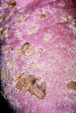 Arsenical keratosis on the sole of a carpenter.
Another type of arsenical keratosis seen in most patients with arsenical cancers manifests as scaly erythematous or pigmented patches on unexposed body areas.
Mee lines (transverse white bands) on the fingernails are seen in acute and chronic arsenic toxicity.
Other skin manifestations of chronic arsenic toxicity include hyperpigmented and hypopigmented macules on the torso and limbs. The pigmented lesions often present as finely freckled or raindrop-like macules distributed bilaterally symmetrically. Diffuse hyperpigmented patches in the intertriginous, genital, and acral areas may be an early sign of chronic arsenic toxicity.
Diagnostics
Laboratory studies
Chemical analysis of arsenic levels in tissue, hair, or nails is possible, but levels may be normal if exposure is not recent. Arsenic levels in hair and nail samples represent exposure in an immediate past period of time. These levels are prone to include contamination of external arsenic. [2]
Blood arsenic level, spot, or 24-hour urine arsenic level adjusted with creatinine are commonly used for detecting arsenic toxicity. However, arsenic depletes rapidly; if the ingestion was not recent, the arsenic levels may be normal in blood and urine. Recent intake of a large volume of seaweed in the diet may affect the arsenic level. [2] Drinking water arsenic levels should be measured if water is the suspected source of exposure.
The complete blood cell count may reveal anemia, leukopenia, and thrombocytopenia. Red blood cells may show cloverleaf nuclei when systemic symptoms are present.
Blood sugar levels may be necessary to rule out arsenic-associated type 2 diabetes. [3]
Renal and liver function tests should be ordered to rule out insidious damage from arsenic toxicity.
In a study of 442 people with skin lesions from arsenic-endemic areas of Bangladesh, Khatun et al found a significant correlation between higher serum periostin levels and degree of arsenical lesions. The investigators also found this correlation with lesion severity and serum immunoglobulin E (IgE) levels and the type 2 cytokines (interleukin-4 [IL-4], IL-5, IL-13, and eotaxin). [4]
In a small study (n=7), patients with arsenical palmar keratosis were noted to have elevated linoleic acid levels in lipid extracts from the lesions when compared to patients with arsenic exposure but no lesions and to a control group with no arsenic exposure. [5]
Imaging studies
Imaging studies are not recommended in a standard workup for arsenical keratoses unless internal malignancy or metastatic disease is suspected after taking a thorough history and completing a physical examination.
Procedures
A skin biopsy may help establish the diagnosis of arsenical keratosis and rule out the diagnosis of skin cancer.
Other tests
Patients with chronic arsenicalism may present with changes on ECG. They may also present with polyneuropathy and changes evident on electromyelography.
Histologic findings
Arsenical keratoses show thick, compact hyperkeratosis and parakeratosis similar to hypertrophic actinic keratoses. Some epidermal keratinocytes may show atypia histologically. [6] The presence of numerous vacuolated keratinocytes and the absence of solar elastosis are suggestive of arsenical keratoses, but these findings are not absolute criteria.
Management
See Treatment for further information.
Surgical removal or destruction (eg, excision, curettage, cryosurgery, dermatome shaving [7] ) is usually the treatment of choice.
Arsenical keratoses show thick, compact hyperkeratosis and parakeratosis similar to hypertrophic actinic keratoses. Some epidermal keratinocytes may show atypia histologically. [6] The presence of numerous vacuolated keratinocytes and the absence of solar elastosis are suggestive of arsenical keratoses, but these findings are not absolute criteria.
Retinoidlike agents decrease the cohesiveness of abnormal hyperproliferative keratinocytes and may reduce the potential for malignant degeneration. [8, 9]
Topical imiquimod has been reported to show some efficacy. [10]
Background
As early as 1888, Hutchinson reported skin cancer in patients who had taken arsenical medications. Numerous reports have since confirmed that ingested arsenic can cause Bowen disease (squamous cell carcinoma in situ); invasive squamous cell carcinoma; basal cell carcinoma of the skin; and (less frequently) internal cancers of the lung, the kidney, the bladder, and the liver. [11]
Arsenic also causes pigment changes and (very frequently) hyperkeratotic lesions of the skin called arsenical keratoses. Arsenical keratoses are the most characteristic skin feature of long-term arsenic exposure.
Other Medscape articles related to arsenic include Neurological Manifestations of Arsenic Intoxication and Arsenic Toxicity in Emergency Medicine.
Pathophysiology
The carcinogenic mechanism of arsenic is not well understood. Arsenic compounds have shown no mutagenicity in standard bacterial and mammalian test systems; however, they can increase the mutagenicity of other DNA-damaging agents. Arsenite impairs nucleotide excision repair, [12] and it may also affect gene expression by increasing or decreasing DNA methylation. [13, 14] The high affinity of arsenic for sulfhydryl groups makes keratin-rich cells (eg, epidermal keratinocytes) a sensitive target for arsenic-induced toxicity.
Arsenic has been shown to alter epidermal keratinocyte differentiation processes, [15] affect cell cycle and apoptosis, induce overexpression of growth factors such as transforming growth factor-α and epidermal growth factor receptor, [16, 17] and enhance proliferation of human keratinocytes. These in vitro findings may have revealed part of the pathogenesis of arsenical keratoses. Whether it is arsenic-induced DNA damage or senescence-associated indirect carcinogenesis, the oncogenesis of arsenic-induced cancers still awaits elucidation. [18, 19]
In the endemic region of West Bengal, India, only 10-30% of people exposed long term to arsenic developed characteristic skin lesions. A 2008 study suggests the individual capacity of DNA repair determines the level of chromosomal aberration, which leads to a susceptibility to develop arsenic-induced premalignant hyperkeratoses. [20]
Etiology
A dose-response relationship exists between the amount of arsenic exposure and the frequency of various skin lesions. However, several studies have shown skin lesions that have developed in persons who consumed drinking water containing arsenic concentrations of less than 50 μg/L. A detail investigation has found that arsenic exposures of greater than 100 μg/L, even 200 μg/L in drinking water, are the cause of the characteristic skin lesions. [21] Three main categories of sources from which patients might have been exposed to arsenic include medicinal, drinking water (environmental), and occupational hazards.
Medicinal arsenic
Inorganic arsenic has been used in medicine for at least 2500 years, particularly since the 18th century, when it was used to treat a great variety of illnesses (eg, acne, diarrhea, gastric ulcer, asthma, malaria, lupus, psoriasis, neurodermatitis, eczema, rheumatism). During the 19th and early 20th centuries, the most popular preparation in the Western world was called Fowler solution, which contained 1% potassium arsenite. [22] In Asia, Chinese proprietary herbal medicine has been the mainstay of remedies for thousands of years and is still popular in that area; it is also available in the United States. Some of these traditional Chinese medicines have been reported to contain high levels of arsenic. [23, 24, 25]
Drinking water
Studies from endemic areas have found that arsenic-contaminated water (mainly well water) is the source of exposure. Cases of arsenic-induced toxicity have been reported in some endemic areas of Chile, Taiwan, India, [26] Bangladesh, and Mexico. An area on the southwest coast of Taiwan has been referred to as the endemic area of Blackfoot disease (a chronic peripheral vascular disease that may progress into gangrenous changes of the lower extremities). The same area also shows a high prevalence of skin cancer. Both conditions are dose related to high arsenic levels in artesian well water that local residents consume daily. In a Taiwanese study, Morales et al [27] reported the lifetime risk of death related to arsenic toxicity is 1 in 100 from consuming 50 μg/L of arsenic in drinking water.
Occupational hazards
Legge reported skin manifestations and lung cancer in sheep dip workers exposed to arsenic as early as 1902. Other reports showed arsenic carcinogenesis in smelter workers, pesticide workers, glass manufacturers, and miners. Burning plywood may cause arsenic-containing fumes to be inhaled. High-tech industries use gallium arsenide to produce semiconductor computer chips.
Epidemiology
Frequency
United States
Arsenical keratosis and arsenic-induced skin cancers are very rare in the United States. Only isolated incidences of cutaneous toxicity from environmental or medicinal exposure have been reported. Several epidemiologic studies of communities exposed to drinking water with higher arsenic levels have not shown any excess incidence of skin cancers.
Documented regions with natural groundwater arsenic contamination include Millard County, Utah; Lane County, Oregon; Lassen County, California; Fallon, Nevada; Fairbanks, Alaska; and New Hampshire. [28] One study suggests persons living in the southwestern United States are at higher risk for being exposed to elevated levels of arsenic in drinking water. [29]
International
Arsenic-induced skin lesions have been noticed in some endemic regions, mainly due to long-term exposure to high levels of arsenic in drinking water. Such regions have been reported in Southern Taiwan, Japan, Vietnam, China, New Zealand, Poland, Hungary, Spain, Canada, Mexico, Chile, Argentina, West Bengal (India), Sri Lanka, Bangladesh, and western Iran. [28, 30]
According to large epidemiologic studies conducted by Yeh et al [31] and Tseng et al [32] in 1968 in Taiwan, among a high-risk population of 40,421 people, arsenic-induced hyperpigmentation was recognized in 18.4% of patients, arsenical keratosis in 7.1%, and skin cancers in 1.5%. Of the 428 people with skin cancer, 72% also presented with arsenical keratosis and 90% had hyperpigmentation.
A study conducted by Fierz [33] in Germany of 262 patients who had been taking Fowler solution for 6-26 years revealed that 40% of these patients developed arsenical keratosis and that 8% had skin cancers.
In a large-scale study in an endemic region, 18,000 persons in Bangladesh and 86,000 persons in West Bengal (India) were clinically examined. Of them, 3695 (20.6%, including 6.11% children) in Bangladesh and 8500 (9.8%, including 1.7% children) in West Bengal had arsenical dermatological features. [34]
An epidemiologic study has been conducted in Kurdistan (western Iran), where the arsenic concentration in drinking water ranges from 42-1500 μg/L. Among 587 villagers examined, 30.7% had skin manifestations of chronic arsenic toxicity. The prevalence rates of Mee lines (transverse bands of leukonychia) on the nails, arsenic keratoses, and pigment disorders among these patients were 86.1%, 77.2%, and 67.8%, respectively. [30]
In a study of 17 people with arsenical lesions from chronic exposure to mine tailings in the Peruvian highlands, 70.6% were men, and farming was the most common occupation. Lesions were most frequently located on the plantar surfaces, followed by palmar and palmoplantar. [35]
Race
Several large studies have focused in regional endemic areas. No global epidemiologic studies have been performed, and no direct predilection has been found among persons of different races.
Sex
In a study in Bangladesh, 430 out of 1481 research subjects older than 30 years who lived in villages with arsenic-contaminated wells were found to have arsenic-induced skin lesions, such as keratosis and hyperpigmentation or hypopigmentation. [36] The prevalence rate is approximately 30%, with a slightly higher prevalence in males than in females. Another study in West Bengal (India) revealed the prevalence of arsenic-induced skin lesions in men was 2- to 3-fold higher than in women. [37]
Age
Immediately consider possible arsenic exposure if skin cancer is found in a relatively young person. The presence of arsenic skin toxicity can be seen in all age groups, although a latency period does occur.
Symptoms of chronic arsenic toxicity develop insidiously after 6 months to 2 years or more of exposure. In the study by Fierz, [33] the minimal latency period from exposure to the development of arsenical keratoses was 2.5 years, and the average latency period for skin cancer was 14 years.
A Japanese study reported the appearance of Bowen disease after 10 years, squamous cell carcinoma after 20 years, and lung cancer after 30 years of apparent arsenic exposure. [38]
In Taiwanese studies, the youngest patients reported to have skin hyperpigmentation, arsenical keratoses, and skin cancer were aged 3, 4, and 24 years, respectively.
Prognosis
Arsenical keratosis is not a fatal disease, but it may persist indefinitely and can become bothersome over time because of pain, bleeding, fissuring, and ulceration. Additionally, some may develop into invasive squamous cell carcinoma. Metastatic arsenic-induced squamous cell carcinoma and arsenic-induced visceral malignancies may result in mortality. [39, 40]
A dose-response relationship exists between arsenic exposure and health effects. Exposure to higher concentrations of arsenic and a longer duration of exposure to arsenic may increase the risk of invasive skin cancers and internal malignancies, which may result in fatality. [41]
-
Arsenical keratosis on the sole of a carpenter.


