Practice Essentials
Porphyrias are metabolic disorders of heme synthesis. Partial enzymic deficiencies result in excessive accumulation and excretion of 5-aminolevulinic acid, porphobilinogen, and/or porphyrins. Porphyria cutanea tarda (PCT) is the most common of the porphyrias in North America and Europe. First described by Waldenström in 1937, this blistering disorder is caused by a deficiency of uroporphyrinogen decarboxylase, an enzyme in heme biosynthesis. [1] Porphyrins accumulate in the liver, are transported in plasma, and are excessively excreted in the urine. Exposure of patients with porphyria cutanea tarda to sunlight results in increased skin fragility, vesicles, bullae, hypertrichosis, hyperpigmentation, sclerodermoid changes, dystrophic calcification, milia, and scarring in a photodistribution. Porphyria cutanea tarda can be inherited or acquired. Treatment options include phlebotomy and antimalarial medications.
Pseudoporphyria describes a bullous photosensitivity that clinically and histologically mimics porphyria cutanea tarda. However, no demonstrable porphyrin abnormalities are present. Pseudoporphyria has been reported in patients with chronic renal failure treated with and without hemodialysis and in those with excessive exposure to ultraviolet A (UV-A) by tanning beds. [2, 3, 4]
Signs and symptoms
Please see Presentation for a full discussion.
Pseudoporphyria is clinically characterized by increased skin fragility; erythema; and the appearance of tense bullae and erosions on sun-exposed skin (see image below), which are identical to those seen in patients with porphyria cutanea tarda.
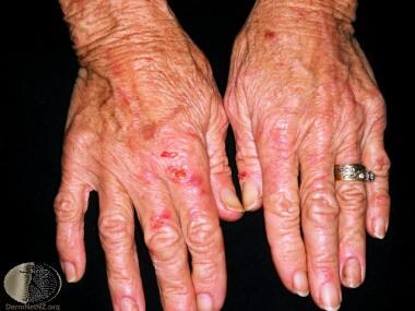 Pseudoporphyria. Courtesy of DermNet New Zealand (http://www.dermnetnz.org/assets/Uploads/reactions/pseudoporphyria1.jpg).
Diagnostics
Of critical importance in the diagnosis of pseudoporphyria is the exclusion of true porphyria. The most important test is a serum/plasma porphyrin assay. If this result is negative, the patient does not have a true porphyria. If the serum/plasma porphyrin assay is unavailable, erythrocytes, urine, and stool may be evaluated for abnormal porphyrin levels. However, such evaluations can represent a diagnostic challenge to complete for patients who may be anuric in the setting of end stage renal disease. [5]
Other causes of photosensitivity, such as connective tissue disease, must be excluded by obtaining a serum antinuclear antibody titer and more specific studies, such as antibodies to Ro, La, ribonucleoprotein, Smith, and double-stranded DNA.
The histologic features of pseudoporphyria are similar to those of porphyria cutanea tarda (PCT) with cell-free subepidermal bullae and festooning of the dermal papillae. [6] The thickness of the blood vessel wall may prove helpful in differentiating pseudoporphyria from porphyria cutanea tarda.
In a comparative histologic study from biopsy samples of patients with porphyria cutanea tarda and pseudoporphyria, Maynard and Peters found thickened blood vessel walls in 11 of 13 patients with porphyria cutanea tarda. In contrast, similar findings in only 1 of 9 patients with pseudoporphyria were present. [7]
Porphyria cutanea tarda and pseudoporphyria have similar, nonspecific direct immunofluorescence findings of granular deposits of immunoglobulins, mostly IgG, and C3 at the basement membrane zone and in the perivascular region. Although direct immunofluorescence is not a useful tool in distinguishing pseudoporphyria from porphyria cutanea tarda, it is helpful in the evaluation of other entities in the differential diagnosis of pseudoporphyria, specifically epidermolysis bullosa acquisita. [8] Epidermolysis bullosa acquisita can be ruled out by the lack of intense, linear immunoreactants at the dermal-epidermal junction. Neither porphyria cutanea tarda nor pseudoporphyria has circulating autoantibodies detected by indirect immunofluorescence study.
Management
The primary treatment of pseudoporphyria is to discontinue the offending agent whenever possible. This may be facilitated by substituting certain medications (eg, tolmetin instead of naproxen for juvenile rheumatoid arthritis, nonthiazide agents for hypertension, nilotinib or dasatinib for imatinib in the setting of chronic myeloid leukemia) for alternative agents that have not been associated with pseudoporphyria. [4, 9, 10, 11] In cases in which substitution or discontinuation is not possible, sun protection has been successfully used as both therapy and prophylaxis against recurrent eruptions. [12, 13, 14] Patients should be adequately educated on this topic. [12] Patients should be adequately educated and counseled to avoid direct sun exposure, wear sun-protective clothing, and apply titanium oxide‒ or zinc oxide‒based sunscreens, which are resistant to wavelengths known to induce porphyric eruptions (400-440 nm). [12] Resolution of the clinical findings may take many months, particularly in drug-induced pseudoporphyria.
In addition to discontinuation of causative agents, some substances have been used in the treatment of pseudoporphyria. N-acetylcysteine (a glutathione precursor) has been reported to improve both dialyzed and nondialyzed forms of chronic renal disease associated pseudoporphyria. [4, 15, 16, 17, 18, 19] Proponents of this therapy have proposed that patients on hemodialysis have decreased levels of glutathione,4,5 (an antioxidant) and that reactive oxygen species may contribute to skin damage. [17] One case series involving 2 patients with pseudoporphyria demonstrated rapid healing of lesions in both patients after the introduction of N-acetylcysteine (with one patient receiving 200 mg four times daily and the other 600 mg twice daily) to treatment, which was otherwise unchanged. [17] It was also noted that discontinuation of the medication in one patient led to a recurrence of blistering. Additional case reports note resolution of blistering after 8 weeks of treatment with dosages of N-acetylcysteine ranging from 600-1200 mg daily. [15, 18] Further investigations evaluating the efficacy are needed to confirm results. However, oral N-acetylcysteine has few, mild adverse effects consisting of nausea, vomiting, and diarrhea and has been suggested to be a safe therapeutic option for pseudoporphyria. [17, 15, 18, 20, 21]
Despite the reported success of N-acetylcysteine, there are cases that demonstrate resistance to this agent. [5, 22] Alternatives that have been documented in the literature include the use of the antimalarials chloroquine and hydroxychloroquine. [19, 22] A case report of pseudoporphyria that was resistant to N-acetylcysteine was noted to have complete resolution after 200 mg of chloroquine administered weekly for 1 month. [22] Improvement in lesions of pseudoporphyria have also been noted with 2 months of daily administration of 10 g of oral glutamine, a nonessential amino acid. [23] This therapy was reportedly well-tolerated with no adverse effects and may be an alternative treatment for cases of pseudoporphyria that are resistant to either N-acetylcysteine or chloroquine. Other case reports have reported improvement with daily administration of 5 mg beta-carotene and 50 mg of green tea extract. [24, 17]
Background
In 1964, Zelickson was first to describe this type of phototoxic reaction in patients after the use of nalidixic acid. [25] The skin lesions were indistinguishable from those observed in patients with porphyria cutanea tarda. Since this initial report, many other drugs have been incriminated in mediating this type of bullous photosensitivity. [26]
Pathophysiology
The precise pathophysiologic mechanism of pseudoporphyria is not fully understood. In 1983, Keane et al developed an animal model for nalidixic acid photosensitivity in CF-1 female mice. [27] Animals injected with nalidixic acid and then exposed to ultraviolet radiation for 10 weeks exhibited more severe cutaneous manifestations than mice treated with sodium chloride solution. Light and electron microscopy demonstrated a subepidermal split beneath the basal lamina at the same level as seen in histologic examination of porphyria cutanea tarda and pseudoporphyria. The authors suggested that a photosensitizing drug might behave in a similar fashion to photoactivated endogenous porphyrins and target similar structures in the skin. Several other authors have corroborated these findings.
Other mechanisms have been proposed to explain the role of ultraviolet or visible light radiation in drug-induced pseudoporphyria. An alternative theory is based on the finding that exogenous photosensitizers are deposited along the endothelium of blood vessels of lesional and nonlesional skin. An immune response targeted against antigens is proposed to develop after phototoxic injury to the dermal microvascular endothelium. Dabski and Beutner proposed a multistep mechanism in which exogenous photosensitizers (drugs) damage the vascular endothelium by the release of proteases after sunlight exposure. [28] Then, immunoglobulin G (IgG) and immunoreactants bind to the damaged endothelium, causing formation of bullae at the level of the lamina lucida as a secondary or tertiary event.
The pathophysiology of pseudoporphyria associated with hemodialysis has not been fully explained. Aluminum hydroxide has been implicated in hemodialysis-associated pseudoporphyria. Aluminum hydroxide is found in dialysis solution and has been shown to produce a porphyrialike disorder after long-term administration in rats.
Etiology
Pseudoporphyria can be induced by a wide range of medications, excessive UV-A exposure, and hemodialysis. [29, 30, 31] Onset of bullae may occur weeks to months after a drug has been initiated. [32]
The list of agents associated with pseudoporphyria will most likely continue to grow. Agents currently associated with pseudoporphyria are as follows [33, 34] :
-
Antipsychotics - Olanzapine [57]
-
Antiarrhythmics - Amiodarone [61]
-
Sulfones - Dapsone
-
Muscle relaxants - Carisoprodol, aspirin [70]
-
Other - Hemodialysis, [73, 74] excessive UV-A, cola, [75] oral contraceptive pills (levonorgestrel and ethinyl estradiol), [76] narrowband UV-B [77] phototherapy (rarely), metformin, [78] diclofenac, [79] country mallow (an herb regional to India [80] ), and oral supplemental chlorophyll, which is sometimes ingested as a purported health food or detoxifying agent [81, 82, 83]
Vitiligo may be associated with pseudoporphyria. [84, 85] Several reports describe patients with vesicles, bullae, and scarring confined to areas of vitiligo on the dorsa of the hands with sparing of normally pigmented skin while taking medications known to cause pseudoporphyria. It is well established that the clinical findings of pseudoporphyria may be precipitated or exacerbated by sunlight. One author suggests that the presence of melanin in healthy skin may be adequate protection to prevent the development of pseudoporphyria in patients with vitiligo.
Epidemiology
Frequency
Pseudoporphyria is not uncommon. Although fewer than 100 cases are documented, pseudoporphyria is most likely underreported in the literature.
Race
Although pseudoporphyria has no predilection toward any one race, it has been shown that fair-skinned children who are highly prone to sunburn are more likely to develop naproxen-induced pseudoporphyria than those children with skin types III or higher. Wallace et al demonstrated that even in the absence of a history of blistering, children with light skin and blue or green eyes are at an increased risk of developing shallow scars on the face while taking naproxen. [86]
Sex
Pseudoporphyria affects males and females equally.
Age
The ages of patients reported with pseudoporphyria range from 2-81 years. It has been reported that the mean age at diagnosis was 50 years in a retrospective study of 20 cases. [87]
Prognosis
The prognosis is good for pseudoporphyria once the offending agent has been discontinued. However, it may take several months for all the skin lesions to resolve, and some can be complicated by infection or prolonged scarring, which has been noted to last up to 5 years. [6, 9]
Patient Education
Educating patients about the causes of pseudoporphyria is important. Patients should avoid solar and tanning salon radiation. If the condition was drug related, patients should avoid medications in a similar class of drugs (eg, other propionic acid NSAIDs) whenever possible.
-
Pseudoporphyria. Courtesy of DermNet New Zealand (http://www.dermnetnz.org/assets/Uploads/reactions/pseudoporphyria1.jpg).


