Practice Essentials
Pityriasis rosea (PR) is a benign rash first described by Gibert in 1860; the name means “fine pink scale.” It is a common skin disorder observed in otherwise healthy people, most frequently children and young adults. Other types of similar skin eruptions include lichen planus, psoriasis, and pityriasis rubra pilaris.
Pityriasis rosea manifests as an acute, self-limiting, papulosquamous eruption with a duration of 6-8 weeks. It evolves rapidly, usually beginning with patch that heralds the eruption, the so-called “herald patch” (see the image below). It may sometimes occur in atypical variants or may mimic other skin disorders, such as tinea corporis and secondary syphilis. [1, 2, 3, 4] Guidelines for diagnosing syphilis (and distinguishing the roseola from pityriasis rosea) have been established. [5] As a rule, pityriasis rosea requires only symptomatic treatment.
 Herald patch. Image courtesy of Drexel Department of Dermatology slide collection.
Etiology
Pityriasis rosea may result from infectious agents, pharmaceuticals, or immunizations.
See Etiology for further information.
Prognosis
Pityriasis rosea is a self-limiting, generally benign disorder for which the prognosis is excellent and the recurrence rate is low (approximately 2%).
Diagnostics
See Workup.
Treatment
Treatment for pityriasis rosea is supportive.
See Treatment and Medication for further information.
Also see 15 Rashes You Need to Know: Common Dermatologic Diagnoses, a Critical Images slideshow, to help identify and treat various rashes.
Pathophysiology
Pityriasis rosea (PR) has often been considered to be a viral exanthem, a view supported by the condition’s seasonal occurrence, its clinical course, the possibility of epidemic occurrence, the presence of occasional prodromal symptoms, and the low rate of recurrence. However, a 4-year recurrence rate of 25.9% was documented in one survey of 212 patients. [6] Oxidative stress may play a role too. [7] One survey noted a significant temporal clustering and seasonal variation, and the incidence of dengue fever significantly correlated. [8]
Pityriasis rosea has been linked to upper tract respiratory infections. An increased incidence is reported among groups with close physical contact (eg, families, students, and military personnel), though the condition does not appear to be highly contagious. The incidence of pityriasis rosea among dermatologists is 3-4 times that among other physicians.
A higher incidence of pityriasis rosea is also noted among patients with decreased immunity (eg, pregnant women and bone marrow transplant recipients). Additionally, ampicillin increases the distribution of the eruption, an effect bearing a striking resemblance to the drug’s effect on the rash of infectious mononucleosis.
Some immunologic data also suggest a viral etiology. [9] A lack of natural killer (NK) cell and B-cell activity in pityriasis rosea lesions has been noted, suggesting a predominantly T-cell mediated immunity in the development of the condition. Increased amounts of CD4 T cells and Langerhans cells are present in the dermis, possibly reflecting viral antigen processing and presentation. Anti−immunoglobulin M (IgM) to keratinocytes has been found in patients with pityriasis rosea; this finding may be associated with the exanthem phase of the presumed viral infection.
The primary plaque is seen on the skin in 50-90% of cases a week or more before the onset of the eruption of smaller lesions. This secondary eruption occurs 2-21 days later in crops following the lines of cleavage of the skin. On the back, this eruption produces a “Christmas tree” pattern (see Presentation).
Atopy, seborrheic dermatitis, and acne vulgaris are more common in patients with pityriasis rosea than in control subjects. Pityriasis rosea during pregnancy may foreshadow premature delivery and fetal demise, especially when it develops within the first 15 weeks of gestation. [10]
Recurrences of pityriasis rosea are generally regarded as rare and are thought by some to indicate a lasting immunity when they do occur. However, they may be more common than previously delineated. [11]
A classification based on pathogenetic mechanisms associated wth the different presentations of pityriasis rosea has been proposed. [12]
Etiology
Infectious agents
An infectious etiology for pityriasis rosea (PR) has been sought for many years. It has been suggested that the condition is precipitated by a viral agent. Accordingly, a number of viruses have been studied with a view to determining whether they are linked to pityriasis rosea.
Picornaviruslike particles have been seen in the tissue of African green monkeys inoculated from human pityriasis rosea lesions. A follow-up study failed to find picornavirus RNA in patients with pityriasis rosea. Serology and polymerase chain reaction (PCR) assay for viral DNA have been negative for Epstein-Barr virus (EBV), parvovirus B19, cytomegalovirus (CMV), human herpesvirus (HHV)-8, [13] HHV-1, and HHV-2 in patients diagnosed with pityriasis rosea. Pityriasis rosea itself was also described in a few patients with coronavirus disease 2019 (COVID-19). [14, 15]
Some initial reports using PCR analysis suggested a role for human herpesvirus (HHV)-7 and HHV-6, but subsequent studies have not been confirmatory. [16, 17, 18, 13, 19, 20, 21, 22, 23, 24, 25, 26, 27, 28] That HHV-7 is frequently found in healthy individuals casts further doubt on its proposed etiologic role. [27] The influenza virus H1N1 has also been proposed as a possible causative pathogen. [29]
Some investigators have suggested that a fungal infection is a more likely cause of pityriasis rosea than a viral infection is. However, no fungus has been isolated as a definite causal agent. No bacterial pathogen, such as Mycoplasma, has been found to be causative either. Thus far, the search for an infectious cause of pityriasis rosea has been unsuccessful.
Vaccines
Vaccine-induced pityriasis rosea and pityriasis rosea-like eruptions have been linked with vaccinations for smallpox, tuberculosis, influenza, papillomavirus, polio, tetanus, diphtheria, pneumococcal, diphtheria-pertussis-tetanus, hepatitis B, and yellow fever, [30] and more recently with the Moderna mRNA-1273 and other COVID-19 vaccines. [31, 32]
Drugs
Pityriasis rosea–like eruptions can also occur in association with many drugs (eg, acetylsalicylic acid, barbiturates, bismuth, captopril, clonidine, gold, imatinib, isotretinoin, ketotifen, levamisole, metronidazole, omeprazole, D-penicillamine, and terbinafine), as well as certain vaccines (eg, bacille Calmette-Guérin [BCG], human papilloma virus, and diphtheria). [2, 33, 30]
Anti−tumor necrosis factor (TNF)-α agents such as adalimumab and etanercept have also been implicated. [34, 35] Pityriasis rosea–like drug eruptions have been reported to be related to use of rituximab, [36] nortriptyline, [37] and clozapine. [38]
Pityriasis rosea–like drug eruptions may be difficult to distinguish from non–drug-induced cases. Drug-induced pityriasis rosea often lasts longer than non–drug-induced pityriasis rosea. Lesions are also thought to be increased in individuals with high stress levels.
Epidemiology
United States statistics
Pityriasis rosea (PR) is very common in the general population, and most cases occur in the spring and winter in temperate climates. The estimated frequency of pityriasis rosea in the United States is approximately 0.13% in females and 0.14% in males, with a 0.3-3% prevalence at dermatologic centers.
International statistics
Worldwide, pityriasis rosea has been estimated to account for 2% of dermatologic outpatient visits. The disease is more common in the spring and the fall in temperate climate zones. However, it may be more frequent in the summer in some other regions. It favors the hot, dry season in Australia, India, and Malaysia.
An increase in the prevalence of pityriasis rosea has been reported in Uganda. No change in the prevalence of pityriasis rosea has been reported in Sweden. It has also been seen in the United Kingdom, Nigeria, Sudan, Brazil, Lagos, Singapore, Turkey, Kuwait, and Hong Kong.
Age-, sex-, and race-related demographics
Pityriasis rosea is observed in people of all age groups, though it is most common in persons aged 10-35 years and rare in infants and elderly persons. The youngest patient reported in the literature was aged 3 months, and the oldest was aged 85 years.
Pityriasis rosea occurs slightly more often in females than in males. [39] The female-to-male ratio is reported as 2:1 or 3:2 in the United States.
No racial predominance is reported. More intensely pigmented Africans tend to have more widespread disease. The lesions in African Americans may lack a rose color, and they may appear darker than the surrounding skin.
Prognosis
Pityriasis rosea (PR) is a self-limiting, generally benign disorder for which the prognosis is excellent and the recurrence rate is low (approximately 2%). Because it is not considered a transmissible disease, patients do not require isolation and may return to work or school. Pityriasis rosea usually lasts for 6-8 weeks, but can last as long as 3-6 months. Protracted cases of severe eczematous or drug-induced pityriasis rosea are referred to as pityriasis rosea perstans.
The main morbidity is from pigmentary changes, which may develop as lesions heal, especially in black people. Both postinflammatory hyperpigmentation and hypopigmentation may occur. However, lesions do not result in scars. Bacterial superinfections may occur, but are rare. In pregnant women, pityriasis rosea is sometimes associated with miscarriage if occurring within the first 15 weeks of pregnancy, premature delivery, or neonatal hypotonia and hyporeactivity. [10, 40]
Patient Education
Patients should be instructed to avoid contact with irritants. In addition, patients and families should be educated regarding the benign and noninfectious nature of the rash and the relatively lengthy course of the disease. Typically, the secondary rash develops over 2 weeks, persists for another 2 weeks, and then fades over another 2 weeks, without the need for treatment, though some lesions have persisted for as long as 3-4 months.
For patient education resources, see the Skin Conditions & Beauty Center, as well as Eczema (Atopic Dermatitis).
-
Herald patch. Image courtesy of Drexel Department of Dermatology slide collection.
-
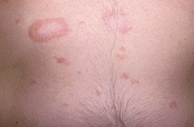
Christmas tree distribution of lesions on trunk. Image courtesy of Drexel Department of Dermatology slide collection.
-
Histopathologic features of pityriasis rosea. Image courtesy of Gary R Kantor, MD, Department of Dermatology, Drexel University, Philadelphia, PA.


