Continually Updated Clinical Reference
 
 
  All Sources     eMedicine     Medscape     Drug Reference     MEDLINE
 
eMedicine - Pigmented Purpuric Dermatitis : Article by

Quick Find
Authors & Editors
Introduction
Clinical
Differentials
Workup
Treatment
Medication
Follow-up
Miscellaneous
Acknowledgments
Multimedia
References

Related Articles
Scurvy


Patient Education
Click here for patient education.


Author: Cedric C Banfield, BSc, MSc, MBBS, MRCP, Consulting Staff, Department of Dermatology, Hinchingbrooke Hospital, UK

Cedric C Banfield is a member of the following medical societies: British Medical Association, Royal College of Physicians, and Royal Society of Medicine

Editors: Jean-Hilaire Saurat, MD, Chair, Professor, Department of Dermatology, University of Geneva, Switzerland; Richard P Vinson, MD, Assistant Clinical Professor, Department of Dermatology, Texas Tech University School of Medicine; Consulting Staff, Mountain View Dermatology, PA; Lester F Libow, MD, Dermatopathologist, South Texas Dermatopathology Laboratory; Joel M Gelfand, MD, MSCE, Medical Director, Clinical Studies Unit, Assistant Professor, Department of Dermatology, Associate Scholar, Center for Clinical Epidemiology and Biostatistics, University of Pennsylvania; Dirk M Elston, MD, Director, Department of Dermatology, Geisinger Medical Center

Author and Editor Disclosure

Synonyms and related keywords: capillaritis, benign pigmented purpura, pigmented purpuric eruptions, Schamberg disease, progressive pigmentary dermatosis, itching purpura of Loewenthal, eczematidlike purpura of Doucas and Kapetanakis, pigmented purpuric lichenoid dermatosis of Gougerot and Blum, lichen aureus, purpura annularis telangiectoides, purpura annularis telangiectodes, Majocchi disease

Background

The pigmented purpuric dermatoses are a group of chronic diseases of mostly unknown etiology that have a very distinctive clinical appearance. They are characterized by extravasation of erythrocytes in the skin with marked hemosiderin deposition.

A number of clinical patterns of pigmented purpuric dermatoses or capillaritis are recognized that may represent different presentations of the same disorder; however, this generally does not influence the treatment or the prognosis. They all show a similar histologic appearance. The term pigmented purpuric dermatoses includes Schamberg disease (ie, progressive pigmentary dermatosis), purpura annularis telangiectodes (Majocchi disease), lichen aureus, itching purpura, eczematidlike purpura of Doucas and Kapetanakis, and the pigmented purpuric lichenoid dermatosis of Gougerot and Blum. Many consider itching purpura and eczematidlike purpura to be variants of Schamberg disease.

Pathophysiology

The etiology is unknown. Venous hypertension, exercise, and gravitational dependency are important cofactors that appear to influence disease presentation. Histologically, a perivascular T-cell lymphocytic infiltrate is centered on the superficial small blood vessels of the skin, which show signs of endothelial cell swelling and narrowing of the lumen. Extravasation of red blood cells with marked hemosiderin deposition in macrophages is also found, and a rare granulomatous variant of chronic pigmented dermatosis has been reported.

Frequency

United States

Pigmented purpuric dermatoses are uncommon.

International

During a 10-month period, the author's United Kingdom hospital-based dermatology practice, which serves a population of 300,000 persons, identified only 10 such cases. Five cases were diagnosed as having lichen aureus, and the remainder had more extensive capillaritis.

Mortality/Morbidity

Typically, the condition is asymptomatic, but pruritus may sometimes be a prominent feature in some cases, especially in patients with itching purpura.

Race

Persons of any race can be affected.

Sex

Pigmented purpuric dermatoses appear to occur more frequently in men than in women.

Age

  • Schamberg disease may occur in persons of any age.
  • Itching purpura and the dermatosis of Gougerot and Blum mainly affect middle-aged men.
  • Lichen aureus and Majocchi disease are predominantly diseases of children or young adults.


History

Patients complain about the appearance of their skin.

  • In Schamberg disease, irregular plaques and patches of orange-brown pigmentation develop on the lower limbs. The lesions are chronic and persist for years. With time, many of the lesions tend to extend, but some may spontaneously clear.
  • In itching purpura, the lesions are much more extensive, and patients typically complain of severe pruritus.

Physical

The hallmark of a pigmented purpuric dermatosis is its characteristic orange-brown, speckled, cayenne pepper–like discoloration.

  • The lower limbs are affected in Schamberg disease, whereas itching purpura is characterized by more generalized skin involvement.
  • In lichen aureus, the eruption is usually a solitary lesion or a localized group of lesions that may affect any part of the body; however, the leg is the most commonly affected area. Linear or quadrantic forms of lichen aureus have been reported.
  • Majocchi disease is characterized by small annular plaques of purpura that contain prominent telangiectasia.
  • Pigmented purpura with lichenoid-type skin change is yet another clinical variant, which Gougerot and Blum first reported.

Causes

The cause is unknown. Rare familial cases of Schamberg disease and Majocchi disease have been reported in the literature, implying a genetic cause in a minority of patients.


Scurvy

Other Problems to be Considered

Pigmented purpuric dermatoses must be distinguished from early cutaneous T-cell lymphoma, purpuric clothing dermatitis, stasis pigmentation, scurvy, leukocytoclastic vasculitis, purpuric generalized lichen nitidus, and drug hypersensitivity reactions (eg, allergy to rituximab, carbamazepine, meprobamate, chlordiazepoxide, furosemide, nitroglycerin, or vitamin B-1). A case of glipizide-induced pigmented purpuric dermatosis has recently been reported. Topical fluorouracil has been implicated in a pigmented purpuric dermatitislike skin eruption. Pharmacologically induced regressed Kaposi sarcoma lesions may be misdiagnosed clinically and histologically as pigmented purpuric dermatitis.


Lab Studies

  • A complete blood cell count is necessary to exclude thrombocytopenia, and coagulation screening helps to exclude other possible causes of purpura.

Imaging Studies

  • Dermoscopy has been reported to be a useful tool for assisting the clinical diagnosis of pigmented purpuric dermatoses.

Other Tests

  • Capillary fragility may be assessed by the Hess test.

Procedures

  • A skin biopsy helps to confirm the diagnosis of a pigmented purpuric eruption and aids in excluding cutaneous T-cell lymphoma, which in its early stages may closely mimic a pigmented purpuric dermatitis both clinically and histologically.

Histologic Findings

Histologically, a perivascular infiltrate of lymphocytes and macrophages is centered on the superficial small blood vessels of the skin. Signs of endothelial cell swelling and narrowing of lumina may be seen (see Media File 4).

The infiltrate is composed of predominantly CD4+ lymphocytes along with occasional CD1a+ dendritic cells. Plasma cells and neutrophils are occasionally present; the latter is not uncommon in lesions of itching purpura. Extravasation of red blood cells with marked hemosiderin deposition in macrophages is typically seen (see Media File 5). However, the degree of hemosiderin deposition may be variable, and it can be minimal in early lesions of itching purpura.

Histochemical staining with Perls stain and Fontana-Masson stain, to demonstrate iron (hemosiderin) and exclude melanin pigment respectively, may be helpful. Hemosiderin deposition in the dermis is more superficial in pigmented purpuric dermatitis than that seen in stasis dermatitis, which is a useful differentiating feature. Mild epidermal spongiosis and exocytosis of lymphocytes may be seen in all variants except lichen aureus, which, in general, tends to show a bandlike infiltrate separated from the epidermis by a thin rim of uninvolved collagen.


Medical Care

No medical intervention is of proven benefit for the treatment of the pigmented purpuric dermatoses.

  • Pruritus may be alleviated by the use of topical corticosteroids and antihistamines.
  • Associated venous stasis should be treated by compression hosiery.
  • Prolonged leg dependency should be avoided.


The goals of pharmacotherapy are to reduce morbidity and to prevent complications.

Drug Category: Corticosteroids

These agents are effective in relieving pruritus. These agents have anti-inflammatory properties and cause profound and varied metabolic effects. They modify the body's immune response to diverse stimuli.

Drug NameHydrocortisone (Westcort)
DescriptionAn adrenocorticosteroid derivative suitable for application to skin or external mucous membranes. Has mineralocorticoid and glucocorticoid effects, resulting in relief of pruritus.
Adult DoseApply sparingly to affected areas bid
Pediatric DoseApply as in adults
ContraindicationsDocumented hypersensitivity; viral, fungal, or tubercular skin infections
InteractionsNone reported
PregnancyC - Safety for use during pregnancy has not been established.
PrecautionsProlonged use, applying over large surface areas, applying potent steroids, and using occlusive dressings may increase systemic absorption of corticosteroids and may cause Cushing syndrome, reversible HPA-axis suppression, hyperglycemia, and glycosuria

Drug NameClobetasol (Temovate)
DescriptionClass I superpotent topical steroid; suppresses mitosis and increases synthesis of proteins that decrease inflammation and cause vasoconstriction.
Adult DoseApply bid for up to 2 wk; not to exceed 50 g/wk
Pediatric DoseNot established
ContraindicationsDocumented hypersensitivity; viral or fungal skin infections
InteractionsNone reported
PregnancyC - Safety for use during pregnancy has not been established.
PrecautionsMay suppress adrenal function in prolonged therapy

Drug NameBetamethasone (Diprolene, Betatrex)
DescriptionFor inflammatory dermatosis responsive to steroids. Decreases inflammation by suppressing migration of polymorphonuclear leukocytes and reversing capillary permeability.
Adult DoseApply thin film bid until response
Pediatric DoseAdminister as in adults
ContraindicationsDocumented hypersensitivity; paronychia; cellulitis; impetigo; angular cheilitis; erythrasma; erysipelas; rosacea; perioral dermatitis; acne
InteractionsNone reported
PregnancyC - Safety for use during pregnancy has not been established.
PrecautionsDo not use in skin with decreased circulation; can cause atrophy of groin, face, and axillae; if infection develops and is not responsive to antibiotic treatment, discontinue until infection is under control; do not use monotherapy to treat widespread plaque psoriasis

Drug Category: Antihistamines

These agents may treat itching by blocking effects of endogenously released histamine.

Drug NameDiphenhydramine (Benadryl, Benylin, Diphen, AllerMax)
DescriptionFor symptomatic relief of pruritus caused by release of histamine in inflammatory reactions.
Adult Dose25-50 mg PO q6-8h prn; not to exceed 400 mg/d
10-50 mg IV/IM q6-8h prn; not to exceed 400 mg/d
Pediatric Dose12.5-25 mg PO tid/qid, or 5 mg/kg/d PO/IV/IM or 150 mg/m2/d PO/IV/IM divided tid/qid; not to exceed 300 mg/d
ContraindicationsDocumented hypersensitivity; MAOIs
InteractionsPotentiates effect of CNS depressants; because of alcohol content, do not give syr dosage form to patient taking medications that can cause disulfiramlike reactions
PregnancyC - Safety for use during pregnancy has not been established.
PrecautionsMay exacerbate angle-closure glaucoma, hyperthyroidism, peptic ulcer, and urinary tract obstruction


Further Outpatient Care

  • A follow-up consultation is required for cases in which initially diagnostic uncertainty exists, particularly to exclude cutaneous lymphoma.
  • Topical steroid treatment if used long term should be monitored for the possible development of adverse effects.

Prognosis

  • Many lesions persist or extend with time. Most eventually resolve spontaneously.


Medical/Legal Pitfalls

  • Failure to perform a skin biopsy in unusual or prolonged cases of pigmented purpuric dermatitis to exclude cutaneous T-cell lymphoma is a pitfall.


The authors and editors of eMedicine gratefully acknowledge the contributions of previous author, John D Wilkinson, MD, MBBS, MRCS, FRCP, to the development and writing of this article.


Media file 1:  
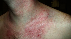
Pigmented purpuric dermatitis affecting the trunk. Some of the lesions show the characteristic orange-brown, speckled, cayenne pepper–like discoloration that is the hallmark clinical sign of a capillaritis. Men are more frequently affected than women. If the lesions are pruritic, then the term itching purpura is sometimes used. Early cutaneous T-cell lymphoma, purpuric clothing contact dermatitis, and drug hypersensitivity reactions should be considered in the differential diagnosis.
Click to see larger pictureClick to see detailView Full Size Image
Media type:  Photo

Media file 2:  Lichen aureus is the name given to localized pigmented purpuric dermatitis or capillaritis. In this patient, the skin on the extensor surface of the elbow is affected.
Click to see larger pictureClick to see detailView Full Size Image
Media type:  Photo

Media file 3:  Histologic features of a skin biopsy sample obtained from a patient with lichen aureus shows extravasation of erythrocytes and a perivascular T-cell infiltrate.
Click to see larger pictureClick to see detailView Full Size Image
Media type:  Photo

Media file 4:  Endothelial cell swelling is a histologic feature of capillaritis. This biopsy sample was obtained from a patient with lichen aureus.
Click to see larger pictureClick to see detailView Full Size Image
Media type:  Photo

Media file 5:  Hemosiderin deposition is seen in dermal macrophages in this biopsy sample obtained from a patient with lichen aureus.
Click to see larger pictureClick to see detailView Full Size Image
Media type:  Photo

Media file 6:  Capillaritis affecting the lower legs is known as Schamberg disease. In Schamberg disease, irregular plaques and patches of orange-brown pigmentation develop on the lower limbs.
Click to see larger pictureClick to see detailView Full Size Image
Media type:  Photo


  • Adams BB, Gadenne AS. Glipizide-induced pigmented purpuric dermatosis. J Am Acad Dermatol. Nov 1999;41(5 Pt 2):827-9. [Medline].
  • Aiba S, Tagami H. Immunohistologic studies in Schamberg''s disease. Evidence for cellular immune reaction in lesional skin. Arch Dermatol. Jul 1988;124(7):1058-62. [Medline].
  • Alexandrescu DT, Dutcher JP, O'Boyle K, et al. Fatal intra-alveolar hemorrhage after rituximab in a patient with non-Hodgkin lymphoma. Leuk Lymphoma. Nov 2004;45(11):2321-5. [Medline].
  • Barnhill RL, Braverman IM. Progression of pigmented purpura-like eruptions to mycosis fungoides: report of three cases. J Am Acad Dermatol. Jul 1988;19(1 Pt 1):25-31. [Medline].
  • Basak PY, Ergin S. Should pentoxifylline be regarded as an effective treatment for Schamberg's disease?. J Am Acad Dermatol. Mar 2001;44(3):548-9. [Medline].
  • Burrows NP, Jones RR. Cell adhesion molecule expression in capillaritis. J Am Acad Dermatol. Nov 1994;31(5 Pt 1):826. [Medline].
  • Champion RH, Burton JL, Burns DA. Purpura. In: Textbook of Dermatology. 6th ed. London, England: Blackwell Science; 1998:. 2149-52.
  • Farrokhzad S, Champion RH. Pigmented purpuric dermatoses. Dermatologica. 1970;140(1):45-53. [Medline].
  • Ghersetich I, Lotti T, Bacci S, et al. Cell infiltrate in progressive pigmented purpura (Schamberg''s disease): immunophenotype, adhesion receptors, and intercellular relationships. Int J Dermatol. Dec 1995;34(12):846-50. [Medline].
  • Hanna S, Walsh N, D'Intino Y, Langley RG. Mycosis fungoides presenting as pigmented purpuric dermatitis. Pediatr Dermatol. Jul-Aug 2006;23(4):350-4. [Medline].
  • Kwon SJ, Lee CW. Figurate purpuric eruptions on the trunk: acetaminophen-induced rashes. J Dermatol. Nov 1998;25(11):756-8. [Medline].
  • Okada K, Ishikawa O, Miyachi Y. Purpura pigmentosa chronica successfully treated with oral cyclosporin A. Br J Dermatol. Jan 1996;134(1):180-1. [Medline].
  • Pantanowitz L, Dezube BJ, Pinkus GS, Tahan SR. Histological characterization of regression in acquired immunodeficiency syndrome-related Kaposi's sarcoma. J Cutan Pathol. Jan 2004;31(1):26-34. [Medline].
  • Puddu P, Ferranti G, Frezzolini A, et al. Pigmented purpura-like eruption as cutaneous sign of mycosis fungoideswith autoimmune purpura. J Am Acad Dermatol. Feb 1999;40(2 Pt 2):298-9. [Medline].
  • Saito R, Matsuoka Y. Granulomatous pigmented purpuric dermatosis. J Dermatol. Aug 1996;23(8):551-5. [Medline].
  • Sardana K, Sarkar R, Sehgal VN. Pigmented purpuric dermatoses: an overview. Int J Dermatol. Jul 2004;43(7):482-8. [Medline].
  • Simon M Jr, Heese A, Gotz A. Immunopathological investigations in purpura pigmentosa chronica. Acta Derm Venereol. 1989;69(2):101-4. [Medline].
  • Tamaki K, Yasaka N, Osada A, et al. Successful treatment of pigmented purpuric dermatosis with griseofulvin. Br J Dermatol. Jan 1995;132(1):159-60. [Medline].
  • Toro JR, Sander CA, LeBoit PE. Persistent pigmented purpuric dermatitis and mycosis fungoides: simulant, precursor, or both? A study by light microscopy and molecular methods. Am J Dermatopathol. Apr 1997;19(2):108-18. [Medline].
  • Wong WK, Ratnam KV. A report of two cases of pigmented purpuric dermatoses treated with PUVA therapy. Acta Derm Venereol. 1991;71(1):68-70. [Medline].
  • Wong WR, Kuo TT, Chen MJ, Chan HL. Granulomatous variant of chronic pigmented purpuric dermatosis: report of two cases. Br J Dermatol. Jul 2001;145(1):162-4. [Medline].
  • Yáñez S, Val-Bernal JF. Purpuric generalized lichen nitidus: an unusual eruption simulating pigmented purpuric dermatosis. Dermatology. 2004;208(2):167-70. [Medline].
  • Zaballos P, Puig S, Malvehy J. Dermoscopy of pigmented purpuric dermatoses (lichen aureus): a useful tool for clinical diagnosis. Arch Dermatol. Oct 2004;140(10):1290-1. [Medline].

Pigmented Purpuric Dermatitis excerpt

Article Last Updated: Sep 8, 2006