Practice Essentials
Pemphigus foliaceus (PF) is generally a benign variety of pemphigus. It is an autoimmune skin disorder characterized by the loss of intercellular adhesion of keratinocytes in the upper parts of the epidermis (acantholysis), resulting in the formation of superficial blisters. It is typified by clinical involvement of healthy-appearing skin that blisters when rubbed (the Nikolsky sign; commonly but incorrectly spelled Nicholsky), a finding named after Dr Piotr Nikolsky, who first described this sign in 1896. [1] Pemphigus foliaceus is characterized by a chronic course, with little or no involvement of the mucous membranes.
Pemphigus foliaceus has the following 6 subtypes: pemphigus erythematosus (PE), pemphigus herpetiformis (PH), endemic pemphigus foliaceus, endemic pemphigus foliaceus with antigenic reactivity characteristic of paraneoplastic pemphigus (but with no neoplasm), immunoglobulin A (IgA) pemphigus foliaceus, and drug-induced pemphigus foliaceus. See Pemphigus Erythematosus; Pemphigus Herpetiformis; Pemphigus, Paraneoplastic (PNP); and Pemphigus, IgA for more information.
Signs and symptoms
The bullae usually start on the trunk. The course of the disease is long-term, with the patient's general health being satisfactory. Spontaneous remission sometimes occurs, but the lesions can persist for several years. A unique clinical pattern may occur in children, with individual lesions appearing as arcuate, circinate, or polycyclic. [2] Eyelid skin involvement without conjunctival changes occurs occasionally in patients with pemphigus foliaceus. [3]
The primary lesions are small, superficial blisters; however, these flaccid bullae are difficult to find because they are transient and transform into erosions. Typical pemphigus foliaceus has scaly, crusted erosions on an erythematosus base confined mainly to so-called seborrhoic areas (eg, face, scalp, upper part of the trunk).
The Nikolsky sign is the finding that physical trauma can shear the pathologic epidermis of the skin of patients with pemphigus foliaceus, resulting in clinical lesions. The Nikolsky sign should probably be regarded as a moderately sensitive but highly specific tool for the diagnosis of pemphigus. [4]
The erosions can become numerous, showing a tendency to generalize. Occasionally, erythrodermia develops. Thus, pemphigus foliaceus may be evident as a an erythroderma or a psoriasiform erythroderma. [5, 6] Atrophic changes of the nails and the hair are sometimes evident. The erosions may be accompanied by a burning sensation and local pain. In contrast to PV, in pemphigus foliaceus, little or no involvement of the mucous membranes occurs. Note the images below.
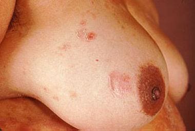 Middle-aged American woman of Mexican lineage with superficial bullae characteristic of pemphigus foliaceus.
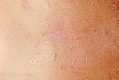 Pemphigus foliaceus. Middle-aged American woman of Mexican lineage with superficial bullae formation.
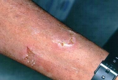 A 41-year-old woman of Puerto Rican origin with a 9-year history of pemphigus foliaceus, often with erythroderma flares.
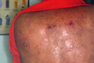 A 41-year-old woman of Puerto Rican origin with a 9-year history of pemphigus foliaceus, often with erythroderma flares.
IgA pemphigus foliaceus begins as pruritic, flaccid vesicles in an annular pattern.
PH commences as intensely pruritic, grouped papules and vesicles suggestive of dermatitis herpetiformis. Erythematous patches with peripheral vesicles may be present. Sometimes, oral erosions are seen.
PE starts as erythematous patches with border vesiculation, often in a butterfly distribution on the cheeks and the forehead, with similar patches on the sternal and interscapular skin. Crusted plaques may appear in the healing phase.
PNP is a subset of pemphigus combining the clinical features of pemphigus vulgaris (PV) variably associated with those of erythema multiforme, bullous pemphigoid, and lichen planus. Chorzelski and associates [7] in 1999 described a most unusual case of PNP with the immunopathologic findings of pemphigus foliaceus. The clinical pattern appears to be correlated with that of the antibody profile; therefore, patients with antibodies directed against desmoglein 1 tend to have the clinical features of pemphigus foliaceus.
Diagnostics
Also see Histologic Findings.
Immunofluorescence using both direct techniques and indirect techniques is the most reliable method to diagnosis pemphigus. [8] Because of the rare occurrence of pemphiguslike antibodies, pemphigus cannot be diagnosed by indirect immunofluorescence (IIF) alone and must be confirmed by direct immunofluorescence (DIF). With the use of 2 appropriate substrates (ie, monkey esophagus [or human skin] and guinea pig esophagus and standardized conjugates), in IIF, PV and pemphigus foliaceus patterns are different; PV stains throughout the epidermis, and pemphigus foliaceus stains only in the upper epidermis, whereas, with DIF, the patterns are similar. With a DIF study, cell surface immune deposits are often present throughout the entire epidermis in both pemphigus foliaceus and PV. Using DIF on telogen hair outer root sheath may be beneficial for diagnosis and follow-up, irrespective of the presence of scalp lesions. [9]
Immunologic examination with DIF testing shows IgG in the intercellular space, mainly in the upper parts of the epidermis; an IIF study documents the presence of circulating pemphigus antibodies, especially with a guinea pig esophagus used as a substrate. One IIF study suggested that using both a monkey esophagus and the human skin increases the sensitivity and aids in distinguishing pemphigus foliaceus from PV. In PH, IgG deposits are evident in the upper epidermis, with circulating IgG to the epidermal cell surface. The subcorneal pustular dermatosis type of IgA pemphigus foliaceus has IgA deposition on the upper epidermal cell surfaces and circulating IgA antibodies to the epidermal cell surfaces. Desmogleins 1 and 3 are the major cell surface target molecules in patients with PH. In the unusual instance when PV becomes pemphigus foliaceus, or vice versa, the clinical alteration is associated with a shift in the antidesmoglein autoantibody profile.
Other methods, such as ELISA [10] and immunoblot assays, [11] can be used, but they require highly purified antigens to give similar results. The sensitivity for PV and pemphigus foliaceus antibodies is more than 98% in at least the renowned laboratory of Jarzabek-Chorzelska and associates, [8] with their many decades of experience. Histologic examination is useful, but it is not the preferred method for diagnosing pemphigus foliaceus because it cannot replace a highly reliable DIF method.
Another less experienced laboratory found ELISA to be superior to an IIF study for serodiagnosis of pemphigus foliaceus at various stages of disease activity. [12]
Pemphigus foliaceus arising during the administration of D-penicillamine was described in an elderly patient in whom withdrawal of D-penicillamine resulted in improvement of the skin lesions and ELISA scores for anti–desmoglein 1 antibodies revealed a rapid decline. [13]
The sensitivity and specificity of a novel multisubstrate immunofluorescence technique called BIOCHIP was evaluated in 35 patients, 21 with pemphigus vulgaris and 14 with pemphigus foliaceus, and the technique was found to be beneficial in differentiating these two types of pemphigus. [14]
Management
Also see Medication.
Present information is probably inadequate to ascertain the optimal therapy for pemphigus foliaceus (PF), including the optimal glucocorticoid dose, the role of adjuvant immunosuppressive medications, and long-term adverse events to improve the risk-to-benefit ratio. [15] Therapy for pemphigus foliaceus is usually less aggressive than that of PV because of lower morbidity and mortality rates. [16]
First results indicate that nonsteroidal treatment of pemphigus is possible. Mestinon may be used to slow down progression of the disease and to treat mild cases with chronic lesions on limited areas. Antimalarial therapy may be effective monotherapy in some patients. However, a major obstacle in comparing therapeutic outcomes is the lack of generally accepted definitions and measurements for the clinical evaluation of patients with pemphigus. [17] Common terms and endpoints of pemphigus are needed to accurately measure and assess disease extent, activity, severity, and therapeutic response.
Topical glucocorticosteroids may be sufficient in cases of limited involvement. [18]
In more extensive cases (similar to PV), adjuvant immunosuppressants, including systemic corticosteroids, azathioprine, mycophenolate mofetil, cyclophosphamide, and cyclosporin A, may be necessary. [19, 20]
In some cases, such as PE, combined therapy is beneficial with the use of corticosteroids and sulfones or antimalarial agents.
Topical treatment with antibiotics and corticosteroids, such as topical clobetasol cream or ointment 0.05% twice a day, is helpful. Other vehicles that may be useful are creams, foams, liquids (for scalp lesions), and aerosols. Antibiotics, such as minocycline 50 mg daily, may be effective. Nicotinamide 1.5 g/d and tetracycline 2 g/d have also been reported to be beneficial in a small number of patients. Antibiotics and nicotinamide are purported to have anti-inflammatory effects. [21]
Photoprotection is appropriate for some patients because UV-B may trigger acantholysis and cause a flare-up of the disease.
Successful anti-CD20 antibody treatment has also been described. [22]
Plasmapheresis is another therapeutic option in patients with recalcitrant disease. It may decrease autoantibody titers in some patients and favorably influence the clinical outcome, especially in patients with otherwise therapy-resistant pemphigus foliaceus. It is often used in conjunction with cytostatic agents, such as cyclophosphamide or azathioprine, to reduce a predictable rebound increase in autoantibody synthesis. Potential complications, including the need for maintaining venous access, a bleeding tendency, electrolyte shifts, pulmonary edema, fever, chills, hypotension, and septicemia, should be considered. [23]
Amagai et al reported that a single cycle of intravenous immunoglobulin at 400 mg/kg/d for 5 days is effective and safe for patients with pemphigus that is relatively resistant to systemic steroid therapy. [24] Toth and Jonkman also reported on successful therapy with intravenous immunoglobulin (low dose). [25]
Background
Pierre Louis Alphee Cazenave, founder of the first journal dedicated entirely to dermatology, documented the first description of pemphigus foliaceus in 1844 in this journal. The description was of a 47-year-old woman who consulted him at l'Hopital Saint Louis in Paris for a generalized eruption of several years' duration. Nikolsky described lateral extension of the preexisting erosion due to lifting up the collarette (and when applying a lateral pressure to the clinically intact skin), whereas Asboe-Hansen described extension of the intact blister due to pressure that is applied to its roof.
Senear and Usher originally described PE in 1926 as an unusual type of pemphigus with features of lupus erythematosus. PE (also known as Senear-Usher syndrome) is best viewed as a localized form of pemphigus foliaceus. Chorzelski et al [26] determined its immunopathology in 1968.
Another pemphigus foliaceus variant with pruritic, flaccid vesicles in an annular pattern has been characterized as IgA pemphigus foliaceus, with antibodies of IgA class providing the basis for diagnosis.
Jablonska and associates [27] coined the term pemphigus herpetiformis for the pemphigus foliaceus variant that often begins as small clusters of pruritic papules and vesicles mimicking dermatitis herpetiformis.
Endemic pemphigus foliaceus, or fogo selvagem (formerly known as Brazilian pemphigus foliaceus because it is evident mainly in the river valleys of rural Brazil), has also been described in Columbia, El Salvador, Paraguay, Peru, and Tunisia. Fogo selvagem (Portuguese for wild fire) displays immunopathologic findings of pemphigus and a distinctive epidemiology suggestive of a disorder triggered by an infectious insect-borne agent (see Fogo Selvagem). [28] A focus of endemic pemphigus foliaceus also exists in El Bagre, Columbia and shares features with Senear-Usher syndrome but occurs in an endemic fashion. [29, 30] It also may have autoantibodies to optic nerve sheath envelope cell junctions, the clinical significance of which is unknown. [31] Heterogeneous antigenic reactivity was observed as in paraneoplastic pemphigus but with no evidence of association with neoplasia. This endemic pemphigus disease in El Bagre had immunologic features similar to pemphigus foliaceus or erythematosus.
Chorzelski et al in 1999 [7] described paraneoplastic pemphigus with cutaneous and serologic features of pemphigus foliaceus in a patient with an underlying lymphoma. The authors are not aware of any similar patients with these highly unusual findings.
Drug-induced pemphigus foliaceus is mostly associated with penicillamine, nifedipine, or captopril, medications with a cysteinelike chemical structure.
A transition from pemphigus vulgaris (PV) to pemphigus foliaceus, or vice versa, is not likely. However, in the experience at the Medical University of Warsaw, PV in the remission period may resemble pemphigus foliaceus. About 7% of patients with pemphigus foliaceus may have the initial features of PH. This figure was 35% in patients with endemic pemphigus foliaceus in Tunisia (see Pemphigus Vulgaris). The pivotal contributions of Jean-Claude Bystryn have been summarized. [32]
Pathophysiology
Superficial blisters in pemphigus foliaceus are induced by immunoglobulin G (IgG) (mainly IgG4 subclass) autoantibodies directed against a cell adhesion molecule, desmoglein 1 (160 kd), expressed mainly in the granular layer of the epidermis. Desmoglein 1 is also a major autoantigen in cases of PH, suggesting that most cases of both PE and PH are clinical variants of pemphigus foliaceus. The mechanism of acantholysis induction by specific autoantibodies may involve phosphorylation of intracellular proteins associated with desmosomes. Complement activation does not play a pathogenic role in pemphigus foliaceus. A polyclonal mixture in pemphigus foliaceus patients of anti-Dsg1 IgG antibodies enhances pathogenic activity for blister formation, with not only pathogenic antibodies, but also nonpathogenic antibodies, contributing to blister formation. [33]
Antibodies against desmoglein 3 are also present in patients with paraneoplastic pemphigus (PNP), a severe condition associated with various antibodies against different components of the cell adhesion complex. Other target antigens, including the acetylcholine receptor, have also been postulated to be relevant in the pathogenesis of pemphigus foliaceus. [34, 35, 36, 37]
Cholinergic control of epidermal cohesion may be important. [38] The regulation of keratinocyte cell-to-cell and cell-matrix adhesion is an important biological function of cutaneous acetylcholine. Progress in therapy of pemphigus using cholinergic drugs supports this concept.
Precipitating factors include medications and ultraviolet light radiation. It has been suggested that both enhanced autoantibody epidermal binding and preferential neutrophil adhesion to UV-irradiated epidermis contribute to acantholysis development in photo-induced pemphigus foliaceus. Radiation therapy may also flare pemphigus foliaceus in those with it. [39, 40, 41]
Endemic pemphigus foliaceus seems to have an environmental cause. The prevalence of antibodies against desmoglein 1 is high in people residing in endemic areas of Brazil, with disease onset preceded by a sustained antibody response due to an as yet unknown environmental factor.
Genetic factors predispose to the development of pemphigus foliaceus. The HLA-DRB1*01:02 allele and the HLA-DRB1*01-DQA1*01-DQB1*05 haplotype had the strongest linkage among 86 pemphigus foliaceus patients from southeastern Brazil. [42] The role of genetic factors is evident in fogo selvagem in which a strong association exists with some human leukocyte antigen DRB1 (HLA-DRB1) haplotypes, including DRB1*0404, 1402, 1406, and 1401. In France, persons with DRB1*0102 and 0404 are at an increased risk of pemphigus foliaceus.
Pemphigus trigger factors have been meticulously analyzed by Ruocco and Ruocco, [43] who have delineated an exhaustive list and stressed the need to detect environmental provoking or precipitating factors. As a superb memory device to facilitate thorough patient evaluation, Ruocco [44] has cleverly observed that PEMPHIGUS should encourage the physician to consider pesticides (PE), malignancy (M), pharmaceuticals (P), hormones (H), infectious agents (I), gastronomy (G), ultraviolet light (U), and stress (S).
Rarely, a change in pemphigus subtype may occur, accompanied by qualitative and quantitative changes in the anti-Dsg autoantibody profile as detected using antigen-specific enzyme-linked immunosorbent assays (ELISAs). Thus, the antibody profile defines the clinical phenotypes of pemphigus, and that intermolecular "epitope spreading" may be the immunological mechanism underlying a shift between pemphigus foliaceus and PV. [45, 46]
It has been suggested that polymorphisms in the 2q33 and 3q21 chromosomal regions influence susceptibility to pemphigus foliaceus. [47]
Scientific evidence links interactions between thymoma function, thymoma, and pemphigus, often associating loss of self-tolerance and the presence of autoimmunity. [48] The association of pemphigus foliaceus and thymoma is noteworthy.
Endemic pemphigus foliaceus in Tunisia has merited analysis. Unlike in North America and Japan, in Tunisia IgG autoantibodies against Dsg1 are commonly detected in healthy individuals. [49] In a Tunisian study, these IgG autoantibodies against Dsg1 were generally against pre-Dsg1 and/or C-terminal domains of Dsg1 in healthy Tunisians.
Long noncoding RNA variants may play a pivotal role in the immunopathogenesis of both endemic and sporadic forms of pemphigus foliaceus. [50]
Etiology
Endemic pemphigus foliaceus, or fogo selvagem, seems to be induced by a viral infection transmitted by insects. Pemphigus vulgaris in a recipient with pemphigus foliaceus in a donor after allogeneic peripheral blood stem cell transplantation between two siblings has been described. [51] Pemphigus foliaceus patients may have antibody responses against other keratinocyte adhesion molecules in addition to the main target autoantigen, Dsg1, and anti-Dsg1 monoclonal antibodies cross-reacted with an environmental antigen, LJM11, a sand fly saliva protein. [46]
In some patients, pemphigus foliaceus may be precipitated by extensive UV exposure or burns and by various drugs (eg, penicillamine, [52, 53] inhibitors of angiotensin convertase, nonsteroid anti-inflammatory agents). [54] Pemphigus foliaceus induced by bucillamine has been described. [55]
A topical ectoparasiticide containing fipronil, amitraz, and S-methoprene has been described as causing pemphigus foliaceus in dogs. [56]
Pemphigus foliaceus was described following COVID-19 vaccination. [57, 58]
Herbal supplements containing blue algae have also been linked with the induction of pemphigus foliaceus. [59]
Epidemiology
Frequency
The incidence of pemphigus foliaceus varies depending on the population studied. Pemphigus foliaceus is rare and sporadic worldwide. In contrast to PV, no predominance of pemphigus foliaceus is found in Jews and in people of Mediterranean descent. An increased incidence of pemphigus foliaceus was noted in Tunisian women (6.6 cases per million per year), whereas, in Western Europe, the incidence of pemphigus foliaceus is about 0.5-1 case per million per year. [60]
Endemic pemphigus foliaceus, or fogo selvagem, occurs with a high frequency in central and southwestern Brazil and in Colombia. The Terena reservation in Brazil has a prevalence of 3.4% of the population. In endemic regions of Brazil, as many as 50 cases per million per year are seen. [61] Other foci may be present in the Maghreb; one was described in Morocco.
Race
Pemphigus foliaceus has been described in all races.
Sex
In general, the prevalence of pemphigus foliaceus in men and women is about equal; however, in the Sousse region of Tunisia, an overwhelming predominance of women are affected. The peak incidence of endemic pemphigus foliaceus in women aged 25-34 years in the Sousse region of Tunisia is 15.5 cases per million per year. In El Salvador, a similar female and age predisposition may also be evident.
Age
The mean patient age at onset of pemphigus foliaceus is about 50-60 years; however, it may occur at any age, from infancy onward. [5] Fogo selvagem often occurs in children, young adults, and genetically related family members. The mean patient age at onset is about 20-30 years. The peak incidence of endemic pemphigus foliaceus occurs in women aged 25-34 years in the Sousse region of Tunisia. An increased incidence in genetically related family members does not appear to exist.
Prognosis
Pemphigus foliaceus tends to persist for months to years. Pemphigus foliaceus may coexist with thymoma, myasthenia gravis, lupus erythematosus, and other autoimmune bullous diseases.
-
Middle-aged American woman of Mexican lineage with superficial bullae characteristic of pemphigus foliaceus.
-
Pemphigus foliaceus. Middle-aged American woman of Mexican lineage with superficial bullae formation.
-
A 41-year-old woman of Puerto Rican origin with a 9-year history of pemphigus foliaceus, often with erythroderma flares.
-
A 41-year-old woman of Puerto Rican origin with a 9-year history of pemphigus foliaceus, often with erythroderma flares.
-
Histologic view shows the typical pattern of a detached stratum corneum without bullae formation. Pigmentary incontinence is prominent in the dermis, reflecting the patient's 9-year history of recurrent superficial bullae.


