Background
Parapsoriasis describes a group of clinically variable cutaneous diseases that can be characterized by scaly patches or slightly elevated papules and/or plaques dispersed on the trunk or proximal extremities, with some lesions that may have a resemblance to psoriasis—hence the nomenclature. However, this description includes several skin diseases that likely arise from a variable degree of lymphocytic inflammation, which may have nonspecific histopathology to histology, with abnormal lymphocytes seen in mycosis fungoides. Some authors have suggested that the term parapsoriasis should be avoided because it does not have well-defined histopathological criteria, and, further, that while dermatologists use the term as a “clinical hypothesis”, dermatopathologists “do not consider parapsoriasis a histopathological diagnosis”. [1] With the range of histology detected, treatments are variable but therapies that affect T cells can be effective.
In 1902, Brocq initially described the following three major entities that fit the description of parapsoriasis:
-
Pityriasis lichenoides (acuta and chronica)
-
Small plaque parapsoriasis
-
Large plaque parapsoriasis (parapsoriasis en plaque)
Pityriasis lichenoides (acuta and chronica)
Pityriasis lichenoides variants describe scaly dermatoses with necrotic papules that are clinically and histologically different from parapsoriasis. These diseases generally are benign and undergo spontaneous resolution but, at times, may have a protracted course (see Pityriasis Lichenoides for further discussion).
Large plaque and small plaque parapsoriasis
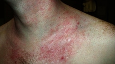
Current terminology of parapsoriasis refers to 2 disease processes that are caused by T-cell–predominant infiltrates in the skin. These disease processes are large plaque parapsoriasis and small plaque parapsoriasis.
As the nomenclature and description of the disease spectrum under the descriptive term parapsoriasis evolved, the primary focus has been on the distinction of whether the disorder progresses to mycosis fungoides (MF) or cutaneous T-cell lymphoma (CTCL). Small plaque parapsoriasis is a benign disorder that rarely if ever progresses. Large plaque parapsoriasis is more ominous in that many patients progress to MF/CTCL. [2] Other cases have given rise to lymphomatoid papulosis. [3]
Controversy exists currently in the classification of large plaque parapsoriasis because some believe it is equivalent to the earliest stage of CTCL, the patch stage. [4, 5, 6] A meta-analysis from 2016 supports a risk of transformation to early CTCL, and newer technology may allow for earlier diagnosis. [7, 8]
The duration of parapsoriasis can be variable. Small plaque disease lasts several months to years and can spontaneously resolve. Large plaque disease is chronic, and treatment is recommended because it may prevent progression to CTCL.
El-Darouti et al reported on a 7-year study of a hypopigmented disorder that the researchers believe should be classified as a new variant of parapsoriasis en plaque. [9]
No clear etiology for small plaque or large plaque parapsoriasis is known, and no specific association has been made with contact exposure or infections.
For more information, see the topic Psoriasis.
Pathophysiology
The initiating cause of parapsoriasis is unknown, but the diseases likely represent different stages in a continuum of lymphoproliferative disorders from chronic dermatitis stimulated by activated T cells to frank malignancy of cutaneous T-cell lymphoma (CTCL).
Small plaque parapsoriasis
Small plaque parapsoriasis likely is a reactive process of predominantly CD4+ T cells. Genotypic pattern observed in small plaque parapsoriasis is similar to that observed in chronic dermatitis, and the pattern of clonality of T cells is consistent with the response of a specific subset of T cells that have been stimulated by an antigen. Multiple dominant clones can be detected by polymerase chain reaction (PCR) of T-cell receptor gene usage, which supports a reactive process. Lymphocytes do not show histologic atypia to suggest malignant transformation. Southern blot analysis of T-cell receptor genes from parapsoriasis does not identify a dominant clone of T cells.
Some physicians believe that small plaque parapsoriasis is an abortive T-cell lymphoma; however, no clear distinguishing evidence, such as genetic changes (eg, TP53 mutations) observed in other malignancies, exists to support this contention. [10] Nevertheless, a hint to the verity of this hypothesis is the recent identification of increased telomerase activity in T cells from CTCL at low-grade stages, high-grade lymphoma, and parapsoriasis, which is activity not exhibited in normal T cells. A better understanding is likely to develop from further molecular characterization. [11]
Large plaque parapsoriasis
Large plaque parapsoriasis is a chronic inflammatory disorder, and the pathophysiology has been speculated to be long-term antigen stimulation. This disorder is associated with a dominant T-cell clone, one that may represent up to 50% of the T-cell infiltrate. If the histologic appearance is benign, without atypical lymphocytes, classification of large plaque parapsoriasis is made. If atypical lymphocytes are present, many would classify such patients as having patch stage CTCL.
In one study, human herpesvirus type 8 was detected in up to 87% of skin lesions of large plaque parapsoriasis. This is the first association of a specific infectious agent with large plaque parapsoriasis, and the significance is unclear. Further studies are important to determine the significance of this finding. [12]
The close relationship between large plaque parapsoriasis and mycosis fungoides is highlighted by the detection of TOX expression, a new marker that has been described to be frequently detected in the abnormal T cells in mycosis fungoides. In one study, large plaque parapsoriasis has expression of TOX similar to that of mycosis fungoides. [13]
Epidemiology
There are no accurate statistics on the incidence and frequency of parapsoriasis, but digitate dermatoses may be underreported because of the lack of symptoms and subtle presentation. Patients may underreport the frequency of large plaque parapsoriasis when it is asymptomatic and subtle. Thus, the prevalence of large plaque parapsoriasis is not known and may be greater than the reported incidence of mycosis fungoides, which is approximately 3.6 cases per million population per year.
Mortality has not been reported for small plaque parapsoriasis. Morbidity is limited to symptoms, which are minimal. For large plaque parapsoriasis, mortality may be associated with progression to MF (cutaneous T-cell lymphoma [CTCL]). The patch stage of MF represents the early stages of CTCL, and the 5-year survival rate is greater than 90%. Long-term survival is the same as that from a matched controlled population. [14]
Small plaque parapsoriasis is associated with male predominance. The male-to-female ratio is 3:1. A slight asymmetry favoring male dominance for large plaque parapsoriasis may exist.
For both small plaque parapsoriasis and large plaque parapsoriasis, presentation most frequently is in middle age; peak incidence is in the fifth decade of life.
Prognosis
Small plaque parapsoriasis may persist in a stable pattern for years to decades and then resolve spontaneously. A small number of cases may progress to mycosis fungoides (MF).
Large plaque parapsoriasis remains indolent for many years. The disease may progress to cutaneous T-cell lymphoma (CTCL) with transformation of lymphocytes from benign small size to larger atypical lymphocytes. The 5-year survival rate, however, still remains high and is greater than 90%.
-
Small plaque parapsoriasis.
-
Small plaque parapsoriasis.
-
Large plaque parapsoriasis.


