Practice Essentials
Onychomycosis is a fungal infection of the toenails or fingernails that may involve any component of the nail unit, including the matrix, bed, or plate. Onychomycosis can cause pain, discomfort, and disfigurement and may produce serious physical and occupational limitations, as well as reducing quality of life. [1] See the image below.
 Proximal subungual onychomycosis. Proximal leukonychia. Image courtesy of Dr Antonella Tosti.
Signs and symptoms
Patients with onychomycosis may present with the following:
-
Initially, complaints about the appearance of the nail, with no physical symptoms
-
As the disease progresses, interference with standing, walking, and exercising
-
Paresthesia, pain, discomfort, and loss of dexterity
-
Loss of self-esteem and inhibited social interaction
Onychomycosis has 5 main subtypes, as follows:
-
Distal lateral subungual onychomycosis (DLSO)
-
White superficial onychomycosis (WSO)
-
Proximal subungual onychomycosis (PSO)
-
Endonyx onychomycosis (EO)
-
Candidal onychomycosis
Patients may have a combination of these subtypes. Total dystrophic onychomycosis, the most advanced form of any subtype, presents as a thickened, opaque, and yellow-brown nail. Presentation varies by subtype.
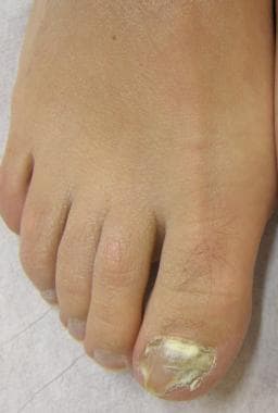
Features of DLSO are as follows:
-
Subungual hyperkeratosis and onycholysis, which is usually yellow-white in color
-
Yellow streaks and/or yellow onycholytic areas in the central portion of the nail plate
Features of WSO are as follows:
-
Confined to the toenails
-
Small, white, speckled or powdery patches on the surface of the nail plate
-
The nail becomes roughened and crumbles easily
-
Molds produce a deep variety of WSO characterized by a larger and deeper nail plate invasion
Features of PSO are as follows:
-
An area of leukonychia in the proximal nail plate that moves distally with nail growth
-
In PSO caused by molds, leukonychia is typically associated with marked periungual inflammation
Features of EO are as follows:
-
Milky white discoloration of the nail plate
-
No evidence of subungual hyperkeratosis or onycholysis
Features of candidal onychomycosis are as follows:
-
Develops in patients with chronic mucocutaneous candidiasis or immunodepression
-
Affects several or all digits
-
Total onychomycosis associated with periungual inflammation
-
The digits often take on a bulbous or drumstick appearance
See Clinical Presentation for more detail.
Diagnosis
Direct microscopy of a 20% potassium hydroxide (KOH) preparation in dimethyl sulfoxide (DMSO) can screen for fungi. The technique is as follows:
-
Before obtaining a specimen, the nails must be clipped and cleansed with an alcohol swab to remove bacteria and debris
-
The preparation does not require heating or prolonged incubation if DMSO is a component of the KOH solution
-
In DLSO, obtain a specimen from the nail bed by curettage; the onycholytic nail plate should be removed, and the sample should be obtained at a site most proximal to the cuticle, where the concentration of hyphae is greatest
-
In PSO, the overlying nail plate must initially be pared with a No. 15 blade; then, a sample of the ventral nail plate may be taken
-
In WSO, a No. 15 blade may be used to remove a specimen from the nail surface
-
In suspected candidal onychomycosis, specimens should be taken from the affected nail bed closest to the proximal and lateral edges
-
Nail fragments must be small enough for examination under low power
-
Large pieces of nail plate may be pulverized prior to microscopy by using a hammer or a nail micronizer
-
Counterstains, such as chlorazol black E or Parker blue-black ink, may be used to accentuate the hyphae
Fungal culture can identify the species of organism and guide therapy. [2] Culture techniques are as follows:
-
Two types of growth medium should be used, one for dermatophytes and one for nondermatophytes
-
Medium with cycloheximide (dermatophyte test medium [DTM], Mycosel, or Mycobiotic) selects for dermatophytes
-
Cultures should be obtained from pulverized nail scrapings or clippings while the patient has abstained from antifungal medication for at least 2 weeks
-
The specimen should be kept at room temperature with the cap placed loosely over the inoculated medium
See Workup for more detail.
Management
Medications for onychomycosis can be administered topically or orally. A combination of topical and systemic treatment increases the cure rate. Adjunctive surgical measures may also be used.
Topical therapy for onychomycosis is as follows:
-
Ciclopirox olamine 8% nail lacquer solution
-
Amorolfine or bifonazole/urea (available outside the United States)
-
Efinaconazole 10% topical solution (the first FDA-approved topical triazole for toenail onychomycosis)
-
Tavaborole 0.5% topical solution, an oxaborole solution (boron-containing compound)
-
Can be used in WSO and DLSO limited to the distal nail
-
Should be limited to cases involving less than half of the distal nail plate or for patients unable to tolerate systemic treatment
-
Topical treatments may be useful to prevent recurrence in patients cured with systemic agents
Oral therapy for onychomycosis is as follows:
-
Terbinafine
-
Itraconazole
-
Fluconazole and posaconazole [5] are off-label alternatives
-
Systemic treatment is always required in PSO and in DLSO involving the lunula region
Nonpharmacologic approaches include the following:
-
Laser treatment
-
Photodynamic therapy
-
Mechanical, chemical, or surgical nail avulsion
-
Chemical removal with a 40-50% urea compound in patients with very thick nails
-
Removal of the nail plate as an adjunct to oral therapy
Laser treatment can be combined with topical antifungals. [6, 7]
See Treatment and Medication for more detail.
Background
Onychomycosis (OM) refers to a fungal infection that affects the toenails or the fingernails. Onychomycosis may involve any component of the nail unit, including the nail matrix, nail bed, or nail plate. Onychomycosis is not life threatening, but it can cause pain, discomfort, and disfigurement and may produce serious physical and occupational limitations. Psychosocial and emotional effects resulting from onychomycosis are widespread and may have a significant impact on quality of life. [8]
The main subtypes of onychomycosis are distal lateral subungual onychomycosis (DLSO), white superficial onychomycosis (WSO), proximal subungual onychomycosis (PSO), endonyx onychomycosis (EO), and candidal onychomycosis. Patients may have a combination of these subtypes. Total dystrophic onychomycosis refers to the most advanced form of any subtype.
Special concerns
HIV disease
Onychomycosis in patients who are immunocompromised is associated with increased severity and morbidity. Lesions may appear atypical and require more aggressive management compared with the healthy population. Proximal subungual (ie, proximal subungual onychomycosis) involvement is much more prevalent in patients with HIV infection than in those without HIV infection. In this population, white superficial onychomycosis is more commonly caused by T rubrum, rather than T mentagrophytes.
Diabetes [9]
The diabetic foot may lead to serious complications associated with onychomycosis. Peripheral neuropathy and sensory loss may lead to increased trauma without pain in patients with diabetes. Bacterial colonization and vascular insufficiency may exacerbate the problem and may lead to serious sequelae.
Elderly age [10]
Onychomycosis in elderly people is complicated by diseases (eg, poor vision, arthritis) that prevent optimal foot care. Nail changes are much more common in elderly persons and often involve the fingernails and the toenails. The potential for drug-drug interactions is more evident and must be addressed before initiating oral therapy.
Pathophysiology
The pathogenesis of onychomycosis depends on the clinical subtype. In distal lateral subungual onychomycosis, the most common form of onychomycosis, the fungus spreads from plantar skin and invades the nail bed via the hyponychium. Inflammation occurring in these areas of the nail apparatus causes the typical physical signs of distal lateral subungual onychomycosis. In contrast, white superficial onychomycosis is a rarer presentation caused by direct invasion of the surface of the nail plate. In proximal subungual onychomycosis, the least common subtype, fungi penetrate the nail matrix via the proximal nail fold and colonize the deep portion of proximal nail plate. Endonyx onychomycosis is a variant of distal lateral subungual onychomycosis in which the fungi infect the nail via the skin and directly invade the nail plate. Total dystrophic onychomycosis involves the entire nail unit (see the image below).
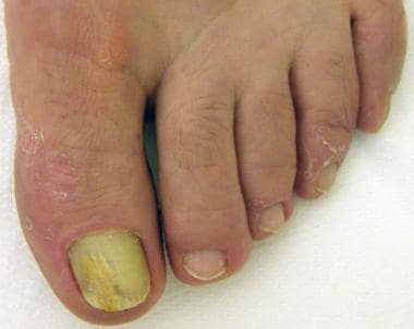 Distal subungual onychomycosis. Onycholysis and yellow streak. Image courtesy of Dr Antonella Tosti.
Nail invasion by Candida is not common because the yeast needs an altered immune response as a predisposing factor to be able to penetrate the nails. Despite the frequent isolation of Candida from the proximal nail fold or the subungual space of patients with chronic paronychia or onycholysis, in these patients Candida is only a secondary colonizer. In chronic mucocutaneous candidiasis, the yeast infects the nail plate and eventually the proximal and lateral nail folds.
Etiology
Onychomycosis is caused by 3 main classes of fungi: dermatophytes, yeasts, and nondermatophyte molds. Dermatophytes are by far the most common cause of onychomycosis. Two major pathogens are responsible for approximately 90% of all onychomycosis cases. Trichophyton rubrum accounts for 70% and Trichophyton mentagrophytes accounts for 20% of all cases. Onychomycosis caused by nondermatophyte molds (Fusarium species, Scopulariopsis brevicaulis,Aspergillus species) is becoming more common worldwide, accounting for up to 10% of cases. [11] Onychomycosis due to Candida is rare. [12]
T rubrum is the most common pathogen in distal lateral subungual onychomycosis. Proximal subungual onychomycosis due to T rubrum infection is typical of immunosuppressed patients . Additionally, Proximal subungual onychomycosis with periungual inflammation is usually caused by molds
White superficial onychomycosis is usually caused by T mentagrophytes; nondermatophyte molds cause deep white superficial onychomycosis.
Candida albicans nail infection is observed in premature children, in immunocompromised patients, and in persons with chronic mucocutaneous candidiasis.
Risk factors for onychomycosis include family history, increasing age, poor health, prior trauma, warm climate, participation in fitness activities, immunosuppression (eg, HIV, drug induced), communal bathing, and occlusive footwear. Biomechanical problems with repetitive microtraumas to the nails cause onycholysis and other nail dystrophies that favor nail invasion by fungi.
Epidemiology
United States
The recent proliferation of fungal infections in the United States can be traced to the large immigration of dermatophytes, especially Trichophyton rubrum, from West Africa and Southeast Asia to North America and Europe. [13]
International
The incidence of onychomycosis has been reported to be 2-13% in North America. [14] A multicenter survey in Canada showed the prevalence of onychomycosis at 6.5%. [15] Onychomycosis accounts for half of all nail disorders, and onychomycosis is the most common nail disease in adults. Toenails are much more likely to be infected than fingernails. Thirty percent of patients with a cutaneous fungal infection also have onychomycosis. The incidence of onychomycosis has been increasing, owing to such factors as diabetes, immunosuppression, and increasing age. [13]
Studies in the United Kingdom, Spain, and Finland found prevalence rates of onychomycosis to be 3-8%.
Race
Onychomycosis affects persons of all races.
Sex
Onychomycosis affects males more commonly than females. However, candidal infections are more common in women than in men.
Age
Studies indicate that adults are 30 times more likely to have onychomycosis than children. Onychomycosis has been reported to occur in 2.6% of children younger than 18 years but as many as 90% of elderly people.
Prognosis
The goals for antifungal therapy are mycological cure and a normal looking nail. Mycological cure can be evaluated at the end of treatment, while clinical cure requires several more months owing to slow nail growth. [16]
Clinical trials have repeatedly demonstrated higher efficacy for terbinafine compared with other antifungal treatments. [17, 18] A meta-analysis of 18 studies on terbinafine, 6 studies on pulse itraconazole, and 3 studies on fluconazole for onychomycosis showed a mycological cure rate of 76%, 63 %, and 48 % respectively. [19]
Yellow streaks along the lateral margin of the nail and/or presence of yellow onycholytic areas in the central portion of the nail (dermatophytoma) are associated with a poor response to treatment.
Residual nail changes persist in most patients as a result of the frequent association of onychomycosis with traumatic toenail dystrophies.
Onychomycosis caused by molds, particularly Fusarium species, are often not responsive to systemic therapy.
Recurrence (relapse or reinfection) of onychomycosis is not uncommon, with reported rates ranging from 10-53%. [20]
Fungal infections of the fingernails have a much more favorable prognosis than toenail infections.
Patient Education
Patients should be educated about the use of appropriate footwear, especially in high-exposure areas such as communal bathing facilities and health clubs.
Following treatment, patients must be advised that nails may not appear normal for up to 1 year, and prophylactic antifungal therapy may be required to prevent reinfection of the skin and the nails. Patients may use topical terbinafine cream twice daily for 1-2 weeks for early tinea pedis or a 1-week pulse of itraconazole (200 mg PO bid) at the first signs of onychomycosis. [21]
For patient education resources, see Nail Psoriasis and Onychomycosis.
-
Distal subungual onychomycosis. Onycholysis and yellow streak. Image courtesy of Dr Antonella Tosti.
-
Distal subungual onychomycosis. Subungual hyperkeratosis onycholysis and yellow streak. Image courtesy of Dr Antonella Tosti.
-
Proximal subungual onychomycosis. Proximal leukonychia. Image courtesy of Dr Antonella Tosti.
-
White superficial onychomycosis. Image courtesy of Dr Antonella Tosti.
-
Candidal onychomycosis in a patient with chronic mucocutaneous candidiasis. Total onychomycosis and paronychia. Image courtesy of Dr Antonella Tosti.
-
Dermoscopy of distal subungual onychomycosis showing irregular margin of the onycholytic area with spikes projecting into the proximal nail plate, reported as the "aurora borealis" pattern. Handyscope at 20X.


