Practice Essentials
Nevus araneus, also known as spider angioma or spider nevus, is a common benign vascular lesion present in 10-15% of healthy adults and young children. [1, 2] They may appear as a solitary or multiple lesions. [2] In particular, when multiple lesions are present, liver disease, [3] arteriovenous malformations (AVM), [4] estrogen therapy, esophageal and gastric varices, [3] and thyrotoxicosis should be considered. The name stems from its physical appearance, which is characterized by a central red arteriole, or punctum, representing the body of the spider, surrounded by a radial pattern of thin-walled capillaries, resembling legs (see the image below).
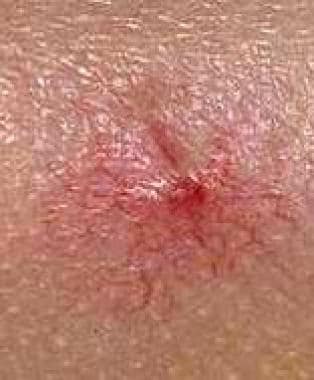 A spider nevus consists of a central arteriole with radiating thin-walled vessels. Compression of the central vessel produces blanching and temporarily obliterates the lesion. When released, the threadlike vessels quickly refill with blood from the central arteriole. The ascending central arteriole resembles a spider's body, and the radiating fine vessels resemble multiple spider legs.
Nevus araneus lesions range in size from 1-10 mm in diameter. Compression of the central vessel with a slide (diascopy) results in blanching and temporary obliteration of the lesion, which is followed by rapid return of blood flow upon release. [1] Pulsations may occasionally be felt upon compression of the punctum. [5] In adults, these lesions are most frequently found on exposed areas of the body, such as the face, neck, upper trunk (above the nipple line), and arms. In children, the backs of the hands and fingers are commonly affected. [1, 5]
Signs and symptoms
Spider angioma (nevus araneus) is asymptomatic and acquired. The following inquiries may be helpful:
-
Ask female patients if they are pregnant, using hormonal supplements, or taking oral contraceptives.
-
Inquire about patient history of alcohol abuse.
-
Ask patients if they are taking medications that may result in liver damage.
Spider angiomas (nevi araneus) are red with a small central arteriole, or punctum, surrounded by thin-walled vessels radiating in a stellate pattern. [1, 5] Sometimes, the punctum is the main finding, without the "legs" of the spider. The lesion measures 1-10 mm in diameter. [1]
Application of pressure to the lesion with a slide (diascopy) causes blanching and temporary obliteration. This is followed by rapid refilling from the central arteriole upon release of pressure. Occasionally, pulsation of the punctum is noted. [1, 5]
Lesions most commonly occur in exposed areas of the skin, including the face, neck, upper trunk, and arms in adults. In children, lesions are common on the fingers and hands. [1, 5] Note the images below.
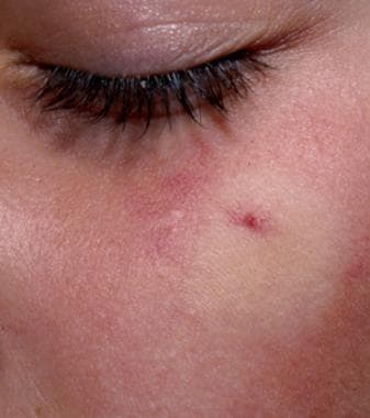 The spider angioma has been compressed and is refilling rapidly from the central vessel.
A spider nevus consists of a central arteriole with radiating thin-walled vessels. Compression of the central vessel produces blanching and temporarily obliterates the lesion. When released, the threadlike vessels quickly refill with blood from the central arteriole. The ascending central arteriole resembles a spider's body, and the radiating fine vessels resemble multiple spider legs.
The spider angioma has been compressed and is refilling rapidly from the central vessel.
A spider nevus consists of a central arteriole with radiating thin-walled vessels. Compression of the central vessel produces blanching and temporarily obliterates the lesion. When released, the threadlike vessels quickly refill with blood from the central arteriole. The ascending central arteriole resembles a spider's body, and the radiating fine vessels resemble multiple spider legs.
Examine the patient for signs of pregnancy, including abdominal enlargement, weight gain, palmar erythema, and/or edema. [6]
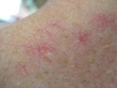
Patients with significant internal disease may exhibit numerous prominent lesions over the trunk and face, as shown in the image below. [1]
Perform a comprehensive abdominal examination with special attention to the liver and spleen. Examine patients for stigmata of liver disease, including ascites, palmar erythema, changes in body fat and hair distribution, muscle and gonadal atrophy, splenomegaly, and leukonychia. [1, 7, 8]
No significant complications are associated with spider angioma (nevus araneus); however, cosmetic issues may be of significant concern to some patients or to parents.
The presence of spider nevi may indicate more severe liver dysfunction in cirrhotic patients. [9]
Diagnostics
Evaluate patients with extensive spider angioma (nevus araneus) lesions for underlying liver disease or for pregnancy, depending on the clinical context. Dermoscopy may prove useful in the diagnosis of spider angioma. [10]
Confirm the diagnosis of spider angioma by observing the classic refill pattern from the central vessel outwards. This refill pattern is seen following compression and release of the lesion. Usually, no other testing is required.
In the rare cases when the diagnosis of spider angioma (nevus araneus) is questionable, consider skin biopsy to exclude basal cell carcinoma or other conditions, particularly if the lesion is enlarging.
Histologic findings
The 5 basic components of the spider angioma (nevus araneus) are (1) an arterial net, (2) a central arteriole, (3) a thin-walled ampulla, (4) efferent spider vessels, and (5) capillaries. [1]
The central ascending arteriole ends in a thin-walled ampulla just below the epidermis. This ampulla feeds small arterial branches that radiate in an outward fashion into the superficial dermis. Glomus cells have been reported in the wall of the central arteriole. [1]
Management
In children, treatment usually is not necessary, and while some lesions resolve spontaneously, others may be permanent. [1] Spider angiomas (nevi araneus) that regress do so over the course of several years.
In young women, lesions often resolve spontaneously within 6 weeks to 9 months after the birth of a child or after discontinuing oral contraceptives. [6]
Numerous lesions associated with liver disease may improve upon treatment of the underlying condition. Reports have described regression after liver transplantation. [11]
Surgical care
Electrodesiccation and laser treatment both can be effective for bothersome facial spider angiomas. Although the risk of a small scar may be slightly higher with electrodesiccation, good results generally are achieved with either intervention. Although cherry angiomas are different vascular lesions, a rater-blinded randomized controlled study of treatment of those showed very little difference between the results of electrodesiccation versus the pulsed dye or KTP laser. [12] Usually, disappearance of the spider angioma follows electrodesiccation. Recurrences are common.
To perform electrodesiccation, move the blood out of the spider by pressing firmly on the lesion. With continuous pressure, slightly move the finger to one side to expose the central arteriole. Then, gently electrodesiccate the central arteriole. If the arteriole is destroyed, radiating capillaries may not fill. Incompletely destroyed lesions may recur. Vigorous desiccation may cause a pitted scar.
Currently available laser systems may eliminate the lesion completely or achieve only partial clearing. [13, 14, 15, 16, 17, 18, 19, 20] In one study, the rate of initial clearing with the 585-nm pulsed dye laser was 95%. The mean follow-up was 37.9 months. Of the 73% of patients who responded to the follow-up survey, 50 (36%) had experienced recurrence of the lesion. [13] Recurrence appears to be related to the deeper arteriolar component of the lesion, which remains patent. Another study demonstrated that the KTP 532-nm laser cleared or markedly improved 99% of 128 patients with spider angiomas, as well as other superficial vascular lesions, with only minimal adverse effects. [14] As described by Yang et al, multiwavelength treatment with a pulsed dye laser followed by neodymium-doped yttrium aluminum garnet (Nd:YAG) laser treatment has produced excellent results in Asian patients with Fitzpatrick skin type IV. [21]
Local anesthesia prior to therapy is optional in adults but advisable in children. Intradermal injection of 0.1-0.2 mL physiological saline solution produces brief complete anesthesia of the site and does not sting on injection. This represents a viable alternative to lidocaine. The central vascular papule has very few nerve endings. Rather than intradermal anesthesia injection, a 30-gauge needle can be inserted directly into the central papule. Anesthesia is flushed into the spider angioma, producing less pain.
See Laser Treatment of Acquired and Congenital Vascular Lesions and Laser Treatment of Benign Pigmented Lesions.
Pathophysiology
Vascular malformations can be classified into 6 categories: hamartomas, malformations, dilatations of preexisting vessels, hyperplasias, benign neoplasms, and malignant neoplasms. [5] Spider angiomas (nevus araneus) are not vascular proliferations; they occur as a result of the dilation of preexisting vessels. [1, 5]
While most lesions are unrelated to internal disease, spider angiomas (nevus araneus) have been associated with thyrotoxicosis, [7] and frequently occur in the presence of estrogen-excess states, such as pregnancy or during the use of oral contraceptives. Resolution of lesions in this context is common 6-9 months postpartum or after discontinuation of oral contraceptive medication. [6]
Spider angiomas (nevus araneus) are also associated with liver disease, liver failure, and cirrhosis. [11, 22, 23] In fact, the spider angioma is rumored to have received its name from barmaids in New York, who used the lesion as a marker of liver disease in their customers. [7] When associated with liver disease, spider angiomas may be numerous, large in size, [24] and appear in atypical locations [25] ; other findings may be present, including palmar erythema, muscle atrophy, gynecomastia, ascites, jaundice, splenomegaly, [7] leukonychia, onychomycosis, and longitudinal nail striations. [8] The number of lesions may be indicative of the extent of hepatic fibrosis. [23]
Etiology
The exact etiology of the spider nevus (nevus araneus) is unclear. Estrogen-excess states such as pregnancy and liver disease have been associated with spider angiomas for many years. This hypothesis is partially based on the hormone’s dilating effects on endometrial spiral arterioles during pregnancy. [26] Additionally, other biologic substances, including vascular endothelial growth factor (VEGF), [27] basic fibroblastic growth factor (bFGF), [28] substance P, and endogenous vasodilators, [26] have been implicated in the pathogenesis of spider angioma (nevus araneus). One study demonstrated that although the ratio of estrogen to testosterone in the serum of cirrhotic patients did, in fact, vary inversely with liver function, the numbers never reached statistical significance. [26]
In the context of liver disease, spider nevi are found more commonly in alcoholic cirrhotics versus nonalcoholic cirrhotics, [29] as well as disease secondary to alcohol abuse versus that caused by viral hepatitis. [30]
Children with liver disease rarely have large numbers of spider angiomas. Although the finding of 5 or more spider angiomas is more common in persons with liver disease, many healthy children also have one or more of these lesions. [2]
Epidemiology
Frequency
United States
Young children and pregnant women most frequently exhibit spider angioma (nevus araneus) lesions. In pregnant women, palmar erythema may also be present. [6] Spider angiomas are common in otherwise healthy children and are present in 10-15% of healthy adults and young children. [1]
International
The frequency of spider angiomas (nevus araneus) is presumed to be similar to that in the United States.
Race
No racial predilection is reported for spider angioma (nevus araneus), but lesions are more apparent in light-skinned patients.
Sex
Spider angiomas (nevus araneus) are more common in women than in men, although a definitive study documenting this is not available. Young children of both sexes and pregnant women frequently exhibit lesions. [2, 6]
Age
One study demonstrated that 38% percent of healthy, school-aged children (ages 5-15 y) had at least one spider nevus (nevus araneus), while most had 1-4 lesions. The trend was an increasing number of lesions with increasing age. [2] Spider angiomas also are common in women of childbearing age in association with pregnancy or oral contraceptive use. [6]
Prognosis
Spider angiomas (nevus araneus) are asymptomatic benign lesions. When extensive, they may be associated with significant underlying internal pathology, such as liver disease. [11, 22, 23] Spider angiomas (nevus araneus) also may produce significant cosmetic concerns in some patients.
When not associated with underling pathology or pregnancy, spider angioma (nevus araneus) lesions may be permanent or may regress over a number of years. Recurrence is not uncommon after treatment. [13, 14, 15, 16, 17, 18, 19, 20]
Although a finding of 5 or more spider nevi is common in children with liver disease, this quantity can also be present in many healthy children. [2] In association with liver disease, lesions may resolve after liver transplantation. [11]
Spider nevi occurring in the context of pregnancy or oral contraceptive use may regress postpartum or after discontinuation of medicine. [6] They may also regress after death or in situations resulting in severe hypotension. [1]
-
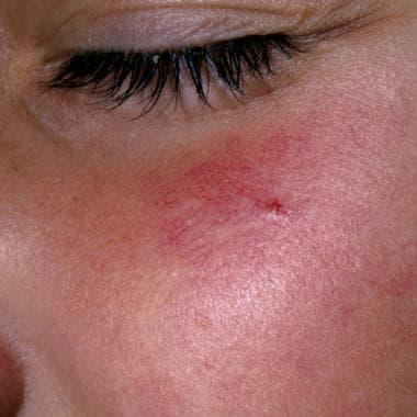
Large spider angioma on the left cheek of a child.
-
The spider angioma has been compressed and is refilling rapidly from the central vessel.
-
A spider nevus consists of a central arteriole with radiating thin-walled vessels. Compression of the central vessel produces blanching and temporarily obliterates the lesion. When released, the threadlike vessels quickly refill with blood from the central arteriole. The ascending central arteriole resembles a spider's body, and the radiating fine vessels resemble multiple spider legs.
-
Multiple spider angiomas in a patient with cirrhosis.


