Continually Updated Clinical Reference
 
 
  All Sources     eMedicine     Medscape     Drug Reference     MEDLINE
 
eMedicine - Malignant Melanoma : Article by

Quick Find
Authors & Editors
Introduction
Clinical
Differentials
Workup
Treatment
Medication
Follow-up
Miscellaneous
Multimedia
References

Related Articles
Atypical Mole (Dysplastic Nevus)

Basal Cell Carcinoma

Blue Nevi

Cherry Hemangioma

Dermatofibroma

Halo Nevus

Keloid and Hypertrophic Scar

Keratoacanthoma

Lentigo

Metastatic Carcinoma of the Skin

Nevi of Ota and Ito

Nevi, Melanocytic

Seborrheic Keratosis

Spitz Nevus

Squamous Cell Carcinoma

Vitiligo


Patient Education
Cancer and Tumors Center

Procedures Center

Skin Cancer Overview

Skin Cancer Causes

Skin Cancer Symptoms

Skin Cancer Treatment

Skin Biopsy Introduction

Skin Biopsy Preparation

Mole Removal Introduction

Mole Removal Preparation


Author: Susan M Swetter, MD, Director, Pigmented Lesion and Cutaneous Melanoma Clinic, Associate Professor, Department of Dermatology, Stanford University Medical Center, Veterans Affairs Palo Alto Health Care System

Susan M Swetter is a member of the following medical societies: American Academy of Dermatology, American Medical Association, American Society of Clinical Oncology, Eastern Cooperative Oncology Group, Pacific Dermatologic Association, Society for Investigative Dermatology, and Women's Dermatologic Society

Editors: Günter Burg, MD, Professor and Chairman Emeritus, Department of Dermatology, University of Zürich School of Medicine, Switzerland; Michael J Wells, MD, Associate Professor, Department of Dermatology, Texas Tech University Health Sciences Center; John G Albertini, MD, Consulting Staff, Dermatologic Surgery, The Skin Surgery Center; Catherine Quirk, MD, Clinical Assistant Professor, Department of Dermatology, Brown University; Dirk M Elston, MD, Director, Department of Dermatology, Geisinger Medical Center

Author and Editor Disclosure

Synonyms and related keywords: melanoma, skin cancer, cutaneous melanoma, malignancy of pigment-producing cells, malignant melanoma, malignancy of melanocytes, invasive cutaneous melanoma, melanoma in situ, metastatic melanoma, acral melanoma, changing mole, superficial spreading melanoma, nodular melanoma, lentigo maligna melanoma

Background

Melanoma is a malignancy of pigment-producing cells (melanocytes) located predominantly in the skin, but also found in the eyes, ears, GI tract, leptomeninges, and oral and genital mucous membranes. Melanoma accounts for only 4% of all skin cancers; however, it causes the greatest number of skin cancer–related deaths worldwide. Early detection of thin cutaneous melanoma is the best means of reducing mortality.

For more information, see Medscape's Skin Cancer Resource Center.

Pathophysiology

The sequence of events in which normal melanocytes transform into melanoma cells, referred to as melanomagenesis, is poorly understood. It likely involves a multistep process of progressive genetic mutations that (1) alter cell proliferation, differentiation, and death and (2) impact susceptibility to the carcinogenic effects of ultraviolet radiation.1 Recent data suggest multiple pathways of melanoma pathogenesis, with melanomas in sun-protected skin (trunk) developing in association with a high nevus count and intermittent ultraviolet radiation as opposed to those developing on sun-exposed skin in patients with low nevus counts and chronic sun exposure.2, 3 Primary cutaneous melanoma may develop in precursor melanocytic nevi (ie, common, congenital, and atypical/dysplastic types), although more than 60% of cases are believed to arise de novo (ie, not from a preexisting pigmented lesion).

The development of melanoma is multifactorial and appears to be related to multiple risk factors, including fair complexion, excessive childhood sun exposure and blistering childhood sunburns, an increased number of common and dysplastic moles, a family history of melanoma, the presence of a changing mole or evolving lesion on the skin, and, importantly, older age.4, 5, 6

Frequency

United States

The incidence of melanoma has more than tripled in the white population during the last 20 years, and melanoma currently is the sixth most common cancer in the United States. Approximately 59,940 Americans (33,910 men and 26,030 women) will develop invasive cutaneous melanoma in 2007, with an estimated additional 48,290 or more cases of melanoma in situ.3 The current lifetime risk for developing invasive melanoma is 1 case per 60 Americans, a 2000% increase since 1930. This risk rises to 1 case per 32 Americans if noninvasive melanoma in situ is included.

International

Melanoma incidence has continued to increase worldwide, with the highest incidence in Australia and New Zealand. The most recent analysis of global cancer statistics, from 2002, demonstrated a prevalence of 37.7 cases per 100,000 men and 29.4 cases per 100,000 women in Australia and New Zealand, compared with 6.4 cases per 100,000 men and 11.7 cases per 100,000 women in North America.7

Mortality/Morbidity

While melanoma accounts for roughly 4% of all skin cancers, it is responsible for more than 74% of skin cancer deaths. In the United States, one person each hour dies from metastatic melanoma. Treatment of melanoma in its early stages provides the best opportunity for cure.

  • United States: An estimated 8110 deaths will occur in 2007 (5220 men and 2890 women). Analysis of US Surveillance, Epidemiology, and End Results (SEER) data from 1969-1999 demonstrated a disproportionate burden of melanoma deaths among middle-aged and older white men. While melanoma mortality rates have fallen 39% in women and 29% in men aged 20-44 years over this period, they have increased 66% in men aged 45-64 years and 157% in older men (>65 y).8 Incidence data generally parallel mortality data and have shown a 3-fold increase in middle-aged men and a 5-fold increase in older men over a similar period. Encouragingly, a stable-to-reduced melanoma rate has been noted in younger age groups in the United States, which may be a result of primary prevention campaigns aimed at reducing excessive sun exposure over the past 30 or more years, although the full impact of primary prevention strategies on melanoma incidence and mortality will not be apparent for several decades.
  • Worldwide: Individuals with cutaneous melanoma have higher survival rates in developed countries (91% in US SEER registries and 81% in Europe) than in developing countries (approximately 40%). Increased educational efforts in developed areas result in earlier diagnosis, treatment, and potential cure of thinner lesions. Worldwide, 160,000 new cases of melanoma were estimated to occur in 2002, with 41,000 deaths reported.7

Race

Melanoma is primarily a malignancy of white individuals. African American persons develop melanoma approximately one twentieth as frequently as white persons, and the prevalence in Hispanic persons is approximately one sixth of that in white persons. However, mortality rates are higher in African Americans and Hispanics, who are more likely to have acral melanoma and advanced disease at presentation.

Sex

In the United States, invasive melanoma has a higher female predilection from birth to age 39 years (1 in 467 women compared with 1 in 775 men). However, from age 40 years and older, melanoma in men predominates, occurring in 1 in 49 men compared with 1 in 73 women over a lifetime.9 Worldwide, of the 160,000 new cases estimated to have occurred in 2002, women were affected slightly more than men (male-to-female ratio, 0.97:1). Conversely, of the estimated 41,000 worldwide deaths in 2002, more occurred in men than in women (male-to-female ratio 1.2:1).

Age

The median age at melanoma diagnosis is 53 years; however, it is the most common cancer in women aged 25-29 years and is second only to breast cancer in women aged 30-34 years. The most striking differences in melanoma incidence and mortality occur in individuals older than 65 years, although modest differences in age-specific incidence and mortality are notable in persons older than 50 years.8

Older individuals are both more likely to acquire and to die from melanoma; thus, elderly persons should be a primary target for secondary melanoma prevention, including early detection and screening.10 Treatment options in elderly persons may also be limited because of comorbid medical conditions, an inability to tolerate adverse medication effects or toxicity, the increased likelihood of drug interactions, and potential exclusion from clinical trials based on age criteria.10


History

A new or changing mole or blemish is the most common warning sign for melanoma. Variation in color and/or an increase in diameter, height, or asymmetry of borders of a pigmented lesion are noted by more than 80% of patients with melanoma at the time of diagnosis. Symptoms such as bleeding, itching, ulceration, and pain in a pigmented lesion are less common but warrant an evaluation. Again, because the majority of cutaneous melanoma arises de novo (ie, not in association with a precursor nevus), the wholesale removal of melanocytic nevi is not warranted for melanoma prevention. However, individuals with numerous moles (common or dysplastic) or a family history of melanoma should be educated regarding the importance of skin self-examination for early detection of skin cancer.

Information regarding the changes noted in the ABCDE criteria listed below is relevant to the patient's history. Physician and patient education regarding the warning signs of early melanoma (particularly the superficial spreading subtype) has been achieved successfully through the use of the ABCDE criteria for a changing mole,11, 12 which are as follows:

  • Asymmetry: Half the lesion does not match the other half.
  • Border irregularity: The edges are ragged, notched, or blurred.
  • Color variegation: Pigmentation is not uniform and may display shades of tan, brown, or black; white, reddish, or blue discoloration is of particular concern.
  • Diameter: A diameter greater than 6 mm is characteristic, although some melanomas may have smaller diameters; any growth in a nevus warrants an evaluation.
  • Evolving: Changes in the lesion over time are characteristic; this factor is critical for nodular or amelanotic (nonpigmented) melanoma, which may not exhibit the classic criteria above.

The ABCDEs have the greatest diagnostic accuracy when used in combination. Lesions exhibiting these features should be considered potential melanoma, although severely atypical nevi may be difficult to distinguish clinically. More recent use of the "ugly duckling" warning sign, wherein skin examination is focused on recognition of a pigmented or clinically amelanotic lesion that simply looks different from the rest, may assist with detection of lesions that lack the classic ABCDE criteria (eg, nodular, amelanotic, or desmoplastic melanomas).13, 14

Physical

Four major clinicopathologic (or histogenetic) subtypes of primary cutaneous melanoma have been identified. These include superficial spreading melanoma, nodular melanoma, lentigo maligna melanoma, and acral lentiginous melanoma. Distinction among the subtypes is based on histologic growth pattern (predominantly junctional in lentiginous types vs buckshot scatter in superficial spreading vs nodular), anatomic site, and degree of sun damage. The pattern of sun exposure varies between the types (sustained in lentigo maligna vs intermittent in superficial spreading). Whether the melanoma subtype affects the overall prognosis remains controversial. However, molecular analysis has demonstrated different patterns of cell death, oncogene expression, gene amplification, and BRAF mutation frequency among the 4 main histogenetic types.15, 16, 17

With the exception of nodular melanoma, the growth patterns of the other subtypes are characterized by a preceding in situ (radial growth) phase that lacks the biologic potential to metastasize and may last from months to years before dermal invasion occurs. While all in situ melanoma may not necessarily progress to invasive melanoma, complete excision is recommended to prevent invasion and effect cure.

Superficial spreading melanoma characteristics are as follows:

  • It is most common on the trunk in men and women and on the legs in women; this subtype is most commonly seen in individuals aged 30-50 years. See Media File 1.
  • Superficial spreading melanoma manifests as a flat or slightly elevated brown lesion with variegate pigmentation (ie, black, blue, pink, or white discoloration).
  • It is generally greater than 6 mm in diameter.
  • Irregular asymmetric borders are characteristic.
  • Histologically, it is characterized by buckshot (pagetoid) scatter of atypical melanocytes within the epidermis.

Nodular melanoma characteristics are as follows:

  • This subtype occurs in 15-30% of patients.
  • It is seen most commonly on the legs and trunk.
  • Rapid growth occurs over weeks to months; this subtype is responsible for most thick melanomas.18, 19
  • It manifests as a dark brown-to-black papule or dome-shaped nodule, which may ulcerate and bleed with minor trauma; it may be clinically amelanotic (ie, not pigmented).
  • It tends to lack the typical ABCDE melanoma warning signs and, thus, may elude early detection. More commonly, it exhibits elevation, ulceration with bleeding, or both at presentation.
  • Histologically, it lacks a radial growth phase.

Lentigo maligna melanoma characteristics are as follows:

  • The incidence of lentigo maligna subtypes (in situ and invasive) appears to be rising in the United States.20
  • It is typically located on the head, neck, and arms (chronically sun-damaged skin) of fair-skinned older individuals (average age 65 y). See Media File 2.
  • It grows slowly over 5-20 years.
  • The in situ precursor lesion is usually large (>1-3 cm in diameter), present for a minimum of 10-15 years, and demonstrates macular pigmentation ranging from dark brown to black, although hypopigmented (white) areas are common within lentigo maligna. Dermal invasion (progression to lentigo maligna melanoma) is characterized by the development of raised blue-black nodules within the in situ lesion.
  • Histologically, it is characterized by a predominantly junctional confluent proliferation of melanocytes and extension along adnexal structures. Solar elastosis is typically prominent.

Acral lentiginous melanoma characteristics are as follows:

  • This is the least common subtype of melanoma (2-8% of melanoma cases in white persons).
  • It accounts for 29-72% of melanoma cases in dark-skinned individuals (ie, African American, Asian, and Hispanic persons) and, because of delays in diagnosis, may be associated with a worse prognosis.21, 22
  • Acral lentiginous melanoma occurs on the palms, on the soles, or beneath the nail plate (subungual variant). See Media File 3.
  • Subungual melanoma may manifest as diffuse nail discoloration or a longitudinal pigmented band within the nail plate.
  • It must be differentiated from a benign junctional melanocytic nevus of the nail bed, which has a similar appearance.
  • Pigment spread to the proximal or lateral nail folds is termed the Hutchinson sign, which is a hallmark for acral lentiginous melanoma.

Rare melanoma variants (<5% of melanomas) include (1) desmoplastic/neurotropic melanoma, (2) mucosal (lentiginous) melanoma,23 (3) malignant blue nevus, (4) melanoma arising in a giant congenital nevus, and (5) melanoma of soft parts (clear cell sarcoma).

Amelanotic melanoma (<5% of melanomas) characteristics are as follows:

  • This type is nonpigmented and, clinically, appears pink or flesh-colored, often mimicking basal cell or squamous cell carcinoma or a ruptured hair follicle.
  • It occurs most commonly in the setting of the nodular melanoma subtype or melanoma metastasis to the skin, presumably because of the inability of these poorly differentiated cancer cells to synthesize melanin pigment.

Melanoma can occur on any skin or mucosal surface, although a history of cutaneous melanoma does not appear to increase the risk of developing primary intraocular or mucosal melanoma. Melanoma occurs most commonly on the trunk in white males and the lower legs and back in white females. In African American, Hispanic, and Asian persons, the most common site is the plantar foot, followed by subungual, palmar, and mucosal sites.

Desmoplastic melanoma typically occurs in conjunction with (or in sites typical for) lentiginous types of melanoma (lentigo maligna and acral lentiginous melanoma).

Causes

Superficial spreading melanoma tends to occur at sites of intermittent, intense sun exposure (ie, on trunk in males and legs and back in females). Lentigo maligna melanoma is more prevalent on the chronically sun-damaged skin of the head, neck, and arms. The disease shows an increased worldwide incidence in fair-complexioned individuals living in sunny climates and nearer the equator, suggesting a causative role for ultraviolet radiation.

Primary risk factors for or clinical warning signs of melanoma include the following:

    • Changing mole (most important clinical warning sign)
    • Clinical atypical/dysplastic nevi (particularly >5-10)
    • Large numbers of common nevi (>100)
    • Large (giant) congenital nevi (>20 cm diameter in an adult)
    • Previous melanoma
    • Sun sensitivity/history of excessive sun exposure
    • Melanoma in first-degree relative(s)
    • Prior nonmelanoma skin cancer (basal cell and squamous cell carcinoma)24
    • Male sex
    • Age older than 50 years
    • Presence of xeroderma pigmentosum or familial atypical mole melanoma syndrome: These 2 genodermatoses confer a 500- to 1000-fold greater relative risk of developing melanoma.

A fair-skin phenotype (blue/green eyes, blond or red hair, light complexion, sun sensitivity) and the occurrence of blistering sunburn(s) in childhood and adolescence are universal risk factors for melanoma. Individuals with these traits have been the focus of preventive efforts worldwide.

Pregnancy or hormonal therapy with oral contraceptives or hormone replacement does not appear to be a risk factor for melanoma development.25, 26, 27, 28, 29


Atypical Mole (Dysplastic Nevus)
Basal Cell Carcinoma
Blue Nevi
Cherry Hemangioma
Dermatofibroma
Halo Nevus
Keloid and Hypertrophic Scar
Keratoacanthoma
Lentigo
Metastatic Carcinoma of the Skin
Nevi of Ota and Ito
Nevi, Melanocytic
Seborrheic Keratosis
Spitz Nevus
Squamous Cell Carcinoma
Vitiligo

Other Problems to be Considered

Chronic paronychia (for subungual melanoma)
Subungual hematoma (for subungual melanoma)
Melanonychia striata (for subungual melanoma)
Traumatized nevus (for superficial spreading or nodular subtypes)


Lab Studies

The most important aspects of the initial workup for patients with cutaneous melanoma are a careful history, review of systems, and physical examination.

  • Sentinel lymph node biopsy (SLNB) is generally indicated for pathologic staging of the regional nodal basin(s) for primary tumors greater than or equal to 1 mm depth and when certain high-risk histologic features (eg, ulceration, extensive regression, high mitotic rate, angiolymphatic invasion) are present in thinner melanomas.
  • Published data have shown that baseline and surveillance laboratory studies (eg, lactate dehydrogenase [LDH] level, liver function tests), chest radiography (CXR), and other imaging studies (eg, CT scanning, positron emission tomography [PET] scanning, bone scanning, MRI) are not typically beneficial for stage I/II (cutaneous) melanoma patients without signs or symptoms of metastasis.30, 31, 32, 33
  • A metastatic workup should be initiated if physical findings or symptoms suggest disease recurrence or if the patient has documented nodal metastasis based on results from the SLNB. 

Practice guidelines developed by the National Comprehensive Cancer Network support the concept that most melanoma recurrences are diagnosed clinically. The current guidelines state that baseline or surveillance CXR and LDH studies may be performed for stages IB to IIC invasive cutaneous melanoma on an "optional" basis every 3-12 months at the discretion of the clinician. Advanced imaging studies should be obtained as clinically indicated for documentation or for suspected metastasis. Current recommendations do not indicate that baseline or surveillance studies are necessary in patients with melanoma in situ (stage 0) or stage IA disease (<1 mm thickness).34

While abnormal laboratory test results are rarely the sole indicator of metastatic disease, serum LDH levels have been incorporated into the American Joint Committee on Cancer (AJCC) 2002 melanoma staging guidelines for the classification of stage IV (distant) disease. Elevated LDH levels are associated with worse survival in this subgroup. Serum S-100 protein levels may also be useful as a tumor marker in patients with metastatic disease, but this practice is not widely used in the United States.35

Imaging Studies

Studies have confirmed that extensive radiologic studies such as CT scanning, MRI, PET scanning, ultrasonography, and bone scanning have an extremely low yield in asymptomatic patients with primary cutaneous melanoma (AJCC stages I and II) and are generally not indicated. However, maintaining a low threshold for obtaining symptom-directed tests is important.

Baseline metastatic staging for melanoma patients with primary tumors greater than 1 mm in depth may include CXR, which typically is repeated every 6-12 months for routine surveillance (optional in the absence of signs or symptoms of metastatic disease).34

Procedures

The criterion standard for melanoma diagnosis is histopathologic examination of clinically suggestive skin or mucosal lesions. An excisional biopsy (or deep saucerization technique) with narrow margins is preferred when possible. In the case of lentigo maligna, a broad, paper-thin shave biopsy or multiple smaller biopsies may be the best techniques. The biopsy report should generally include the following:  

  • Tumor thickness (Breslow depth)
  • Presence of ulceration
  • Anatomic level of invasion (Clark level)
  • Presence of mitoses
  • Presence of regression
  • Lymphatic/vessel invasion or vascular involvement
  • Host response (tumor-infiltrating lymphocytes)

Immunohistochemical staining for lineage (S-100, homatropine methylbromide 45 [HMB-45], melan-A/Mart-1) or proliferation markers (proliferating cell nuclear antigen, Ki67) may be helpful in some cases for histologic differentiation from melanoma simulators. Additionally, evidence of lack of maturation with HMB-45 staining and patchy, rather than diffuse, staining with S-100A6 may be helpful for distinguishing spitzoid melanoma from Spitz nevus.

Generally, when an excisional biopsy is performed, 1-3 mm of normal skin surrounding the pigmented lesion should be removed to provide accurate diagnosis and histologic microstaging. Wider margins (>1 cm) could theoretically disrupt afferent cutaneous lymphatic flow and affect the ability to identify the sentinel node(s) accurately in patients eligible for this staging procedure. Some data, however, suggest that accurate mapping is possible after wider excision.

Superficial shave biopsies of suggestive pigmented lesions are discouraged because partial removal of the primary melanoma may not provide an accurate measurement of tumor thickness, which is the most important histologic prognostic factor for cutaneous melanoma. As noted above, a very important exception to this rule is the lentigo maligna subtype of melanoma in situ. In the case of lentigo maligna, the risk of misdiagnosis is high if small, deep biopsy specimens are taken. Taking large, deep biopsy specimens can be disfiguring. The best diagnostic biopsy technique in this case is often a broad shave biopsy that extends into at least the papillary dermis so that microinvasive melanoma can be excluded.

Histologic Findings

Superficial spreading melanoma has an in situ (radial growth) phase characterized by increased numbers of intraepithelial melanocytes, which (1) are large and atypical, (2) are arranged haphazardly at the dermoepidermal junction, (3) show upward (pagetoid) migration, and (4) lack the biologic potential to metastasize. Lentigo maligna melanoma and acral lentiginous melanoma demonstrate predominant in situ growth at the dermoepidermal junction and with little tendency for the pagetoid scatter of cells.

Dermal invasion confers metastatic potential, although the greatest risk occurs in the setting of a vertical growth (tumorigenic) phase.36, 37 Tumorigenicity is characterized by a distinct population of melanoma cells with evidence of proliferation (mitoses, MIB-1 staining) and nuclear pleomorphism within the dermis and, possibly, the subcutaneous fat. Lateral intraepidermal extension of melanoma cells occurs in all subtypes except nodular melanoma. Failure of melanocyte maturation and dispersion as the tumor extends downward into the dermis is characteristic of melanoma. Some investigators have defined a vertical growth phase as (1) any dermal nest larger than the largest junctional nest or (2) invasion into either the reticular dermis or band of solar elastosis.

Tumor thickness, as defined by the Breslow depth, is the most important histologic determinant of prognosis and is measured vertically in millimeters from the top of the granular layer (or base of superficial ulceration) to the deepest point of tumor involvement. Increased tumor thickness confers a higher metastatic potential and a poorer prognosis.38, 39 Analysis of worldwide data has shown that the presence of ulceration microscopically, defined as the loss of epidermis overlying the melanoma, is the next most important histologic determinant of patient prognosis and, when present, should be used to up-stage patients with melanoma.40 The Clark level is a measurement of tumor invasion anatomically and appears to affect prognosis only in thinner (<1 mm depth) melanomas.

Staging

The melanoma staging system initially developed in 1983 by the AJCC and the International Union Against Cancer (UICC) divided melanoma into 4 stages and incorporated tumor thickness and anatomic level of invasion for stages I and II (localized cutaneous disease), with the later recommendation to follow Breslow depth over Clark level when any discordance arose. Stage III disease involved the regional lymph nodes; stage IV disease included distant skin, subcutaneous, nodal, visceral, skeletal, or CNS metastasis.

Major revisions in the 2002 AJCC/UICC melanoma staging system were made based on a critical analysis of prior versions of the staging protocol.40 The AJCC formed an international multidisciplinary Melanoma Staging Committee and established a new clinicopathologic database of more than 17,000 patients worldwide to test the validity of the proposed staging changes.41, 42, 43 Several important modifications in the 2002 AJCC staging system include the incorporation of histologic ulceration and number of lymph nodes involved (instead of size) to better stratify metastatic risk and patient prognosis.44 In the revised staging system, the Clark level is included only in thin primary tumors (<1 mm depth, stages IA and IB) because its prognostic value is minimal in thicker primary melanoma. Microscopic regional lymph node metastasis as detected by SLNB is differentiated from macroscopic nodal metastasis.

Overall survival (OS) in the staging Table below is based on worldwide AJCC data.42 The next iteration of the AJCC melanoma staging system is anticipated in 2009.

AJCC 2002 Revised Melanoma Staging
StageTNM ClassificationHistologic/Clinical Features5-Year Survival Rate, %
0Tis N0 M0Intraepithelial/in situ melanoma100
IAT1a N0 M0£1 mm without ulceration and level II/III>95
IBT1b N0 M0
T2a N0 M0
£1 mm with ulceration or level IV/V
1.01-2 mm without ulceration
89-91
IIAT2b N0 M0
T3a N0 M0
1.01-2 mm with ulceration
2.01-4 mm without ulceration
77-79
IIBT3b N0 M0
T4a N0 M0
2.01-4 mm with ulceration
³4 mm without ulceration
63-67
IICT4b N0 M0>4 mm with ulceration45
IIIAT1-4a N1a M0
T1-4a N2a M0
Single regional nodal micrometastasis, nonulcerated primary
2-3 microscopic positive regional nodes, nonulcerated primary
63-69
IIIBT1-4b N1a M0
T1-4b N2a M0
T1-4a N1b M0
T1-4a N2b M0
T1-4a/b N2c M0
Single regional nodal micrometastasis, ulcerated primary
2-3 microscopic regional nodes, nonulcerated primary
Single regional nodal macrometastasis, nonulcerated primary
2-3 macroscopic regional nodes, no ulceration of primary
In-transit met(s)* and/or satellite lesion(s) without metastatic lymph nodes
46-53

30-50

IIICT1-4b N2a M0
T1-4b N2b M0
Any T N3 M0
Single macroscopic regional node, ulcerated primary
2-3 macroscopic metastatic regional nodes, ulcerated primary
4 or more metastatic nodes, matted nodes/gross extracapsular extension, or in-transit met(s)/satellite lesion(s) and metastatic nodes
24-29
IVAny T any N M1a
Any T any N M1b
Any T any N M1c
Distant skin, subcutaneous, or nodal mets with normal LDH levels
Lung mets with normal LDH
All other visceral mets with normal LDH or any distant mets with elevated LDH
7-19

*Met is metastasis.


Medical Care

Numerous adjuvant therapies have been investigated for the treatment of localized cutaneous melanoma following complete surgical removal. No survival benefit has been demonstrated for adjuvant chemotherapy, nonspecific (passive) immunotherapy, radiation therapy, retinoid therapy, vitamin therapy, or biologic therapy.45 Adjuvant interferon (IFN) alfa-2b is the only adjuvant therapy approved by the US Food and Drug Administration for high-risk melanoma (currently defined as stages IIB, IIC, and III), which is associated with a 40-80% chance of relapse and death. Various experimental melanoma vaccines also show promise in the adjuvant setting.

Interferon alfa trials

In the United States, 3 prospective, multicenter, randomized, controlled trials have been conducted to assess the effect of adjuvant high-dose IFN alfa-2b on relapse-free survival (RFS) and OS rates in patients with high-risk melanoma (primary tumors >4 mm depth and regional nodal disease). The Eastern Cooperative Oncology Group (ECOG) trial 1684 showed an 11% increase (26% to 37%) in RFS rates at 5 years in the IFN-alfa treatment group compared with the observation arm. Similarly, this trial showed an increase in 5-year OS rates from 37% to 46% (median OS 2.78 to 3.82 y) in the treatment arm compared with observation.46

The confirmatory Intergroup trial (ECOG 1690) again showed an increase in the estimated 5-year RFS rates from 35% in the observation arm to 44% in the high-dose IFN-alfa arm. No significant benefit in the RFS rate was associated with low-dose IFN. Importantly, no difference in the OS rate was seen in the IFN-treated groups (high- or low-dose) compared with the observation arm.47 Despite further data analysis that suggested postrelapse salvage therapy with an IFN-alfa–containing regimen may have confounded the OS results (ie, "crossover effect"), the ECOG 1690 trial is largely viewed as a negative study for high-dose IFN effects on OS.

The most recent Intergroup trial (ECOG 1694) compared the use of standard high-dose IFN alfa with GM2 ganglioside vaccine (GMK). The study was closed prematurely due to a significant benefit observed for IFN alfa over GMK for both RFS and OS rates. Hazard ratio analysis revealed that the likelihood of disease relapse and death in patients treated with high-dose IFN was reduced by one third compared with GMK.48

A pooled analysis of the 3 ECOG/Intergroup trials (with median follow-up ranging from 2.1-12.6 y) revealed that RFS, but not OS, was significantly prolonged for patients treated with high-dose IFN versus observation.49 The authors concluded there is "strong evidence for improved RFS and evidence for moderate improvement in OS based on two prospective randomized studies (E1684 and E1694), but not in the pooled analysis" and called for further analysis of predictors of both response and relapse to improve the therapeutic value of high-dose IFN therapy.

In any case, the potential benefits of high-dose IFN must be weighed against its substantial tolerability and toxicity issues, including the yearlong duration of therapy, commonly associated flulike symptoms, and potential for significant adverse reactions.

Data from 2006 suggest that high-dose IFN-induced autoimmunity, as manifested clinically by new-onset vitiligo, and/or serologically by the development of autoantibodies (antithyroid, antinuclear, and anticardiolipin), is associated with prolonged RFS and OS in melanoma patients.50 The apparent prognostic significance of autoimmunity during high-dose IFN treatment warrants further study.

Melanoma vaccines

Melanoma vaccines are a theoretically attractive alternative to chemotherapy or immunotherapy with systemic cytokines because they are typically associated with relatively little toxicity (eg, fatigue, myalgias, local inflammatory skin reactions). Melanoma vaccines are a type of specific active immunotherapy based on melanoma cell expression of certain HLA- and tumor-associated antigens. Numerous melanoma-associated antigens have been identified, and which of these are the most important in eliciting the necessary cytotoxic and humoral responses to kill melanoma cells remains unclear. In addition, HLA haplotype restriction (mainly to the A2 allele) limits the use of peptide vaccines in many patients. Most current trials for melanoma vaccines are for advanced disease (stages III and IV); trials aimed at prevention are not yet available.

Vaccine types include whole cell preparations, cell lysates, gangliosides, peptides/proteins, dendritic cell vaccines, and DNA vaccines. Melanoma vaccines may be (1) autologous (killed cell and recombinant types), allogeneic, shed from tumor, defined antigen-directed, or genetically engineered and (2) either polyvalent or univalent in nature. Enhanced delivery systems, such as dendritic cell preparations, DNA-plasmid vectors, and intranodal infusion, are under active study to enhance immunogenicity and host response. Biologic response modifiers such as granulocyte macrophage colony-stimulating factor, interleukin (IL)–2, IL-12, and IFN gamma are often integrated into vaccine strategies. As yet, no large, phase 3 randomized trial has demonstrated a survival advantage for vaccine-treated melanoma patients; however, multiple studies are in progress.51

Surgical Care

Surgery is the primary mode of therapy for localized cutaneous melanoma.

Surgical margins for primary melanoma   

The narrowest efficacious margins for cutaneous melanoma have yet to be determined. Surgical margins of 5 mm are currently recommended for melanoma in situ, and margins of 1 cm are recommended for melanomas up to 1 mm in depth (low-risk primaries).52 In some settings, tissue sparing may be critical and Mohs margin-controlled excision may be appropriate.

Randomized prospective studies show that 2-cm margins are appropriate for tumors of intermediate thickness (1-4 mm Breslow depth), although 1-cm margins have been proven effective for tumors of 1- to 2-mm thickness.53, 54 Margins of 2 cm are recommended for cutaneous melanomas greater than 4 mm in thickness (high-risk primaries) to prevent potential local recurrence in or around the scar site.

A 2004 prospective study of melanoma greater than or equal to 2 mm thickness (median depth 3 mm) from the United Kingdom suggests that narrower margins (1 cm) result in higher locoregional recurrence compared with wider margins (3 cm), although no difference was noted in melanoma-specific survival between the 2 groups.55 However, this study has been criticized for combining satellite, in-transit, and regional nodal recurrences as the primary end point and by excluding SLNB (which would have demonstrated existing occult regional nodal metastasis at the time of wide local excision). Likewise, because a 2-cm margin is as efficacious as a 4-cm margin for melanomas of 1-4 mm depth, a 3-cm margin is unlikely to prove more beneficial than a 2-cm margin.

A well-conducted retrospective study of high-risk primary melanomas (>4 mm thickness, median depth 6 mm) showed that excisional margins greater than 2 cm have no effect on local recurrence, disease-free relapse, or OS rates; therefore, a 2-cm margin is likely appropriate in this subgroup.56

Mohs micrographic surgery has also been proposed for cutaneous melanoma and has the advantage of providing visualization of 100% of peripheral and deep margins microscopically. While studies have shown no increased local recurrence for Mohs surgery compared with historical controls, much of the data stem from thinner tumors with a lower risk of local recurrence and metastasis.57 Mohs surgery may have certain "niche" indications, including melanomas located the head, neck, hands, or feet. Mohs surgery may prove useful in completely removing subclinical tumor extension in certain subtypes of melanoma in situ, such as lentigo maligna and acral lentiginous melanoma in situ.

Elective lymph node dissection   

Prophylactic lymph node dissection for primary cutaneous melanoma of intermediate thickness initially was believed to confer a survival advantage on patients with tumors of 1-4 mm in depth. Subsequently, prospective randomized clinical trials have shown no survival benefit for elective lymphadenectomy for melanomas of varying thicknesses on the extremities and marginal, if any, benefit for nonextremity melanomas.58, 59

The 10-year follow-up data from 2 of the trials conducted by the World Health Organization and the Melanoma Intergroup now suggest a survival benefit for certain subsets of patients studied. In particular, patients in the World Health Organization trial who had occult metastasis detected at the time of wide local excision and immediate elective node dissection had a significantly better 5-year survival rate (48%) compared with those who underwent delayed (therapeutic) lymph node dissection when lymphadenopathy became apparent clinically (27%).60 However, the differences in OS rates for all patients who had delayed lymph node dissection were not statistically significant compared with the immediate node dissection group.

SLNB/dissection   

Lymphatic mapping and sentinel node biopsy have effectively solved the dilemma of whether to perform regional lymphadenectomy (in the absence of clinically palpable nodes) in patients with thicker melanomas (>1 mm in depth).

SLNB for cutaneous melanoma was developed in the early 1990s to allow a selective approach to identifying individuals with occult regional nodal metastasis through localization of the first-draining, or sentinel, node. The success of the technique is based on the concept that cutaneous lymphatic flow is well-delineated in melanoma and that the histology of the sentinel node is characteristic of the entire lymph node basin (ie, a negative sentinel node obviates the need for further lymph node dissection). Both of these concepts were borne out in initial and subsequent studies of the staging technique.61

Preoperative radiographic mapping (lymphoscintigraphy) and vital blue dye injection around the primary melanoma or biopsy scar (at the time of wide local excision/reexcision) is performed to identify and remove the initial draining regional node(s).

The sentinel node is examined for the presence of micrometastasis using both routine histology and immunohistochemistry; if present, a therapeutic or completion lymph node dissection (CLND) is performed. A negative sentinel node biopsy result prevents the morbidity associated with an unnecessary lymphadenectomy.

Sentinel node status (positive or negative) is the most important prognostic factor for recurrence and is the most powerful predictor of survival in melanoma patients. In a study of 612 patients with cutaneous melanoma (stage I/II), negative results from SLNB were associated with a nearly 60% increase in 3-year disease-free survival compared with positive SLNB results.62 Current AJCC 2002 melanoma staging and National Comprehensive Cancer Network clinical practice guidelines advocate pathologic staging of the regional lymph nodes for cutaneous melanoma of greater than 1 mm depth along with microstaging of the primary melanoma as the most complete means of staging.34, 43

While SLNB certainly enhances metastatic staging for patients with intermediate-thickness and deeper primary melanomas and provides a more accurate determination of the patient's prognosis, its therapeutic role has yet to be established.63 The results of the Multicenter Selective Lymphadenectomy Trial (MSLT), the Florida Melanoma Trial, and the Sunbelt Melanoma Trial should help to determine whether SLNB provides a therapeutic benefit in patients with cutaneous melanoma.

The third of 5 planned analyses of the MSLT-1 has been published.64 This interim analysis of the subset of 1269 patients with intermediate-thickness melanoma (1.2-3.5 mm) demonstrated no overall (melanoma-specific) survival differences in the group that underwent SLNB at the time of primary excision of the melanoma versus the group that underwent wide local excision alone. However, immediate lymphadenectomy in the setting of a positive sentinel lymph node was associated with improved 5-year survival compared with delayed CLND in patients who developed macroscopic nodal metastasis following primary excision alone (72% vs 52%, respectively). The risk of death was reduced by  one half (hazard ratio, 0.51; 95% confidence interval, 0.32-0.81; P = .004) in the node-positive subset of patients who underwent immediate versus delayed CLND for regional nodal metastasis. OS rates did not differ.

Longer follow-up with continued analysis of this MSLT-1 and other important SLNB trials will help to elucidate the potential therapeutic benefit of early removal of micrometastasis in the regional nodal basin. See The Role of Sentinel Node Biopsy in Skin Cancer for further information.

Consultations

  • Surgical oncologist
    • For sentinel node biopsy, typically performed at the time of wide local excision and following preoperative lymphoscintigraphy
    • For surgical treatment of regional lymph node disease and soft tissue and/or in-transit recurrence (stage III disease)
    • For palliative surgical treatment of visceral and CNS metastasis
  • Medical oncologist
    • To discuss adjuvant therapy with IFN alfa, experimental melanoma vaccines, or other clinical trials: Patients should be referred to a medical oncologist or melanoma specialist soon after the melanoma diagnosis and treatment in order to optimize the chances for appropriate adjuvant therapy or clinical trial entry.
    • To discuss and initiate treatment of metastatic melanoma (stage IV) with chemotherapy, high-dose IL-2, concurrent biochemotherapy, or clinical trials, as indicated clinically
  • Nuclear medicine specialist
    • For preoperative lymphoscintigraphy if SLNB is performed
    • For PET scan interpretation
  • Pathologist/dermatopathologist
    • For accurate histologic microstaging of primary melanoma
    • For evaluation of nodal tissue from SLNB for micrometastasis
    • For confirmation of the diagnosis of disseminated disease
  • Radiation oncologist
    • For adjuvant treatment of resected regional nodal metastasis with extracapsular extension or resected intransit metastasis
    • For palliative treatment of distant metastatic disease, particularly bony metastasis or brain involvement (whole brain radiotherapy or stereotactic radiosurgery)
  • Neurosurgeon - For evaluation for resectable brain metastasis


High-dose IFN alfa-2b is the only adjuvant therapy approved by the US Food and Drug Administration for high-risk resected melanoma, defined as deep primaries greater than 4 mm in Breslow depth (AJCC stage IIB) and regional lymph node metastasis (stage III). Various trials of low-dose IFN have shown no benefit in disease-free relapse or OS rates.65 Similarly, multiple melanoma vaccine trials are in progress, predominantly for stage III and IV disease, but they have not demonstrated an OS advantage to date.

Drug Category: Immunomodulatory agents

Enhance host immunity for cancer surveillance and eradication.

Drug NameInterferon alfa-2b (Intron A)
DescriptionProtein product manufactured by recombinant DNA technology. Produced naturally by cells in the body to combat infections and tumors. Mechanism of antitumor activity is not clearly understood; however, direct antiproliferative effects against malignant cells and modulation of host immune response may play important roles.
Generally initiated within 56 d of surgery and typically administered by medical oncologists.
Adult Dose20 million IU/m2 IV 5 d/wk for 4 wk (induction phase), followed by 10 million IU/m2 SC 3 times/wk for 48 wk (maintenance phase)
Pediatric DoseNot established
ContraindicationsDocumented hypersensitivity; anaphylactic sensitivity to mouse immunoglobulin (IgG), egg protein, or neomycin; autoimmune hepatitis
InteractionsPotential risk of renal failure when administered concurrently with IL-2; theophylline may increase toxicity by reducing clearance; cimetidine may increase antitumor effects; zidovudine and vinblastine may increase toxicity; coadministration with ribavirin may cause worsening of mental depression, anger, and hostility
PregnancyC - Fetal risk revealed in studies in animals but not established or not studied in humans; may use if benefits outweigh risk to fetus
PrecautionsOrgan and bone marrow transplant recipients generally should not receive IFN because of potential graft rejection; adverse reactions to high-dose therapy include fatigue, neutropenia/leukopenia, fever, myalgia, anorexia, vomiting/nausea, increased SGOT (hepatotoxicity reported), headache, chills, depression, diarrhea, alopecia, altered taste sensation, dizziness/vertigo, and anemia; caution in brain metastases, severe hepatic or renal insufficiencies, seizure disorders, multiple sclerosis, or compromised CNS
May cause bone marrow suppression, including thrombocytopenia/aplastic anemia; rhabdomyolysis, cardiomyopathy/myocardial infarction, and/or cerebrovascular accident may occur; functional visual loss and hearing loss reported; other adverse effects include hypertriglyceridemia, injection site necrosis, nephrotic syndrome/renal failure, pancreatitis, and pulmonary infiltrate/pneumonia/pneumonitis; may increase risk of moderate-to-severe fever and other flulike symptoms
Revised product labeling boxed warning (since 2002), as follows: "Alpha interferons cause or aggravate fatal or life-threatening neuropsychiatric, autoimmune, ischemic, and infectious disorders. Patients should be monitored closely with periodic clinical and laboratory evaluations. Patients with persistently severe or worsening signs or symptoms of these conditions should be withdrawn from therapy. In many, but not all cases, these disorders resolve after stopping therapy."


Further Outpatient Care

Patients should be monitored regularly after a diagnosis of cutaneous melanoma, particularly in the setting of thicker tumors, because most metastases occur in the first 1-3 years after treatment of the primary tumor. Annual skin examinations are recommended for life because an estimated 5% of patients with a history of melanoma develop new primary melanoma, generally within the first 3 years following diagnosis.66 The risk of new primary melanoma increases in the setting of multiple clinical atypical/dysplastic nevi, family history of melanoma, and atypical mole syndrome or familial atypical mole-melanoma syndrome.

The diagnosis of recurrent/metastatic disease and new primary melanoma depends on a routine evaluation schedule that varies according to the following:  

  • Tumor depth
  • The presence of histologic ulceration
  • Lymph node status
  • Results of the examination of the melanoma scar
  • Results of the examination of regional and distant lymph node basins
  • The presence of hepatosplenomegaly upon abdominal examination
  • Mole pattern and examination findings from the entire cutaneous surface for new primaries

Complications

Metastasis may occur locally (within or around the primary site), in the regional lymph node basins, or distally in the following sites: 

  • Remote skin (away from the melanoma scar)
  • Remote lymph node(s)
  • Viscera
  • Skeletal
  • CNS sites

Disease relapse is seen most commonly in the skin, subcutaneous tissue, and lymph nodes.

Prognosis

Prognosis is multifactorial and primarily depends on (1) tumor thickness, (2) the presence or absence of histologic ulceration, and (3) lymph node involvement (most important).

  • Cutaneous melanoma (stages I and II)
    • Thin primaries (£1 mm) are associated with a 5-year survival rate of 91-95%, depending on the presence or absence of histologic ulceration and a Clark level of greater than III.
    • Intermediate-thickness melanoma (1.01-4 mm) is associated with a 5-year survival rate of 63-89%, depending on ulceration and the thickness (1.01-2 mm, 2.01-4 mm) of the primary tumor.
    • Patients with high-risk tumors (>4 mm) have a 5-year survival rate of 67% without ulceration, compared with 45% with an ulcerated primary.
    • Ulceration significantly reduces survival at each tumor stage, even when regional lymph nodes are involved.
  • Stage III disease
    • Regional lymph node metastasis is associated with a 5-year survival rate of 13-69%, depending on the number of nodes involved, microscopic or macroscopic (matted nodes/gross extracapsular extension) disease, and ulceration of the primary melanoma. In-transit metastasis/satellite lesions are associated with a 30-50% 5-year survival rate, with a significantly worse prognosis in the setting of concomitant regional nodal metastasis (10-30%).
    • A pooled analysis of high-dose adjuvant IFN-alfa trial results from the United States has shown significantly improved disease-free survival for stage III disease; modest improvement in OS has been observed in 2 prospective randomized studies. Melanoma vaccines/biologic response modifiers show promise in prolonging disease-free survival and OS rates in melanoma patients.
  • Stage IV disease
    • Prognosis for distant metastatic disease is extremely poor, with median survival of only 6-9 months and 5-year survival rates of 7-19%, depending on the site(s) of metastasis. In general, patients with soft tissue, nodal, and isolated lung metastases have slightly better prognoses than those with other visceral metastases and/or elevated LDH levels. However, survival beyond 1 year occurs in only a minority of stage IV patients.
    • Systemic chemotherapy is the mainstay of treatment, despite low response rates (<20%), which also tend to be of short duration.
    • Biochemotherapy, using standard chemotherapeutic agents with biologic response modifiers such as IL-2, IFN alfa, or granulocyte macrophage colony-stimulating factor, has shown limited success in the management of unresectable stage IV melanoma and is under further investigation. High-dose IL-2 alone, or combined with histamine dihydrochloride, has also shown promise in patients with advanced disease.44
    • As with regional nodal disease, numerous trials are investigating the use of melanoma vaccines (with or without biologic response modifiers) in the treatment of disseminated disease. The hope is that data from the many phase 3 trials in progress worldwide will show improvement in survival for patients with advanced melanoma.

Despite advances in the treatment of metastatic disease, detection and treatment of cutaneous melanoma in its thin, early phase remains the best chance for cure.

Patient Education

Educate patients with a history of melanoma regarding the following: 

  • Sun-protective measures (including sun-protective clothing and sunscreens)
  • Skin self-examinations for new primary melanoma
  • Possible recurrence within the melanoma scar
  • Screening of first-degree relatives, particularly if they have a history of atypical moles

For excellent patient education resources, visit eMedicine's Cancer and Tumors Center and Procedures Center. In addition, see eMedicine's patient education articles Skin Cancer, Skin Biopsy, and Mole Removal.


Medical/Legal Pitfalls

Clinical or histopathologic misdiagnosis of melanoma or a delay in the clinical diagnosis and skin biopsy may result in thicker tumors with an increased risk of metastasis.

Consensus indicates that skin biopsy results from pigmented lesions suggestive of melanoma should be assessed by a pathologist experienced in the interpretation of melanocytic lesions and a dermatopathologist, whenever possible.


Media file 1:  Superficial spreading melanoma, left breast, 1.3-mm Breslow depth.
Click to see larger pictureClick to see detailView Full Size Image
Media type:  Photo

Media file 2:  Lentigo maligna melanoma, right lower cheek. Centrally located erythematous papule represents invasive melanoma with surrounding macular lentigo maligna (melanoma in situ).
Click to see larger pictureClick to see detailView Full Size Image
Media type:  Photo

Media file 3:  Acral lentiginous melanoma (1-mm Breslow depth), left sole. Diagnostic punch biopsy site is located superiorly.
Click to see larger pictureClick to see detailView Full Size Image
Media type:  Photo

Media file 4:  Malignant melanoma. Courtesy of Hon Pak, MD.
Click to see larger pictureClick to see detailView Full Size Image
Media type:  Photo

Media file 5:  
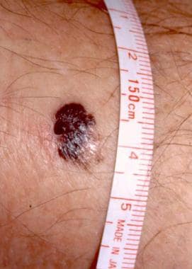
Cutaneous melanoma with characteristic asymmetry, irregular borders, and color variation. Courtesy of Wendy Brick, MD.
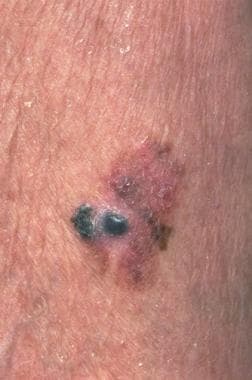
Click to see larger pictureClick to see detailView Full Size Image
Media type:  Photo


  1. Demierre MF, Nathanson L. Chemoprevention of melanoma: an unexplored strategy. J Clin Oncol. Jan 1 2003;21(1):158-65. [Medline].
  2. Whiteman DC, Watt P, Purdie DM, Hughes MC, Hayward NK, Green AC. Melanocytic nevi, solar keratoses, and divergent pathways to cutaneous melanoma. J Natl Cancer Inst. Jun 4 2003;95(11):806-12. [Medline].
  3. Maldonado JL, Fridlyand J, Patel H, Jain AN, Busam K, Kageshita T, et al. Determinants of BRAF mutations in primary melanomas. J Natl Cancer Inst. Dec 17 2003;95(24):1878-90. [Medline].
  4. Sober AJ, Fitzpatrick TB, Mihm MC, Wise TG, Pearson BJ, Clark WH, et al. Early recognition of cutaneous melanoma. JAMA. Dec 21 1979;242(25):2795-9. [Medline].
  5. Rhodes AR, Weinstock MA, Fitzpatrick TB, Mihm MC Jr, Sober AJ. Risk factors for cutaneous melanoma. A practical method of recognizing predisposed individuals. JAMA. Dec 4 1987;258(21):3146-54. [Medline].
  6. Williams ML, Sagebiel RW. Melanoma risk factors and atypical moles. West J Med. Apr 1994;160(4):343-50. [Medline].
  7. Parkin DM, Bray F, Ferlay J, Pisani P. Global cancer statistics, 2002. CA Cancer J Clin. Mar-Apr 2005;55(2):74-108. [Medline].
  8. Geller AC, Miller DR, Annas GD, Demierre MF, Gilchrest BA, Koh HK. Melanoma incidence and mortality among US whites, 1969-1999. JAMA. Oct 9 2002;288(14):1719-20. [Medline].
  9. Jemal A, Siegel R, Ward E, Murray T, Xu J, Thun MJ. Cancer statistics, 2007. CA Cancer J Clin. Jan-Feb 2007;57(1):43-66. [Medline].
  10. Swetter SM, Geller AC, Kirkwood JM. Melanoma in the older person. Oncology (Williston Park). Aug 2004;18(9):1187-96; discussion 1196-7. [Medline].
  11. Friedman RJ, Rigel DS, Kopf AW. Early detection of malignant melanoma: the role of physician examination and self-examination of the skin. CA Cancer J Clin. May-Jun 1985;35(3):130-51. [Medline].
  12. Abbasi NR, Shaw HM, Rigel DS, Friedman RJ, McCarthy WH, Osman I, et al. Early diagnosis of cutaneous melanoma: revisiting the ABCD criteria. JAMA. Dec 8 2004;292(22):2771-6. [Medline].
  13. Grob JJ, Bonerandi JJ. The 'ugly duckling' sign: identification of the common characteristics of nevi in an individual as a basis for melanoma screening. Arch Dermatol. Jan 1998;134(1):103-4. [Medline].
  14. Gachon J, Beaulieu P, Sei JF, Gouvernet J, Claudel JP, Lemaitre M, et al. First prospective study of the recognition process of melanoma in dermatological practice. Arch Dermatol. Apr 2005;141(4):434-8. [Medline].
  15. Miracco C, Santopietro R, Biagioli M, Lazzi S, Nyongo A, Vatti R, et al. Different patterns of cell proliferation and death and oncogene expression in cutaneous malignant melanoma. J Cutan Pathol. May 1998;25(5):244-51. [Medline].
  16. Bastian BC, Kashani-Sabet M, Hamm H, Godfrey T, Moore DH 2nd, Bröcker EB, et al. Gene amplifications characterize acral melanoma and permit the detection of occult tumor cells in the surrounding skin. Cancer Res. Apr 1 2000;60(7):1968-73. [Medline].
  17. Sasaki Y, Niu C, Makino R, Kudo C, Sun C, Watanabe H, et al. BRAF point mutations in primary melanoma show different prevalences by subtype. J Invest Dermatol. Jul 2004;123(1):177-83. [Medline].
  18. Richard MA, Grob JJ, Avril MF, Delaunay M, Thirion X, Wolkenstein P, et al. Melanoma and tumor thickness: challenges of early diagnosis. Arch Dermatol. Mar 1999;135(3):269-74. [Medline].
  19. Demierre MF, Chung C, Miller DR, Geller AC. Early detection of thick melanomas in the United States: beware of the nodular subtype. Arch Dermatol. Jun 2005;141(6):745-50. [Medline].
  20. Swetter SM, Boldrick JC, Jung SY, Egbert BM, Harvell JD. Increasing incidence of lentigo maligna melanoma subtypes: northern California and national trends 1990-2000. J Invest Dermatol. Oct 2005;125(4):685-91. [Medline].
  21. Cress RD, Holly EA. Incidence of cutaneous melanoma among non-Hispanic whites, Hispanics, Asians, and blacks: an analysis of california cancer registry data, 1988-93. Cancer Causes Control. Mar 1997;8(2):246-52. [Medline].
  22. Byrd KM, Wilson DC, Hoyler SS, Peck GL. Advanced presentation of melanoma in African Americans. J Am Acad Dermatol. Jan 2004;50(1):21-4; discussion 142-3. [Medline].
  23. Rogers RS 3rd, Gibson LE. Mucosal, genital, and unusual clinical variants of melanoma. Mayo Clin Proc. Apr 1997;72(4):362-6. [Medline].
  24. Friedman GD, Tekawa IS. Association of basal cell skin cancers with other cancers (United States). Cancer Causes Control. Dec 2000;11(10):891-7. [Medline].
  25. Lederman JS, Lew RA, Koh HK, Sober AJ. Influence of estrogen administration on tumor characteristics and survival in women with cutaneous melanoma. J Natl Cancer Inst. May 1985;74(5):981-5. [Medline].
  26. Hannaford PC, Villard-Mackintosh L, Vessey MP, Kay CR. Oral contraceptives and malignant melanoma. Br J Cancer. Mar 1991;63(3):430-3. [Medline].
  27. Driscoll MS, Grin-Jorgensen CM, Grant-Kels JM. Does pregnancy influence the prognosis of malignant melanoma?. J Am Acad Dermatol. Oct 1993;29(4):619-30. [Medline].
  28. Smith MA, Fine JA, Barnhill RL, Berwick M. Hormonal and reproductive influences and risk of melanoma in women. Int J Epidemiol. Oct 1998;27(5):751-7. [Medline].
  29. Schwartz JL, Mozurkewich EL, Johnson TM. Current management of patients with melanoma who are pregnant, want to get pregnant, or do not want to get pregnant. Cancer. May 1 2003;97(9):2130-3. [Medline].
  30. Weiss M, Loprinzi CL, Creagan ET, Dalton RJ, Novotny P, O'Fallon JR. Utility of follow-up tests for detecting recurrent disease in patients with malignant melanomas. JAMA. Dec 6 1995;274(21):1703-5. [Medline].
  31. Johnson TM, Bradford CR, Gruber SB, Sondak VK, Schwartz JL. Staging workup, sentinel node biopsy, and follow-up tests for melanoma: update of current concepts. Arch Dermatol. Jan 2004;140(1):107-13.