Continually Updated Clinical Reference
 
 
  All Sources     eMedicine     Medscape     Drug Reference     MEDLINE
 
eMedicine - Linear IgA Dermatosis : Article by

Quick Find
Authors & Editors
Introduction
Clinical
Differentials
Workup
Treatment
Medication
Follow-up
Multimedia
References

Related Articles
Epidermolysis Bullosa Acquisita


Patient Education
Click here for patient education.


Author: Peter A Klein, MD, Staff Physician, Department of Dermatology, University Hospital, State University of New York at Stony Brook

Coauthor(s): Jeffrey P Callen, MD, Professor of Medicine, Chief, Division of Dermatology, University of Louisville School of Medicine

Editors: Russell Hall, MD, Chief, Professor, Department of Internal Medicine, Division of Dermatology, Duke University; Richard P Vinson, MD, Assistant Clinical Professor, Department of Dermatology, Texas Tech University School of Medicine; Consulting Staff, Mountain View Dermatology, PA; Jeffrey J Miller, MD, Associate Professor, Department of Dermatology, Penn State University, Milton S Hershey Medical Center; Catherine Quirk, MD, Clinical Assistant Professor, Department of Dermatology, Brown University; William D James, MD, Paul R Gross Professor of Dermatology, University of Pennsylvania School of Medicine; Vice-Chair, Program Director, Department of Dermatology, University of Pennsylvania Health System

Author and Editor Disclosure

Synonyms and related keywords: linear immunoglobulin A dermatosis, LAD, linear IgA bullous disease, LABD, chronic bullous disease of childhood, chronic bullous dermatosis of childhood, CBDC, linear immunoglobulin A bullous disease

Background

Linear immunoglobulin A (IgA) dermatosis (LAD) is an autoimmune subepidermal vesiculobullous disease that may be idiopathic or drug-induced. Children and adults are affected, with disease of the former historically referred to as chronic bullous dermatosis of childhood. The clinical presentation is heterogeneous and appears similar to other blistering diseases, such as bullous pemphigoid and dermatitis herpetiformis.

Pathophysiology

LAD is an autoimmune disease histopathologically characterized by the linear deposition of IgA at the basement membrane zone (BMZ). Antibody deposition leads to complement activation and neutrophil chemotaxis, which eventuates in loss of adhesion at the dermal-epidermal junction and in blister formation. Disease in children is immunologically identical to that of adults. The mechanism of loss of self-tolerance to target antigens is unknown.

Within the dermal-epidermal junction, different antigenic target sites, including the lamina lucida, the sublamina densa, or both locations simultaneously, have been identified. The best-characterized antigen is a 97-kd protein extracted from human epidermis that binds IgA antibodies from sera of patients with LAD. Sera that binds the 97-kd antigen localizes to the lamina lucida of salt-split skin. Originally thought to be a unique protein of the lamina lucida, recent work reveals that the 97-kd protein may represent a portion of the extracellular domain of the 180-kd bullous pemphigoid antigen (BPAg2).

The same patient sera have been shown to bind a 120-kd antigen in the BMZ. The 97- and 120-kd antigens may represent cleaved fragments of BPAg2, which exist as such in vivo or are produced by proteolytic digestion in vitro. These smaller molecules could also be alternative splicing products of the same BPAg2 gene. Because antibodies that bind the 97- and 120-kd antigens do not recognize the 180-kd BPAg2, the former may express unique epitopes distinct from those of the parent protein.

A recent case series reported patients with sera not reactive against the 97-kd antigen but rather against both bullous pemphigoid antigens. Of 11 patients, a 230-kd antigen (BPAg1) was recognized in 6 patients and BPAg2 was recognized in 5 patients. The authors suggest that an IgA-specific immune response may occur against bullous pemphigoid antigens in LAD. These results are provocative given that the 97-kd LAD antigen may represent a portion of the extracellular domain of BPAg2.

A 285-kd target antigen has been identified in the lamina lucida and the sublamina densa; this antigen is recognized by circulating antibodies in some patients with LAD, but it has not been further characterized. A 250-kd dermal antigen corresponding to collagen VII of anchoring fibrils has also been reported as a target antigen in some patients.

LAD illustrates the importance of identifying the target antigen. In cases where type VII collagen is the molecule against which the antibody response is directed, patients are less likely to be responsive to treatment. Thus, viewing this condition as a subset of epidermolysis bullosa acquisita is better. Similarly, patients with antibodies directed against the bullous pemphigoid antigens may be classified as having bullous pemphigoid but with an IgA response rather than an IgG response. Until more patients are reported whose antibody response is detailed to the molecular level and until this definition becomes clinically available, these heterogeneous patients will continue to be grouped into a single category, LAD.

Frequency

United States

The prevalence of LAD in Utah has been estimated as 0.6 per 100,000 adults. The prevalence in children has not been reported.

International

The incidence of adult LAD in southern England has been estimated to be 1 case in 250,000 population per year. The incidence in France has been reported as 0.13 in 250,000 population. Estimates have not been reported for LAD in children.

Mortality/Morbidity

The mean duration of idiopathic LAD of childhood is 3.9 years, ranging from 2.1-7.9 years. Remission has been reported to occur in 64% of children, in most cases within 2 years. Disease of adults is more protracted, with a mean duration of 5.6 years, lasting anywhere from 1-15 years. The remission rate in adults is less than that in children (48%). The disease tends to wax and wane in severity. Drug-induced cases typically resolve quickly once the causative agent is identified and withdrawn. Cutaneous lesions usually heal without scarring.

Lesions of the mucous membranes heal with scarring and pose considerable morbidity. Desquamative gingivitis may secondarily damage teeth. Ocular LAD may be indistinguishable from cicatricial pemphigoid and lead to blindness. Involvement of the pharynx, the larynx, the nose, the rectum, and the esophagus has been reported.

A retrospective study of 12 women with LAD showed improvement during pregnancy, usually by 10 weeks' gestation. Improvement was most marked in the third trimester. The most common agent used for treatment of LAD is dapsone, which is classified as pregnancy class C (uncertain safety; animal studies show an adverse effect, no human studies) by the US Food and Drug Administration. In this series, the authors observed no serious adverse effects from dapsone during 11 pregnancies. Postpartum relapses of LAD were common, occurring in 1 case within 2 hours. The range of time to relapse for the remaining 11 patients was 1-6 months, with 3 patients achieving complete remission at 2 years. Fetal outcome was unaffected by the disease.

Sex

Some case series have reported a slight female preponderance; the female-to-male ratio is 1.6:1.

Age

LAD has a bimodal age of onset. Disease in children commences at ages ranging from 6 months to 10 years, with a mean of 3.3-4.5 years based on 2 case series. Disease of adults ranges from 14-83 years, with a mean of 52 years. Disease is most common in the nonreproductive years. Drug-induced disease is more likely to occur in the older population because this group is often being treated for multiple medical conditions.


History

  • Some patients note a prolonged period of prodromal itching or transient pruritus or burning before lesions appear.
  • Patients with ocular manifestations may complain of pain, grittiness, or discharge.
  • Bullae may be chronic, or lesions may appear acutely, as seen in drug-induced disease. Rash latency in vancomycin-induced cases of LAD ranges from 1-13 days after the first dose. Review of medication exposures and delineation of the drug timeline are crucial in identifying potential inciting agents.

Physical

  • The classic primary lesions of LAD are clear and/or hemorrhagic round or oval vesicles or bullae on normal, erythematous, or urticarial skin. Cutaneous manifestations may also include erythematous plaques, blanching macules and papules, or targetoid erythema multiforme–like lesions. The diagnosis is not dependent on the presence of vesicles and/or bullae.
    • Bullae may be discrete or arranged in a herpetiform pattern, often described as the cluster of jewels sign. Alternatively, vesicles and bullae may be seen at the edge of annular or polycyclic lesions, the appearance of which has been described as the string of beads sign (see Image 1).
    • The distribution of LAD differs between adults and children. Lesions in children are typically localized to the lower abdomen and anogenital areas with frequent involvement of the perineum (see Image 2). Other sites of involvement include the feet, the hands, and the face, particularly the perioral area. In adults, the trunk and the limbs are most commonly affected. In adults, involvement of the perineum and the perioral area is less frequent than in children. Lesions in both children and adults may be distributed symmetrically or asymmetrically. Dermatitis herpetiformis–like involvement of the extensor surfaces of the knees and the elbows is seen infrequently.
    • Crusts, excoriations, erosions, or ulcers may be present.
  • Oral manifestations are common in children and adults with LAD. Oral lesions include vesicles, ulcerations, erythematous patches, erosions, desquamative gingivitis, or erosive cheilitis, and they may precede skin lesions.
  • Both children and adults frequently complain of ocular symptoms, such as grittiness, burning, or discharge. Ophthalmologic findings even in the absence of ocular complaints may include subconjunctival fibrosis, shrinkage of the fornices, symblepharon formation, and cicatricial entropion with trichiasis (see Image 3).

Causes

The list of agents implicated in LAD continues to grow, especially with regard to medications. Many patients report prodromal events, such as illnesses or ingestion of drugs. In absent large series or a preponderance of case reports, only a subset of cases have identifiable causes. Gluten-sensitive enteropathy is not associated with LAD.

  • Seventeen case reports have implicated vancomycin in LAD. Of all reported causative drugs, it is the best-documented agent in the literature. Other potential triggers and their respective number of published case reports (listed in parentheses) include the following: amiodarone (2), ampicillin sodium (1), captopril (2), cefamandole nafate (1), cyclosporine (1), depot sulfonamide (1), diclofenac (3), glibenclamide (1), interferon gamma and interleukin 2 (2), iodine contrast agent (1), lithium carbonate (1), penicillin sodium (2), phenytoin sodium (2), somatostatin (1), sulfamethoxazole/trimethoprim (1), sulfisoxazole (1), topical sodium hypochlorite (1), and vigabatrin (1).
  • Preceding illnesses, such as typhoid, brucella, tuberculosis, antibiotic-treated tetanus, varicella, herpes zoster, Paecilomyces lung infection, gynecologic infections, and upper respiratory infections, have all been reported in association with LAD. The significance of these associations is uncertain. Their potential role in stimulation of the IgA mucosal system has yet to be elucidated.
  • LAD associated with malignancy has been reported in as many as 5% of cases. Lymphoproliferative malignancies, specifically Hodgkin disease, non-Hodgkin lymphoma, and chronic lymphocytic leukemia have been described. LAD has also been reported with solid tumors, such as bladder carcinoma. Other associated malignancies include polycythemia rubra vera, plasmacytoma, multiple myeloma, ocular melanoma, squamous cell carcinoma of the esophagus, breast carcinoma, uterine carcinoma, eccrine carcinoma, metastatic squamous cell carcinoma, colon carcinoma, thyroid carcinoma, retroperitoneal carcinoma, metastatic hypernephroma, and hydatidiform mole. The validity of the association between LAD and malignancy remains to be proven.
  • Because LAD is itself an autoimmune disease, an association with other such disorders is interesting despite proven causality. Cases have been described in association with systemic lupus erythematosus, dermatomyositis, rheumatoid arthritis, polymyalgia rheumatica, hypothyroidism, chronic hepatitis, Crohn disease, ulcerative colitis, multiple sclerosis, and IgA nephropathy. Again, these associations may be coincidental.
  • LAD in children has been reported in association with human leukocyte antigen B8 (HLA-B8), but the significance of this finding is unknown.
  • Pancreatic lipase deficiency has been reported once in association with LAD.


Epidermolysis Bullosa Acquisita


Lab Studies

  • Immunofluorescence studies
    • Direct immunofluorescence study of both perilesional skin and healthy skin typically shows linear deposition of IgA at the BMZ. Linear deposition of C3 may also be seen. Direct immunofluorescence study of salt-split skin reveals IgA deposition on either the dermal side (blister floor) or the epidermal side (blister roof). Some patients demonstrate both linear IgA deposition and immunoglobulin G (IgG) deposition at the BMZ. Immunoglobulin M (IgM) deposition has rarely been reported.
    • Serum should be obtained for indirect immunofluorescence studies. Approximately 50% of patients with LAD have detectable circulating antibody that binds to the BMZ. Sensitivity is greater for immunofluorescence studies performed on salt-split healthy human skin. Circulating antibody titers are typically low (1:10 to 1:20). When present, linear deposition of antibody is observed at the BMZ or at the blister roof or floor in salt-split skin. Children with LAD may demonstrate circulating anti-BMZ antibodies more frequently than adults.
  • In patients with atypical presentations, additional testing, including bacterial culture and Gram stain of blister fluid to rule out bullous impetigo and Tzanck smear to rule out herpes virus infection, may be helpful.

Histologic Findings

Early urticarial papules or plaques reveal neutrophils aligned along the BMZ accompanied by vacuolar change. Neutrophilic microabscesses may be seen in dermal papillae (see Image 4). Fully developed lesions reveal subepidermal blistering with a predominantly polymorphonuclear infiltrate, although mononuclear cells and eosinophils may be present. Obtaining a frozen section of a blister roof may be helpful in some patients to rule out full-thickness epidermal necrosis as seen in toxic epidermal necrolysis.


Medical Care

Bullae do not need special care, as long they remain intact. Ruptured lesions and erosions should be covered with sterile dressings. Infected lesions may be treated with topical mupirocin and sterile dressing changes twice daily.

Consultations

  • Consult a dermatologist.
  • Consult an ophthalmologist. Patients with LAD can have changes, such as fine scarring, in the absence of ocular complaints. Therefore, most, if not all, patients once diagnosed should be seen by an ophthalmologist.


Large, randomized, placebo-controlled, double-blind studies have not been performed for the treatment of LAD in children or adults. Most cases have been reported to respond to dapsone or sulfapyridine. Some clinicians favor the use of sulfapyridine because of the lower incidence of adverse effects. However, some patients' conditions may not respond to sulfapyridine but do respond to treatment with dapsone. A response may be seen in 48-72 hours. Other reportedly useful medications include prednisone, sulfamethoxypyridazine, colchicine, dicloxacillin, and intravenous immunoglobulin in a single patient with chronic renal failure.

Drug-induced disease may be treated merely by withdrawal of the offending agent. In cases of LAD induced by vancomycin, new lesions stop forming within approximately 2 weeks of withdrawal. Particularly severe cases of drug-induced LAD respond to a short course of oral corticosteroids.

Drug Category: Leprostatic agents

These agents have been shown to be beneficial in the treatment of LAD.

Drug NameDapsone (Avlosulfon)
DescriptionBactericidal and bacteriostatic against mycobacteria; mechanism of action is similar to that of sulfonamides where competitive antagonists of PABA prevent formation of folic acid, inhibiting bacterial growth.
Adult Dose25-100 mg PO qd initial; if blistering is not controlled, use 50-mg increments q1-2wk; most patients require 200 mg PO qd or less
Pediatric Dose1-2 mg/kg PO qd initial; not to exceed 3-4 mg/kg/d
ContraindicationsAbsolute: hypersensitivity
Relative: G-6-PD deficiency (especially in African Americans, persons of Middle Eastern heritage, and Asians), significant cardiopulmonary or hematologic disease, sulfa allergy (cautious use in patients with sulfa allergy may be attempted; cross-reactivity relatively rare and mild)
InteractionsTrimethoprim, probenecid, and folic acid antagonists (eg, pyrimethamine, methotrexate) increase levels; activated charcoal, PABA, and rifampin decrease levels; sulfonamides and hydroxychloroquine may increase hemolysis
PregnancyC - Safety for use during pregnancy has not been established.
PrecautionsAgranulocytosis is a rare complication; screen patients for G-6-PD deficiency, as dapsone causes acute hemolysis in these patients; causes dose-related hemolysis and methemoglobinemia, especially in those with methemoglobin reductase deficiency; less common adverse effects include dose-related hepatitis and peripheral neuropathy (neuropathy is predominately motor; however, sensory defects can occur); reported to cause toxic epidermal necrolysis, particularly in patients infected with HIV; CBC counts are recommended weekly to biweekly for first mo of therapy and monthly to bimonthly thereafter for the next 5 mo; check baseline LFTs and q6mo thereafter

Drug Category: Sulfonamides

These agents exert bacteriostatic action by competitive antagonism of para-aminobenzoic acid (PABA). Microorganisms that require exogenous folic acid and do not synthesize folic acid are not susceptible to the action of sulfonamides.

Drug NameSulfapyridine
DescriptionCompetitive antagonist of PABA. Mechanism of action in LAD is unknown.
Adult Dose500 mg PO bid initial; increase by 1 g q1-2wk until disease is controlled; may require 1-4 g/d
Pediatric Dose35 mg/kg PO bid; not to exceed 100 mg/kg/d
ContraindicationsDocumented hypersensitivity; slow acetylators may require smaller doses or more gradual initial dose adjustment
InteractionsBioavailability of digoxin is reduced by sulfapyridine; interval of 2-3 h between administrations recommended
PregnancyB - Usually safe but benefits must outweigh the risks.
PrecautionsIdiosyncratic reactions (eg, hypersensitivity pneumonitis, a lupuslike syndrome, pancreatitis, toxic hepatitis) may occur; agranulocytosis rarely occurs; both immune and nonimmune hemolytic anemia develop (the latter is more common in G-6-PD–deficient patients); folate deficiency may occur secondary to impaired absorption; nephrolithiasis may occur as with other sulfa drugs; toxic epidermal necrolysis has been reported with medications containing sulfa groups; check CBC count and LFTs monthly for 5 mo then q6wk thereafter

Drug Category: Corticosteroids

These agents have anti-inflammatory properties and cause profound and varied metabolic effects. In addition, these agents modify the body's immune response to diverse stimuli.

Drug NamePrednisone (Deltasone)
DescriptionImmunosuppressant for treatment of autoimmune disorders; may decrease inflammation by reversing increased capillary permeability and suppressing PMN activity. Stabilizes lysosomal membranes and also suppresses lymphocyte and antibody production.
Adult Dose5-60 mg/d PO qd or divided bid/qid; taper over 2 wk as symptoms resolve; 0.5-2 mg/kg/d; taper as condition improves; single morning dose is safer for long-term use, but divided doses have more anti-inflammatory effect
Pediatric Dose4-5 mg/m2/d PO; alternatively, 0.05-2 mg/kg PO divided bid/qid; taper over 2 wk as symptoms resolve
ContraindicationsDocumented hypersensitivity; viral, fungal, tubercular skin, or connective tissue infections; peptic ulcer disease; hepatic dysfunction; GI disease
InteractionsCoadministration with estrogens may decrease prednisone clearance; when used with digoxin, digitalis toxicity secondary to hypokalemia may increase; phenobarbital, phenytoin, and rifampin may increase metabolism of glucocorticoids (consider increasing maintenance dose); monitor for hypokalemia with coadministration of diuretics
PregnancyB - Usually safe but benefits must outweigh the risks.
PrecautionsAbrupt discontinuation of glucocorticoids may cause adrenal crisis; hyperglycemia, edema, osteonecrosis, myopathy, peptic ulcer disease, hypokalemia, osteoporosis, euphoria, psychosis, myasthenia gravis, growth suppression, and infections may occur with glucocorticoid use

Drug Category: Anti-inflammatory agents

These agents modulate events leading to inflammatory reactions.

Drug NameColchicine
DescriptionDecreases leukocyte motility and phagocytosis in inflammatory responses.
Adult Dose0.5-1.2 mg PO initially, followed by 0.5-0.6 q1-2h or 1-1.2 mg q2h until a satisfactory response attained; not to exceed 4 mg/d
Pediatric Dose<12 years: Not established
>12 years: Administer as in adults
ContraindicationsDocumented hypersensitivity; severe renal, hepatic, GI, or cardiac disorders; blood dyscrasias
InteractionsSympathomimetic agent toxicity and effect of CNS depressants are significantly increased with colchicine
PregnancyC - Safety for use during pregnancy has not been established.
PrecautionsRisk of renal failure, hepatic failure, permanent hair loss, bone marrow suppression, numbness or tingling in hands and feet, disseminated intravascular coagulopathy, and decreased sperm count; dose-dependent GI upset is common

Drug Category: Antibiotics

Empiric antimicrobial therapy must be comprehensive and should cover all likely pathogens in the context of the clinical setting.

Drug NameDicloxacillin (Dycill, Dynapen)
DescriptionTreatment of infections caused by penicillinase-producing staphylococci. May use to initiate therapy when staphylococcal infection is suspected.
Adult Dose125-250 mg PO q6h
Pediatric Dose<40 kg: 12.5 mg/kg/d PO q6h
>40 kg: 125 mg PO q6h
ContraindicationsDocumented hypersensitivity
InteractionsDecreases efficacy of oral contraceptives; may decrease effects of anticoagulants; probenecid and disulfiram may increase penicillin levels; tetracyclines may decrease effect of penicillins with concurrent use
PregnancyB - Usually safe but benefits must outweigh the risks.
PrecautionsMonitor PT in patients taking anticoagulant medications; toxicity may increase in patients with renal impairment

Drug Category: Immunoglobulins

These agents are used to improve the clinical and immunologic aspects of the disease. They may decrease autoantibody production and increase solubilization and removal of immune complexes.

Drug NameImmune globulins intravenous (IVIG, Gammagard, Gamimune)
DescriptionFeatures that may be relevant to its efficacy include neutralization of circulating myelin antibodies through anti-idiotypic antibodies; downregulation of proinflammatory cytokines, including IFN-gamma; blockade of Fc receptors on macrophages; suppression of inducer T and B cells; augmentation of suppressor T cells; blockade of the complement cascade; promotion of remyelination; and 10% increase in CSF IgG.
Adult Dose2 g/kg IV over 2-5 d
Pediatric DoseAdminister as in adults
ContraindicationsDocumented hypersensitivity; IgA deficiency; anti-IgE/IgG antibodies
InteractionsNone reported
PregnancyB - Usually safe but benefits must outweigh the risks.
PrecautionsCheck serum IgA before IVIG (use an IgA-depleted product, eg, Gammagard S/D); infusions may increase serum viscosity and thromboembolic events; infusions may increase risk of migraine attacks, aseptic meningitis (10%), urticaria, pruritus, or petechiae (2-30 d postinfusion); increases risk of renal tubular necrosis in elderly patients and in patients with diabetes, volume depletion, and preexisting kidney disease; laboratory result changes associated with infusions include elevated antiviral or antibacterial antibody titers for 1 mo, 6-fold increase in ESR for 2-3 wk, and apparent hyponatremia


Further Outpatient Care

  • Patients should be closely monitored after initiating therapy with dapsone or sulfapyridine.
  • The medication tables (see Medication section above) provide appropriate laboratory follow-up intervals.
  • If patients do not respond to dapsone or sulfapyridine, the diagnosis should be reconsidered.


Media file 1:  Annular lesions demonstrating the string of beads sign.
Click to see larger pictureClick to see detailView Full Size Image
Media type:  Photo

Media file 2:  
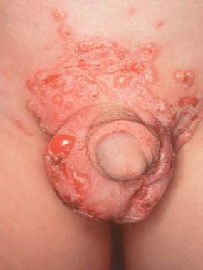
Bullous lesions on the genital area in a child with linear IgA dermatosis.
Click to see larger pictureClick to see detailView Full Size Image
Media type:  Photo

Media file 3:  Persons with linear immunoglobulin A (IgA) dermatosis may present with prominent ocular signs and symptoms.
Click to see larger pictureClick to see detailView Full Size Image
Media type:  Photo

Media file 4:  Neutrophilic microabscesses in linear immunoglobulin A (IgA) dermatosis.
Click to see larger pictureClick to see detailView Full Size Image
Media type:  Photo


  • Aultbrinker EA, Starr MB, Donnenfeld ED. Linear IgA disease. The ocular manifestations. Ophthalmology. Mar 1988;95(3):340-3. [Medline].
  • Bernard P, Vaillant L, Labeille B, et al. Incidence and distribution of subepidermal autoimmune bullous skin diseases in three French regions. Bullous Diseases French Study Group. Arch Dermatol. Jan 1995;131(1):48-52. [Medline].
  • Chan LS, Regezi JA, Cooper KD. Oral manifestations of linear IgA disease. J Am Acad Dermatol. Feb 1990;22(2 Pt 2):362-5. [Medline].
  • Chorzelski TP, Jablonska S, Maciejowska E. Linear IgA bullous dermatosis of adults. Clin Dermatol. Jul-Sep 1991;9(3):383-92. [Medline].
  • Collier PM, Kelly SE, Wojnarowska F. Linear IgA disease and pregnancy. J Am Acad Dermatol. Mar 1994;30(3):407-11. [Medline].
  • Godfrey K, Wojnarowska F, Leonard J. Linear IgA disease of adults: association with lymphoproliferative malignancy and possible role of other triggering factors. Br J Dermatol. Oct 1990;123(4):447-52. [Medline].
  • Jablonska S, Chorzelski TP, Rosinska D, Maciejowska E. Linear IgA bullous dermatosis of childhood (chronic bullous dermatosis of childhood). Clin Dermatol. Jul-Sep 1991;9(3):393-401. [Medline].
  • Klein PA, Callen JP. Drug-induced linear IgA bullous dermatosis after vancomycin discontinuance in a patient with renal insufficiency. J Am Acad Dermatol. Feb 2000;42(2 Pt 2):316-23. [Medline].
  • Leonard JN, Haffenden GP, Ring NP, et al. Linear IgA disease in adults. Br J Dermatol. Sep 1982;107(3):301-16. [Medline].
  • Nousari HC, Kimyai-Asadi A, Caeiro JP, Anhalt GJ. Clinical, demographic, and immunohistologic features of vancomycin- induced linear IgA bullous disease of the skin. Report of 2 cases and review of the literature. Medicine (Baltimore). Jan 1999;78(1):1-8. [Medline].
  • Wojnarowska F, Marsden RA, Bhogal B, Black MM. Chronic bullous disease of childhood, childhood cicatricial pemphigoid, and linear IgA disease of adults. A comparative study demonstrating clinical and immunopathologic overlap. J Am Acad Dermatol. Nov 1988;19(5 Pt 1):792-805. [Medline].
  • Zone JJ, Taylor TB, Meyer LJ, Petersen MJ. The 97 kDa linear IgA bullous disease antigen is identical to a portion of the extracellular domain of the 180 kDa bullous pemphigoid antigen, BPAg2. J Invest Dermatol. Mar 1998;110(3):207-10. [Medline].

Linear IgA Dermatosis excerpt

Article Last Updated: Feb 15, 2007