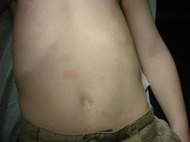
Continually Updated Clinical Reference
 
 
  All Sources     eMedicine     Medscape     Drug Reference     MEDLINE
 
eMedicine - Lichen Spinulosus : Article by

Quick Find
Authors & Editors
Introduction
Clinical
Differentials
Workup
Treatment
Medication
Follow-up
Miscellaneous
Multimedia
References

Related Articles
Keratosis Pilaris


Patient Education
Click here for patient education.


Author: Stephen W White, MD, Clinical Assistant Professor, Department of Dermatology, George Washington University Hospital

Stephen W White is a member of the following medical societies: American Academy of Dermatology, International Society of Dermatology, Society for Investigative Dermatology, and Society for Pediatric Dermatology

Coauthor(s): Christopher R Gorman, MD, Resident Physician, Department of Dermatology, University of Virginia School of Medicine

Editors: James J Nordlund, MD, Professor Emeritus, Department of Dermatology, University of Cincinnati College of Medicine; Richard P Vinson, MD, Assistant Clinical Professor, Department of Dermatology, Texas Tech University School of Medicine; Consulting Staff, Mountain View Dermatology, PA; Rosalie Elenitsas, MD, Associate Professor of Dermatology, University of Pennsylvania School of Medicine; Director, Penn Cutaneous Pathology Services, Department of Dermatology, University of Pennsylvania Health System; Catherine Quirk, MD, Clinical Assistant Professor, Department of Dermatology, Brown University; William D James, MD, Paul R Gross Professor of Dermatology, University of Pennsylvania School of Medicine; Vice-Chair, Program Director, Department of Dermatology, University of Pennsylvania Health System

Author and Editor Disclosure

Synonyms and related keywords: keratosis follicularis spinulosa, lichen pilaris seu spinulosus of Crocker, keratosis follicularis spinosa of Unna

Background

Lichen spinulosus (LS) is an uncommon dermatosis manifested by large patches of follicular papules topped by keratotic spines (see Media Files 1-2). In 1883, Crocker published a description of LS. Since then, few other similar reports were published until 1990, when Friedman presented data on 35 patients with LS. The etiology is unknown. Some minor progress has been made in therapy.

Pathophysiology

The classic lesion of LS is a keratotic plug located within the dilated follicular orifice. Histologically, an inflammatory lymphohistiocytic infiltrate occurs around the follicle and in the dermis. Hyperkeratosis, parakeratosis, and acanthosis are visible in the follicle. Differentiating LS from keratosis pilaris by microscopy may not be possible.

Frequency

United States

Apparently, LS is not a common disorder. This conclusion is based on the paucity of published reports regarding LS. In the past, LS was reported to be associated with the administration of arsphenamine, thallium, gold, and diphtheria toxin. More recently, authors have noted association with HIV and Crohn disease. These associations may reflect the interests of the authors.

International

LS has been reported worldwide. In 1990, Friedman described 35 patients with LS. He and his coworkers in the Philippines examined 7435 people attending a dermatology clinic. The incidence of LS was approximately 5 cases per 1000 population with skin disorders. This prevalence exceeds reports from various American surveys on cutaneous diseases in children and adolescents.

Mortality/Morbidity

LS affects only the skin and is not known to be associated with abnormalities of internal organ systems. Occasionally, a patient with LS complains of pruritus. Otherwise, the disorder mostly is of cosmetic significance. Misdiagnosis can result in inappropriate treatment.

Race

Worldwide distribution suggests no predilection of LS in any ethnic group.

Sex

Case reports suggest an equal distribution in males and females. Friedman's study in the Philippines included 14 males and 21 females.

Age

Reports indicate that LS is a disease that occurs during childhood to young adulthood. Peak incidence appears to occur during adolescence. The condition can persist for decades. In most patients, LS remits spontaneously within 1-2 years. Friedman calculated that in the Philippines, the average age at onset was 16.2 years ± 10.1 years.


History

LS tends to have a sudden onset and is not accompanied by other signs or symptoms. The keratotic papules group into large plaques that can spread rapidly to affect large areas of skin.

Physical

  • Patches and plaques of follicular papules have a diameter that ranges from 2-5 cm.
    • Patches are distributed symmetrically over the integument.
    • Patches affect the neck, buttocks, abdomen, trochanters, knees, and extensor surfaces of the arms.
  • Individual papules are flat to conical.
    • Individual papules usually are small, approximately 1-3 mm in diameter.
    • Papules have a pointed or hairlike horny spine that extends approximately 1 mm around the tip of the follicle.
    • When a patch is rubbed gently with the fingers, it feels similar to a nutmeg grater.

Causes

The cause of LS is unknown. Infection has been postulated, but no data support this hypothesis. Other authors have suggested that LS is part of atopy, but no association of LS with atopy was found in the Philippines. A report notes a family with the condition in 4 generations, an observation that suggests a genetic predisposition.


Keratosis Pilaris

Other Problems to be Considered

Dermatophytosis
Frictional lichenoid eruption
Lichen scrofulosorum
Lymphoproliferative disorders
Hypovitaminosis A phrynoderma


Lab Studies

  • Diagnosis should be made on clinical grounds alone. At present, no laboratory tests are specific or diagnostic.

Histologic Findings

Histologic findings of LS are similar to those observed in keratosis pilaris. In LS, dilated hair follicles are filled with a keratotic plug. An inflammatory lymphocytic infiltrate occurs around the follicle and in the dermis. Hyperkeratosis, parakeratosis, and acanthosis may be present in the follicle.


Medical Care

No cure exists for LS, but some medications ameliorate its clinical manifestations. Because of the horny plug, keratolytics have been used as a treatment. These include salicylic acid, lactic acid, and/or urea in various creams, ointments, gels, and lotions. The literature does not support the use of topical steroids.

Consultations

Consultation with an experienced dermatologist is indicated if any doubt exists concerning the diagnosis.


The goal of treatment is to improve the cosmetic disfigurement caused by the disorder.

Drug Category: Keratolytic agents

Topical lactic acid creams have provided the most successful therapy to date. Salicylic acid gel and urea containing lotions also have been reported to help soften the horny papules. Gentle abrasion with a pad, soft brush, or luffa pad can be tried to remove the horny spines.

Drug NameLactic acid (Lac-Hydrin)
DescriptionContains lactic acid, an alpha-hydroxy acid with keratolytic action, thus facilitating release of comedones. Available in 12% and 5% strengths. The 12% form may cause irritation on the face. Causes disadhesion of corneocytes. Found in a variety of topical emollient lotions.
May be combined with 10-20% urea cream or be used with salicylic acid gel.
Adult DoseApply topically qd/bid
Pediatric DoseApply as in adults
ContraindicationsDocumented hypersensitivity
InteractionsNone reported
PregnancyB - Usually safe but benefits must outweigh the risks.
PrecautionsMay sting or cause pain if applied on broken skin; may cause irritation with erythema, burning, and peeling if applied to face in 12% concentrations

Drug NameSalicylic acid 6% (cream, lotion, or gel)
DescriptionBeta-hydroxy acid reported to soften papules. By dissolving intercellular cement substance, produces desquamation of the horny layer of skin, while not affecting structure of viable epidermis. Comes as a cream, lotion, or gel.
Adult DoseApply topically several times/d
Pediatric DoseTo avoid salicylate toxicity, use care when applying to large areas
ContraindicationsDocumented hypersensitivity; prolonged use in infants and patients with diabetes or impaired circulation; use on moles, birthmarks, warts with hair growth, genital or facial warts, warts on mucous membranes, irritated skin, or infected or reddened areas
InteractionsNone reported
PregnancyC - Safety for use during pregnancy has not been established.
PrecautionsAvoid contact with mucous membranes; immediately flush with water for 15 min if contact with eyes or mucous membranes occurs

Drug NameUrea 40% cream or lotion
DescriptionPromotes hydration and removal of excess keratin.
Adult DoseApply prn to affected area
Pediatric DoseApply as in adults
ContraindicationsDocumented hypersensitivity; viral skin disease
InteractionsNone reported
PregnancyC - Safety for use during pregnancy has not been established.
PrecautionsDo not use near eyes; caution if applied to broken or swollen skin


Complications

  • LS is confined to the skin and has no known associations with internal disorders or genetic syndromes.

Prognosis

  • LS can be ameliorated using emollient keratolytics. Case reports suggest that cure is the result of spontaneous remission over time. Most cases appear to remit within 1-2 years; however, well-documented cases exist that have lasted for decades.


Medical/Legal Pitfalls

  • Failure to diagnose LS correctly may result in years of ineffective treatment.


Media file 1:  Lichen spinulosus on the abdomen.
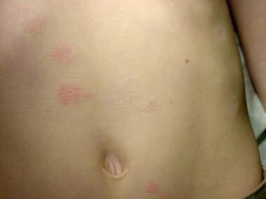
Click to see larger pictureClick to see detailView Full Size Image
Media type:  Photo

Media file 2:  Close-up view.
Click to see larger pictureClick to see detailView Full Size Image
Media type:  Photo


  • Boyd AS. Lichen spinulosus: case report and overview. Cutis. Jun 1989;43(6):557-60. [Medline].
  • Cohen SJ, Dicken CH. Generalized lichen spinulosus in an HIV-positive man. J Am Acad Dermatol. Jul 1991;25(1 Pt 1):116-8. [Medline].
  • Friedman SJ. Lichen spinulosus. Clinicopathologic review of thirty-five cases. J Am Acad Dermatol. Feb 1990;22(2 Pt 1):261-4. [Medline].
  • Kano Y, Orihara M, Yagita A, Shiohara T. Lichen spinulosus in a patient with Crohn''s disease. Int J Dermatol. Sep 1995;34(9):670-1. [Medline].
  • Maiocco KJ, Miller OF. Lichen spinulosus: response to therapy. Cutis. Feb 1976;17(2):294-99. [Medline].

Lichen Spinulosus excerpt

Article Last Updated: Dec 15, 2006