Continually Updated Clinical Reference
 
 
  All Sources     eMedicine     Medscape     Drug Reference     MEDLINE
 
eMedicine - Lentigo : Article by

Quick Find
Authors & Editors
Introduction
Clinical
Differentials
Workup
Treatment
Medication
Follow-up
Miscellaneous
Multimedia
References

Related Articles
Actinic Keratosis

Ephelides (Freckles)

Seborrheic Keratosis


Patient Education
Click here for patient education.


Author: Robert A Schwartz, MD, MPH, Professor and Head of Dermatology, Professor of Medicine, Professor of Pediatrics, Professor of Pathology, Professor of Preventive Medicine and Community Health, UMDNJ-New Jersey Medical School

Robert A Schwartz is a member of the following medical societies: Alpha Omega Alpha, American Academy of Dermatology, American College of Physicians, and Sigma Xi

Coauthor(s): Jason F Okulicz, MD, Assistant Professor of Medicine, Uniformed Services University of the Health Sciences; Fellow, Department of Infectious Disease, Wilford Hall United States Air Force Medical Center; Sergiusz Jozwiak, MD, PhD, Head, Professor, Department of Child Neurology, The Children's Memorial Health Institute of Warsaw, Poland

Editors: Neil Shear, MD, Professor and Chief of Dermatology, Professor of Medicine, Pediatrics and Pharmacology, University of Toronto Faculty of Medicine; Head of Dermatology, Sunnybrook Women's College Health Sciences Center and Women's College Hospital, Canada; Michael J Wells, MD, Associate Professor, Department of Dermatology, Texas Tech University Health Sciences Center; John G Albertini, MD, Consulting Staff, Dermatologic Surgery, The Skin Surgery Center; Glen H Crawford, MD, Assistant Clinical Professor, Department of Dermatology, University of Pennsylvania School of Medicine; Chief, Division of Dermatology, The Pennsylvania Hospital; William D James, MD, Paul R Gross Professor of Dermatology, University of Pennsylvania School of Medicine; Vice-Chair, Program Director, Department of Dermatology, University of Pennsylvania Health System

Author and Editor Disclosure

Synonyms and related keywords: lentigo simplex, solar or actinic lentigo, lentigo senilis, sun spot, liver spot, freckle, ephelides, PUVA lentigines, tanning-bed lentigines, vulvar lentigines, penile lentigines, ink spot lentigines, ink-spot lentigines, reticulated black solar lentigo, oral and labial melanotic macules, Laugier-Hunziker syndrome, Peutz-Jeghers syndrome, radiation lentigo, lentigines profuse, generalized lentigines, agminated lentiginosis, xeroderma pigmentosum, XP, LEOPARD syndrome, multiple lentigines syndrome, Carney syndrome, LAMB syndrome, NAME syndrome, inherited patterned lentiginosis, myxoma syndrome

Background

A lentigo, or lentigines (plural), is a small, sharply circumscribed, pigmented macule surrounded by normal-appearing skin. Histologic findings may include hyperplasia of the epidermis and increased pigmentation of the basal layer. A variable number of melanocytes are present; these melanocytes may be increased in number, but they do not form nests. Lentigines may evolve slowly over years, or they may be eruptive and appear rather suddenly. Pigmentation may be homogeneous or variegated, with a color ranging from brown to black.

Multiple clinical and etiologic varieties exist. The distinction of a lentigo from other melanocytic lesions (eg, nevi, melanoma) and its role as a marker for ultraviolet damage and systemic syndromes is of major significance.

A recent case-controlled study in France comparing 145 adults with multiple solar lentigines on the upper back and 145 matched control subjects found that multiple solar lentigines on the upper back and shoulders of adults may serve as clinical markers of past severe sunburn and may be used to identify a population at higher risk of developing cutaneous melanoma.1

A Medscape CME course related to Peutz-Jeghers Syndrome is Hamartomatous Polyposis Syndromes.

Pathophysiology

Depending on the type of lentigo present, a solitary lesion or multiple lesions that can occur anywhere on the body. Some lentigines have associated systemic manifestations that accompany the skin lesions.

A Japanese microarray analysis evaluation of solar lentigo in 16 adults demonstrated up-regulation of genes related to inflammation, fatty-acid metabolism, and melanocytes and down-regulation of cornified envelope-related genes.2 The researchers suggested solar lentigo may be induced by the mutagenic effect of repeated past UV light exposures, leading to characteristic enhancement of melanin production.

Frequency

United States

In America, solar lentigines are observed in as many as 90% of whites older than 60 years and in 20% of whites younger than 35 years. Psoralen UVA (PUVA) lentigines are noted in almost one half of individuals with psoriasis who receive PUVA therapy for at least 5 years.

Lentigo simplex is the most common form of lentigo, but its frequency has yet to be determined. Alper and Holmes3 noted multiple lentigines in 91 (18.5%) of 492 black newborns and 1 (0.04%) of 2682 white newborns; however, histologic confirmation of these lesions was lacking.

International

Lentigines are observed worldwide. The incidence depends on the type of lesion.

Seborrheic keratosis and lentigo solaris were found to be increased on the driver side of the face in an evaluation of truck drivers in Turkey assessing the effects of UV light.4

Mortality/Morbidity

Lentigines are benign by nature. However, some lentigines are associated with systemic abnormalities, in addition to the dermatologic manifestations.

Race

Solar lentigines are more abundant in fair-skinned whites than in dark-skinned individuals, in whom the disease is distinctly uncommon because they have a greater amount of natural pigment that provides some degree of photoprotection.

  • Inherited patterned lentiginosis can occur in blacks, particularly those with mixed American Indian heritage and those with relatives with red hair.5
  • Ephelides, PUVA lentigines, tanning-bed lentigines, vulvar lentigines, ink-spot lentigines,6 oral and labial melanotic macules, and Laugier-Hunziker syndrome are also more common in people with light skin than in those with dark skin. Ink-spot lentigo occurs in patients of Celtic ancestry.
  • Acral lentigines are more common in dark-skinned individuals, but they may also be present in light-skinned individuals.

Sex

  • PUVA lentigines are more common in men than in women.
  • Tanning-bed lentigines and oral and labial melanotic macules are more common in women than in men.

Age

  • Lentigines can appear in both children and adults; however, children are more likely to have genetically associated lesions such as those of Peutz-Jeghers syndrome.
  • Adults are more likely to acquire lesions due to chronic exposures, which cause solar lentigo for example.


History

The initial appearance of lentigines varies widely and depends on the following:

  • Race
  • History of exposures
  • Genetic predisposition
  • Other factors, depending on the type of lentigo

Physical

The physical appearance and morphology of lentigines depend on the type of lesion.

  • Lentigo simplex (eg, simple lentigo, juvenile lentigo) is the most common form of lentigo.
    • Lentigo simplex is not induced by sun exposure, and it is not associated with systemic disease.
    • Clinically, the lesions are round or oval asymptomatic macules that are 3-15 mm in diameter.
    • Their margins can be either jagged or smooth.
    • Pigmentation is evenly distributed, with a color ranging from brown to black.
    • The lesions are few in number and may occur anywhere on the skin or mucous membranes.
    • The lesions usually appear first in early childhood, but they can also be present at birth or develop later.
  • Solar lentigo (eg, actinic lentigo, senile lentigo, sun spot, liver spot) is the most common benign sun-induced lesion that occurs in sun-exposed areas.
    • Solar lentigo most commonly appears on the face, arms, dorsa of the hands, and upper part of the trunk.
    • The spots initially are smaller than 5 mm in diameter.
    • The surface of the lesions is either flat or depressed, and it may be split by fine wrinkles.
    • The lesions are usually brown, but the color may range from yellow-tan to black. Older lesions are often dark brown or brownish black.
    • Solar lentigines slowly increase in number and in size. Many lesions eventually coalesce to form larger patches.
    • Although these lesions are most common in individuals aged 30-50 years, they are now seen in younger individuals because of their increased exposure to sun tanning and the use of artificial sources of UV light.
    • Although they are often called liver spots, they are not a manifestation of systemic disease.
  • Ink-spot lentigo (ie, reticulated black solar lentigo) can be distinguished by a wiry or beaded, markedly irregular outline; these lesions occur in patients of Celtic ancestry.
    • The benign lesions have a reticulated pattern, and most lesions resemble a spot of ink.
    • The distribution is limited to sun-exposed areas of the body, similar to that of solar lentigo; however, in contrast to solar lentigines, patients usually have only 1 ink-spot lentigo. The most common presentation includes 1 ink-spot lentigo among an extensive number of solar lentigines.
    • These lesions can also be distinguished from the darkly pigmented PUVA lentigo by their more reticulated or beaded pattern and multiple central and peripheral skip areas.
    • Ink-spot lentigines can initially suggest melanoma because of their dark color, irregular border, and limited number; however, further investigation, which may include biopsy, reveals the characteristic features of these benign lesions.
  • PUVA lentigo is a persistent, pale brown macule appearing 6 months or longer after the start of PUVA therapy for psoriasis.
    • The lesions resemble solar lentigines, but they often have more irregular borders and may mimic ephelides.
    • The occurrence of lesions is closely associated with greater cumulative doses of PUVA, and the lesions may occur on all treatment sites. The most common areas include the upper part of the chest and back, groin, buttocks, glans penis, and penile shaft. The axillae, palms, soles, and gluteal cleft are spared.
    • The lesions vary from 3-8 mm in diameter, but stellate lesions can be as large as 3 cm in diameter.
    • The lentigines may persist for 3-6 months after therapy is discontinued. In contrast, stellate lesions can persist longer than 2 years.
  • Radiation lentigo resembles UV-induced lentigo, but it often includes other histopathologic signs of long-term cutaneous radiation damage such as epidermal atrophy, subcutaneous fibrosis, keratosis, and telangiectasias.
    • The presence of a radiation lentigo is considered an indicator of a prior cutaneous exposure to a large single dose of ionizing radiation (eg, exposure during the Chernobyl nuclear accident).
    • These lesions have not been shown to occur after local fractionated radiation therapy.
    • Radiation lentigines are persistent and typically occur 4 months or longer after the initial exposure.
    • The malignant potential of the lesions is unknown; however, the induction of melanoma by ionizing radiation has never been proven.
  • Tanning-bed lentigines7, 8, 9 usually occur in women with a history of tanning-bed use.
    • Tanning-bed lentigines are similar to PUVA lentigines, except psoralens are not involved.
    • The site of tanning-bed lentigines is primarily acral despite whole-body exposure. The most common areas include the anterior aspects of the arms and legs, but they can also occur on the neck and chest.
    • The size of the lesions varies. The lesions are usually 2-5 mm in diameter, with color ranging from dark brown to black.
    • Some lentigines may coalesce and enlarge.
    • Pigmentation of the lesions may be uneven, with irregular or stellate shapes.
    • The lesions can appear abruptly after intense tanning, or they can appear after prolonged (eg, 1 y) regular use of tanning beds.
    • The malignant potential of the lesions is unknown.
  • Oral and labial melanotic macules are similar to each other.
    • Labial lesions almost always occur on the vermilion of the lower lip, and their color ranges from brown to blue to blue-black. Occasionally, variegated pigmentation occurs.
    • The lesions are usually solitary, symmetric, and asymptomatic.
    • Oral lesions can appear on the gingiva, buccal mucosa, palate, and tongue. Usually, oral and labial lesions have a diameter smaller than 4 mm.
    • The individual's age at onset ranges from 25-71 years.
    • The differential diagnosis should include Laugier-Hunziker syndrome and Peutz-Jeghers syndrome. The absence of systemic features and involvement at other sites should assist in differentiating oral and labial melanotic macules from these syndromes.
    • Both oral and labial melanotic macules are benign; however, lesions suggestive of malignancy should be examined at biopsy.
  • Vulvar and penile lentigo are benign lesions similar to labial melanotic macules.
    • In men, the most common sites are the glans penis, corona, corona sulcus, and penile shaft. The lesions vary in color from tan to brown to dark brown, and they have irregular borders and skip areas. Individual lesions may have a diameter as large as 15 mm.
    • In women, the lesions appear anywhere on the genital mucosa as a mottled, pigmented patch with skip areas. The diameter can be 5-15 mm or larger. The lesions may also occur in episiotomy scars after childbirth.
    • Lentigines involving the external genitalia are also reported in LAMB syndrome.
  • Lentigines profusa (ie, generalized lentigines) is characterized by numerous lentigines without signs of associated abnormalities or triggering factors.
    • The clinical appearance of lentigines profusa resembles that of ephelides, but its distribution is widespread.
    • Usual areas of involvement include the extremities, trunk, palms, and genitalia. Mucosal surfaces such as the conjunctiva can also be affected, but the buccal mucosa may be spared.
    • The macules vary from 1 mm to 2 cm in diameter.
    • The color of the macules ranges from dark brown to black.
    • Lentigines profusa appears similar to the cutaneous manifestations of LEOPARD, LAMB, and NAME syndromes; however, a notable exception is the absence of the variety of the physical anomalies and defects associated with these syndromes.
  • Agminated (segmental, unilateral, partial unilateral) lentiginosis is characterized by numerous lentigines confined to a body segment, with a sharp demarcation at the midline.
    • The distribution frequently corresponds to one or more dermatomes. Less commonly, the lesions can be distributed unilaterally, bilaterally, in a checkerboard pattern, or in midline clusters.
    • Usually, the disease appears in early childhood, although the lentigines may also be noted at birth.
    • These lesions have been associated with numerous diseases.
    • Clinically, the lesions appear as circumscribed, tan or dark brown macules on healthy background skin.
  • Xeroderma pigmentosum (XP) is an autosomal-recessive condition involving abnormalities that stem from an inability of cells to repair DNA damage induced by exposure to UV light and certain chemicals (see Xeroderma Pigmentosum).
    • Clinically, patients have skin atrophy and progressive pigmentary changes.
    • Subsequently, neoplastic changes occur on the skin, often in childhood; squamous cell and basal cell carcinomas are the most common malignancies. Other cancers, such as melanoma, may appear as well. All of these neoplastic changes evolve on sun-exposed areas, particularly the head, neck, and face.
    • XP is diagnosed in young children, who are typically healthy. Children should avoid sun exposure because the acceleration of skin changes leads to the formation of neoplasms.
    • Ocular and neurologic defects are also associated with XP.
  • LEOPARD (lentigines, electrocardiographic conduction defects, ocular hypertelorism, pulmonary stenosis, abnormal genitalia, retardation of growth, and deafness) syndrome (ie, multiple lentigines syndrome) is a complex dysmorphogenetic disorder that is transmitted as an autosomal-dominant trait with variable penetrance (see LEOPARD Syndrome).
    • The diagnosis can be difficult because most patients have only 3-5 of the criteria.
    • Lentigines are the most common feature of the syndrome, although they do not have to be present to diagnose LEOPARD syndrome. In the absence of lentigines, the diagnosis can be made if the patient has 3 features of the disease and an immediate relative with the disease.
    • Lentigines are present at birth and increase in number until puberty.
    • The intensity of the pigmentation varies.
    • The lesions are numerous on the neck and trunk, but they can also be widespread and involve the genitalia, palms, soles, and scalp.
    • Lentigines spare some parts of the face and may be limited to one side of the body in some cases.
  • Peutz-Jeghers syndrome is an autosomal-dominant condition with a high degree of penetrance and is characterized by gastrointestinal polyps and pigmented macules (see Peutz-Jeghers Syndrome).
    • The polyps are benign hamartomas that can affect the entire bowel, most characteristically the jejunum. These polyps result in recurrent perirectal bleeding and abdominal pain.
    • Patients are often first seen with bleeding or with intussusception that manifests as an obstruction, abdominal pain, rectal prolapse, vomiting, and/or currant jelly–like stool.
    • Lentigines are brown-to-black-to-blue macules that usually arise in early childhood. Their size ranges from 1-12 mm in diameter.
    • Hyperpigmented macules occur in more than 95% of patients, and they have a characteristic distribution around the mouth, on the lips, and on the buccal mucous membranes; they can also be scattered around the nose and face. Additionally, lesions appear on the fingers and toes on both the palmar and volar surfaces.
    • Lesions are characteristically absent on the flexor and extensor surfaces of the rest of the body.
    • The buccal mucosal macules are important because they persist, whereas the other macules may fade with age.
    • A relationship between the extent of melanosis and the extent of polyposis has not been found.
    • Almost all women have unusual sex cord tumors that are small, bilateral, asymptomatic, and multifocal. Gonadal tumors are also noted in men. Women have an increased risk of breast cancer, either unilateral or bilateral in presentation.
  • Laugier-Hunziker syndrome is characterized by a variable number of pigmented macules that most commonly appear on the lower lip; buccal mucosa; hard palate; and, occasionally, tips of the fingers. Other locations include the labial commissure, tongue, gums, floor of the mouth, neck, thorax, abdomen, nails, and soles (see Laugier-Hunziker Syndrome).
    • The lentigines may be numerous and confluent, but they rarely occur in a linear pattern. The lesions mostly occur on the nails. Their borders are smooth and well defined. The color of the lesions can vary from gray to brown, blue, or black.
    • Although the syndrome has a chronic course without remission, individuals are generally asymptomatic. This syndrome differs from Peutz-Jeghers syndrome because of the absence of intestinal polyps. Laugier-Hunziker syndrome occurs in individuals aged 20-50 years and in both sexes, whereas isolated labial melanotic macules affect younger patients and are usually solitary.
    • Pigmentation of the oral mucosa and nails is reported to occur in patients taking the antiretroviral medication zidovudine. The clinical and drug histories are important in differentiating this phenomenon from Laugier-Hunziker disease.
  • The myxoma syndrome is associated with mucocutaneous lentigines along with various additional abnormalities. Some constellations of abnormalities have been given specific names. They may all be part of a spectrum of manifestations of the same disorder.
    • LAMB (lentigines, atrial myxomas, mucocutaneous myxomas, and blue nevi) syndrome is one of the myxoma syndromes.
      • The lentigines most commonly occur on the lips, face, sclera, and vulva.
      • The lesions are brown and can be as large as 1 cm in diameter.
      • The mucocutaneous myxomas appear as papules or dermal nodules at various sites on the body, including the breasts, shoulders, oral mucosa, and tongue.
      • Cardiac myxomas are rare in children and usually occur in the form of atrial myxomas, which are clinically evident as intermittent embolic episodes and valvular obstructions.
      • An association with benign thyroid nodules is noted.
    • NAME (nevi, atrial myxoma, myxoid neurofibroma, and ephelides) syndrome is another myxoma syndrome.
      • This possible variant of LAMB syndrome involves multiple, flat, pigmented macules.
      • The lesions begin at birth and are accentuated in the summer.
      • The color of the lesions varies from pale to dark brown.
      • The most commonly involved areas are the neck, trunk, and thighs. The palms and soles are sometimes affected.
    • Carney syndrome involves cardiac, cutaneous, and mammary myxomatous masses; lentigines; blue nevi; endocrine disorders; and testicular tumors (see Carney Syndrome).
      • Often, multicentric and bilateral organ involvement is present; this usually occurs in young patients.
      • The cutaneous myxomas are often observed on the eyelids and other sites such as the nipples, scalp, face, oral mucosa, ears, neck, trunk, limbs, and perineum. Cardiac myxomas are either single or multiple and often result in fibrosis or calcification.
      • Two types of pigmented macules exist: blue nevi and lentigines. Lentigines are brown to black and 0.2-2 mm in diameter; they have irregularly shaped, jagged margins. They are widespread throughout the body and may coalesce to form brown patches. The lesions can be found on the face, eyelids, ears, vermilion borders of the lips, conjunctiva, vulva, extremities, and glans penis.
      • Unlike Peutz-Jeghers syndrome, Carney syndrome infrequently involves buccal lesions.
      • Endocrine involvement includes calcifying pigmented neuroectodermal tumors, pituitary adenomas with acromegaly and gigantism, and adrenocortical disease leading to Cushing syndrome.
      • Testicular tumors feature Sertoli cell tumors, Leydig cell tumors, and adrenocortical rest tumors.
      • Mammary involvement includes gynecomastia and myxomatous enlargement of the stroma.
  • Inherited patterned lentiginosis can occur in blacks.
    • This form is characterized by hyperpigmented macules on the face and lips. At times, additional lesions are seen on the elbows, knees, buttocks, and palmoplantar surfaces.
    • Lesions are not present on the oral mucosa, and they are not associated with organ involvement or an apparent risk of cancer.
    • The inheritance pattern appears to be autosomal dominant.
    • Light-skinned African American families, some of whom have a mixed American Indian heritage and those with relatives with red hair, are particularly affected.
  • The nevus spilus is a both a lentigo and melanocytic nevus, a unique neoplasm that has only a slight potential to develop into melanoma.10 It is evident as multiple pigmented macules or papules within a congenital or acquired pigmented patch.

Causes

The cause of lentigo formation depends on the type of lesion.

  • The cause of lentigo simplex is unknown. Case reports have described simple lentigines developing in children after use of topical tacrolimus for atopic dermatitis.
  • Solar lentigo and ink-spot lentigo are associated with sun exposure in fair-skinned people.
  • PUVA lentigines11, 12, 13 are associated with PUVA therapy in patients with psoriasis.
  • Radiation lentigo is caused by local high-dose irradiation.14
  • Genetic factors may be involved in other forms of lentigines, including XP, LEOPARD syndrome, Peutz-Jeghers syndrome, and inherited patterned lentiginosis.


Actinic Keratosis
Ephelides (Freckles)
Seborrheic Keratosis

Other Problems to be Considered

Melanoma
Lentigo Maligna Melanoma

Ephelides (ie, freckles) are light brown macules that occur on the sun-exposed areas of the face, dorsum of the hands, and forearms. These lesions are more common in individuals with light hair, especially red hair, and light skin, but they can also occur in people with dark hair and skin. They usually appear in the summer months, and they may persist throughout life. Examination of the skin with a Wood lamp may reveal ephelides that are not evident in ordinary light. The distribution is symmetric, and the lesions may be either sparse or dense. The color of the lesions tends to deepen after sun exposure, ranging from pale yellow to brown or even black. The borders are well defined and irregular, and the lesions can have a diameter as large as 5 mm. In people with natural ephelides, a single UVB exposure can induce the formation of additional ephelides. See Ephelides (Freckles).


Other Tests

Dermoscopic evaluation of a few lichenoid regressing solar lentigines showed a pattern similar to that of lichenoid regressing seborrheic keratosis.15

Histologic Findings

Lentigo simplex is characterized by a slight-to-moderate elongation of the rete ridges with melanocyte proliferation in the basal layer, increased melanin in both the melanocytes and the basal keratinocytes, and the presence of melanophages in the upper dermis. Lentigines profusa and agminated lentigo are similar in appearance.

Solar lentigines have elongated rete ridges and a proliferation of pigmented basaloid cells, which form buds and strands. Ink-spot lentigines are also similar to solar lentigines, except the rete ridges in ink-spot lentigines appear less blunted and more tortuous. No atypia of the melanocytes is present. The solar lentigo has an increased number of melanophages compared with unaffected skin from the same subject.16 These melanophages were demonstrated to be factor XIIIa-positive dermal dendrocytes.

The melanocytes of PUVA lentigines are increased in number and hypertrophic; they frequently demonstrate cellular atypia. Elongation of the rete ridges and increased pigmentation in the basal cell region is present with transepidermal pigment cell excretion.

Radiation lentigines show increased melanin deposition in basal keratinocytes, cellular or nuclear atypia, increased number of melanocytes, and reduction of rete ridges.

Oral and labial melanotic macules show epithelial hyperplasia with somewhat irregular widening and elongation of the rete ridges. Melanin is increased in the melanocytes and keratinocytes of the basal layer and in the melanophages in the dermal papillae; this increase in melanin indicates pigment incontinence. Vulvar and penile lentigines have similar histologic features except for a slight increase in dendritic melanocytes along the basal layer of the epidermis.

Tanning-bed lentigines resemble PUVA lentigines with the increased density of melanocytes, some of which show mild nuclear atypia. Ultrastructurally, these lesions are similar to those of other forms of lentigo.

Ephelides have an increase in pigment content in the basal cell layer, with neither elongated rete ridges nor increased number of melanocytes.

The myxoma syndromes (ie, LAMB, NAME, and Carney syndromes) show basal layer and, sometimes, spinous layer hyperpigmentation with or without elongation of the rete ridges and melanocytic hyperplasia. Melanocytes may have large dendritic processes, and melanophages may be observed in the upper dermis.

In Peutz-Jeghers syndrome, the lentigines also show marked hyperpigmentation of the basal layer. LEOPARD syndrome involves increased pigment content of the epidermis with an increased number of melanocytes. The melanocytes are filled with melanosomes and are distributed singly or in the form of micronests.


Medical Care

  • Cryosurgery17 is a simple treatment for isolated lentigines. Many consider the first-line therapy for solar lentigines to be ablative therapy with cryotherapy.18
    • This procedure is often successful because of the susceptibility of melanocytes to freezing with liquid nitrogen.
    • Squamous cells resist injury at -20°C, whereas melanocytes freeze at -4 to -7°C.
  • Lasers are effective in the treatment of various lentigines. The recent development of short-pulsed, pigment-specific lasers to selectively destroy the pigment within the solar lentigo has led to significant clinical improvement, a low risk of adverse effects, and high patient acceptance.19 The frequency-doubled, Q-switched Nd:YAG laser, the HGM K1 krypton laser, and the 532-nm diode-pumped vanadate laser are all used with success.20
  • Noninvasive topical creams are also used. After several months of application, tretinoin cream and hydroquinone cream can lighten lentigines.
  • The efficacy and safety of cryotherapy and trichloroacetic acid (TCA) were compared for the treatment of solar lentigo.21 Cryotherapy was more effective than TCA 33% solution in the treatment of solar lentigines of the back of the hands, particularly in lighter-complexioned individuals. For darker-complexioned people, TCA 33% may be preferred, although postinflammatory hyperpigmentation remains a risk for both modalities.

Surgical Care

Treatment of solar lentigines with a focal medium-depth chemical peel may be clinically superior to treatment with cryosurgery, owing to the paucity of adverse effects (eg, hypopigmentation, pain) associated with chemical peels.22


The goal of pharmacotherapy is to reduce morbidity and prevent complications.

Drug Category: Retinoids

Retinoids decrease the cohesiveness of abnormal hyperproliferative keratinocytes and may reduce the potential for malignant degeneration. These agents modulate keratinocyte differentiation. They have been shown to reduce the risk of skin cancer in patients who have undergone renal transplantation.

Drug NameTretinoin 0.025 to 0.1% (Retin-A, Avita)
DescriptionKeratolytic agent. Acts by increasing epidermal cell mitosis and turnover while suppressing keratin synthesis. Important side effect of interest is hypopigmentation, which reduces the appearance of lentigines.
Adult DoseApply cream qhs/bid for several mo; may be followed by the application of hydroquinone cream
Pediatric DoseNot established
ContraindicationsDocumented hypersensitivity
InteractionsCoadministration with benzoyl peroxide, salicylic acid, and resorcinol increases toxicity; avoid topical sulfur, resorcinol, salicylic acid, other keratolytics, abrasives, astringents, spices, and lime; sunlight may partially deactivate (best used overnight)
PregnancyC - Fetal risk revealed in studies in animals but not established or not studied in humans; may use if benefits outweigh risk to fetus
PrecautionsPhotosensitivity may occur with excessive sunlight exposure; caution in eczema; do not apply to mucous membranes, mouth, and angles of nose; adverse effects include blistering, crusting, severe burning or redness, and swelling of skin

Drug Category: Bleaching creams

These agents lighten hyperpigmented skin by inhibiting enzymatic oxidation of tyrosine and by suppressing other melanocyte metabolic processes, thereby further inhibiting melanin production.

Drug NameHydroquinone (Eldopaque-Forte, Solaquin Forte, Lustra)
DescriptionSuppresses melanocyte metabolic processes, especially enzymatic oxidation of tyrosine to 3,4-dihydroxyphenylamine. Exposure to sun reverses effects and causes repigmentation.
Adult DoseApply 4% cream qd/bid for several mo; may be applied after tretinoin application
Pediatric DoseNot established
ContraindicationsDocumented hypersensitivity; sunburns
InteractionsConcurrent use of hydrogen peroxide may result in transient dark staining of skin areas
PregnancyC - Fetal risk revealed in studies in animals but not established or not studied in humans; may use if benefits outweigh risk to fetus
PrecautionsAdverse effects include erythema, stinging, irritation, sensitization, and contact dermatitis in sensitive individuals (especially with initial use and increased frequency of applications); application area should not exceed face, neck, hands, or arms


Deterrence/Prevention

  • The application of sunscreen helps decrease the rate of appearance and the darkening of solar lentigines.
  • Limiting one's exposure to sun tanning and the use of artificial sources of UV light may help prevent solar lentigines.
  • Individuals of Celtic ancestry might limit their exposure to the sun to help prevent ink-spot lentigines.
  • The avoidance of a large single dose of ionizing radiation may prevent radiation lentigines.
  • Children with XP should avoid sun exposure because the acceleration of skin changes leads to the formation of neoplasms.
  • The avoidance of the intense or prolonged, regular use of tanning beds may help in preventing tanning-bed lentigines.

Complications

  • Some lentigines have associated systemic manifestations that accompany the skin lesions (see Physical).

Prognosis

  • Lentigines are benign by nature.
  • In patients in whom lentigines are associated with systemic abnormalities or complications, the prognosis may depend on the severity of the associated conditions.
  • Treatments with cryosurgery, lasers, and/or topical creams have been successful (see Medical Care).

Patient Education

  • Patients should be advised about the risks of sun exposure and the use of tanning beds.


Medical/Legal Pitfalls

  • The failure to perform a thorough investigation is a pitfall, because patients with lentigines may have an associated systemic disease.
  • The failure to distinguish lentigo from other melanocytic lesions (eg, nevi, melanoma) and to recognize its role as a marker for ultraviolet damage and systemic syndromes is a pitfall.


Media file 1:  Thirteen-year-old Greek adolescent with xeroderma pigmentosum.
Click to see larger pictureClick to see detailView Full Size Image
Media type:  Photo

Media file 2:  Ephelides (ie, freckles) on the forearm of a 26-year-old redheaded patient.
Click to see larger pictureClick to see detailView Full Size Image
Media type:  Photo

Media file 3:  
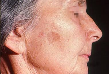
Woman with solar lentigo.
Click to see larger pictureClick to see detailView Full Size Image
Media type:  Photo

Media file 4:  
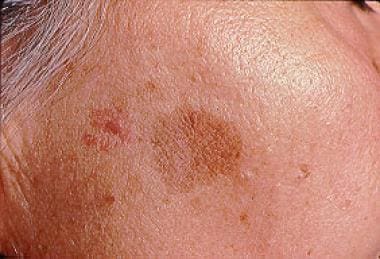
Close-up view of a woman with solar lentigo (same woman as in Media File 3).
Click to see larger pictureClick to see detailView Full Size Image
Media type:  Photo


  1. Derancourt C, Bourdon-Lanoy E, Grob JJ, Guillaume JC, Bernard P, Bastuji-Garin S. Multiple large solar lentigos on the upper back as clinical markers of past severe sunburn: a case-control study. Dermatology. 2007;214(1):25-31. [Medline].
  2. Aoki H, Moro O, Tagami H, Kishimoto J. Gene expression profiling analysis of solar lentigo in relation to immunohistochemical characteristics. Br J Dermatol. Jun 2007;156(6):1214-23. [Medline].
  3. Alper JC, Holmes LB. The incidence and significance of birthmarks in a cohort of 4,641 newborns. Pediatr Dermatol. Jul 1983;1(1):58-68. [Medline].
  4. Kavak A, Parlak AH, Yesildal N, Aydogan I, Anul H. Preliminary study among truck drivers in Turkey: Effects of ultraviolet light on some skin entities. J Dermatol. Mar 2008;35(3):146-50. [Medline].
  5. O'Neill JF, James WD. Inherited patterned lentiginosis in blacks. Arch Dermatol. Sep 1989;125(9):1231-5. [Medline].
  6. Bolognia JL. Reticulated black solar lentigo ('ink spot' lentigo). Arch Dermatol. Jul 1992;128(7):934-40. [Medline].
  7. Roth DE, Hodge SJ, Callen JP. Possible ultraviolet A-induced lentigines: a side effect of chronic tanning salon usage. J Am Acad Dermatol. May 1989;20(5 Pt 2):950-4. [Medline].
  8. Salisbury JR, Williams H, du Vivier AW. Tanning-bed lentigines: ultrastructural and histopathologic features. J Am Acad Dermatol. Oct 1989;21(4 Pt 1):689-93. [Medline].
  9. Williams HC, Salisbury J, Brett J, du Vivier A. Sunbed lentigines. Br Med J (Clin Res Ed). Apr 16 1988;296(6629):1097. [Medline].
  10. Vaidya DC, Schwartz RA, Janniger CK. Nevus spilus. Cutis. Dec 2007;80(6):465-8. [Medline].
  11. Basarab T, Millard TP, McGregor JM, Barker JN. Atypical pigmented lesions following extensive PUVA therapy. Clin Exp Dermatol. Mar 2000;25(2):135-7. [Medline].
  12. Miller RA. Psoralens and UV-A-induced stellate hyperpigmented freckling. Arch Dermatol. Aug 1982;118(8):619-20. [Medline].
  13. Rhodes AR, Harrist TJ, Momtaz-T K. The PUVA-induced pigmented macule: a lentiginous proliferation of large, sometimes cytologically atypical, melanocytes. J Am Acad Dermatol. Jul 1983;9(1):47-58. [Medline].
  14. Peter RU, Gottlober P, Nadeshina N, Krahn G, Plewig G, Kind P. Radiation lentigo. A distinct cutaneous lesion after accidental radiation exposure. Arch Dermatol. Feb 1997;133(2):209-11. [Medline].
  15. Zaballos P, Rodero J, Pastor L, Vives JM, Puig S, Malvehy J. Dermoscopy of lichenoid regressing solar lentigines. Arch Dermatol. Feb 2008;144(2):284. [Medline].
  16. Unver N, Freyschmidt-Paul P, Horster S, Wenck H, Stab F, Blatt T. Alterations in the epidermal-dermal melanin axis and factor XIIIa melanophages in senile lentigo and ageing skin. Br J Dermatol. Jul 2006;155(1):119-28. [Medline].
  17. Leroy D, Dompmartin A, Dubreuil A, Louvet S. Cryotherapy of PUVA lentigines. Br J Dermatol. Dec 1996;135(6):988-90. [Medline].
  18. Ortonne JP, Pandya AG, Lui H, Hexsel D. Treatment of solar lentigines. J Am Acad Dermatol. May 2006;54(5 Suppl 2):S262-71. [Medline].
  19. Bukvic Mokos Z, Lipozencic J, Pasic A, Fattorini I. Laser therapy for solar lentigines: review of the literature and case report. Acta Dermatovenerol Croat. 2006;14(2):81-5. [Medline].
  20. Chan HH, Fung WK, Ying SY, Kono T. An in vivo trial comparing the use of different types of 532 nm Nd:YAG lasers in the treatment of facial lentigines in Oriental patients. Dermatol Surg. Aug 2000;26(8):743-9. [Medline].
  21. Raziee M, Balighi K, Shabanzadeh-Dehkordi H, Robati RM. Efficacy and safety of cryotherapy vs. trichloroacetic acid in the treatment of solar lentigo. J Eur Acad Dermatol Venereol. Mar 2008;22(3):316-9. [Medline].
  22. Sezer E, Erbil H, Kurumlu Z, Tastan HB, Etikan I. A comparative study of focal medium-depth chemical peel versus cryosurgery for the treatment of solar lentigo. Eur J Dermatol. Jan-Feb 2007;17(1):26-9. [Medline].
  23. Atherton DJ, Pitcher DW, Wells RS, MacDonald DM. A syndrome of various cutaneous pigmented lesions, myxoid neurofibromata and atrial myxoma: the NAME syndrome. Br J Dermatol. Oct 1980;103(4):421-9. [Medline].
  24. Barnhill RL, Albert LS, Shama SK, Goldenhersh MA, Rhodes AR, Sober AJ. Genital lentiginosis: a clinical and histopathologic study. J Am Acad Dermatol. Mar 1990;22(3):453-60. [Medline].
  25. Beacham BE. Solar-induced epidermal tumors in the elderly. Am Fam Physician. Jul 1990;42(1):153-60. [Medline].
  26. Ber Rahman S, Bhawan J. Lentigo. Int J Dermatol. Apr 1996;35(4):229-39. [Medline].
  27. Bhawan J, Purtilo DT, Riordan JA, Saxena VK, Edelstein L. Giant and "granular melanosomes" in Leopard syndrome: an ultrastructural study. J Cutan Pathol. 1976;3(5):207-16. [Medline].
  28. Buchner A, Hansen LS. Melanotic macule of the oral mucosa. A clinicopathologic study of 105 cases. Oral Surg Oral Med Oral Pathol. Sep 1979;48(3):244-9. [Medline].
  29. Buchner A, Merrell PW, Hansen LS, Leider AS. Melanocytic hyperplasia of the oral mucosa. Oral Surg Oral Med Oral Pathol. Jan 1991;71(1):58-62. [Medline].
  30. Carney JA, Gordon H, Carpenter PC, Shenoy BV, Go VL. The complex of myxomas, spotty pigmentation, and endocrine overactivity. Medicine (Baltimore). Jul 1985;64(4):270-83. [Medline].
  31. Finan MC, Ray MK. Gastrointestinal polyposis syndromes. Dermatol Clin. Jul 1989;7(3):419-34. [Medline].
  32. Gilchrest BA, Fitzpatrick TB, Anderson RR, Parrish JA. Localization of malanin pigmentation in the skin with Wood's lamp. Br J Dermatol. Mar 1977;96(3):245-8. [Medline].
  33. Gupta G, Williams RE, Mackie RM. The labial melanotic macule: a review of 79 cases. Br J Dermatol. May 1997;136(5):772-5. [Medline].
  34. Hickey JR, Robson A, Barker JN, Smith CH. Does topical tacrolimus induce lentigines in children with atopic dermatitis? A report of three cases. Br J Dermatol. Jan 2005;152(1):152-4. [Medline].
  35. Jones SK, Moseley H, Mackie RM. UVA-induced melanocytic lesions. Br J Dermatol. Jul 1987;117(1):111-5. [Medline].
  36. Jozwiak S, Schwartz RA, Janniger CK. LEOPARD syndrome (cardiocutaneous lentiginosis syndrome). Cutis. Apr 1996;57(4):208-14. [Medline].
  37. Józwiak S, Schwartz RA, Janniger CK, Zaremba J. Familial occurrence of the LEOPARD syndrome. Int J Dermatol. Jan 1998;37(1):48-51. [Medline].
  38. Koch SE, LeBoit PE, Odom RB. Laugier-Hunziker syndrome. J Am Acad Dermatol. Feb 1987;16(2 Pt 2):431-4. [Medline].
  39. Leibowitz E, Janniger CK, Schwartz RA, Lambert WC. Xeroderma pigmentosum. Cutis. Aug 1997;60(2):75-7, 81-4. [Medline].
  40. Lucky AW. Pigmentary abnormalities in genetic disorders. Dermatol Clin. Apr 1988;6(2):193-203. [Medline].
  41. Maize JC. Mucosal melanosis. Dermatol Clin. Apr 1988;6(2):283-93. [Medline].
  42. Martin-Reay DG, Shattuck MC, Guthrie FW Jr. Psammomatous melanotic schwannoma: an additional component of Carney's complex. Report of a case. Am J Clin Pathol. Apr 1991;95(4):484-9. [Medline].
  43. Mehregan AH. Lentigo senilis and its evolutions. J Invest Dermatol. Nov 1975;65(5):429-33. [Medline].
  44. Micali G, Nasca MR, Innocenzi D, Lembo D. Agminated lentiginosis: case report and review of the literature. Pediatr Dermatol. Sep 1994;11(3):241-5. [Medline].
  45. Montagna W, Hu F, Carlisle K. A reinvestigation of solar lentigines. Arch Dermatol. Oct 1980;116(10):1151-4. [Medline].
  46. Pique E, Aguilar A, Farina MC, Gallego MA, Escalonilla P, Requena L. Partial unilateral lentiginosis: report of seven cases and review of the literature. Clin Exp Dermatol. Jul 1995;20(4):319-22. [Medline].
  47. Rees JR, Ross FG, Keen G. Lentiginosis and left atrial myxoma. Br Heart J. Aug 1973;35(8):874-6. [Medline].
  48. Rhodes AR, Melski JW, Sober AJ, Harrist TJ, Mihm MC Jr, Fitzpatrick TB. Increased intraepidermal melanocyte frequency and size in dysplastic melanocytic nevi and cutaneous melanoma. A comparative quantitative study of dysplastic melanocytic nevi, superficial spreading melanoma, nevocellular nevi, and solar lentigines. J Invest Dermatol. May 1983;80(5):452-9. [Medline].
  49. Rhodes AR, Silverman RA, Harrist TJ, Perez-Atayde AR. Mucocutaneous lentigines, cardiomucocutaneous myxomas, and multiple blue nevi: the "LAMB" syndrome. J Am Acad Dermatol. Jan 1984;10(1):72-82. [Medline].
  50. Rhodes AR, Stern RS, Melski JW. The PUVA lentigo: an analysis of predisposing factors. J Invest Dermatol. Nov 1983;81(5):459-63. [Medline].
  51. Roenigk HH Jr. Treatment of the aging face. Dermatol Clin. Apr 1995;13(2):245-61. [Medline].
  52. Roewert HJ, Ackerman AB. Large-cell acanthoma is a solar lentigo. Am J Dermatopathol. Apr 1992;14(2):122-32. [Medline].
  53. Tadini G, D'Orso M, Cusini M, Alessi E. Oral mucosa pigmentation: a new side effect of azidothymidine therapy in patients with acquired immunodeficiency syndrome. Arch Dermatol. Feb 1991;127(2):267-8. [Medline].
  54. Todd MM, Rallis TM, Gerwels JW, Hata TR. A comparison of 3 lasers and liquid nitrogen in the treatment of solar lentigines: a randomized, controlled, comparative trial. Arch Dermatol. Jul 2000;136(7):841-6. [Medline].
  55. Tsao H. Update on familial cancer syndromes and the skin. J Am Acad Dermatol. Jun 2000;42(6):939-69; quiz 970-2. [Medline].
  56. Uhle P, Norvell SS Jr. Generalized lentiginosis. J Am Acad Dermatol. Feb 1988;18(2 Pt 2):444-7. [Medline].
  57. Veraldi S, Cavicchini S, Benelli C, Gasparini G. Laugier-Hunziker syndrome: a clinical, histopathologic, and ultrastructural study of four cases and review of the literature. J Am Acad Dermatol. Oct 1991;25(4):632-6. [Medline].
  58. Voron DA, Hatfield HH, Kalkhoff RK. Multiple lentigines syndrome. Case report and review of the literature. Am J Med. Mar 1976;60(3):447-56. [Medline].
  59. Wilson PD, Kligman AM. Experimental induction of freckles by ultraviolet-B. Br J Dermatol. Apr 1982;106(4):401-6. [Medline].

Lentigo excerpt

Article Last Updated: May 1, 2008