Background
Infantile digital fibromatosis is a benign asymptomatic nodular proliferation of fibrous tissue occurring almost exclusively on the dorsal and lateral aspects of the fingers or the toes, as is shown in the image below. [1] Reye first described infantile digital fibromatosis in 1965 as a recurring digital fibrous tumor. [2]
Pathophysiology
The etiology of infantile digital fibromatosis is unknown. Defective organization of actin filaments in myofibroblasts has been hypothesized. It has been suggested that possible deregulation of the normal bone morphogenetic protein (a member of the transforming growth factor-β superfamily) mediated apoptotic pathway may explain the location of these lesions at the sites of digital septation. Transforming growth factor-β1 also mediates myofibroblast differentiation from fibroblasts. [3, 4]
Epidemiology
Frequency
Infantile digital fibromatosis is rare, with approximately 250 cases reported worldwide.
Sex
Males and females are equally affected by infantile digital fibromatosis.
Age
Most nodules appear in the first few months of life; one third are congenital, and 75-80% are noted during the first year of life. Reports of infantile digital fibromatosis developing in older children and adults are rare.
Prognosis
The prognosis for infantile digital fibromatosis is excellent. Infantile digital fibromatosis is benign, without evidence of malignant transformation or metastases. There may be single or multiple nodules. Infantile digital fibromatosis lesions tend to spontaneously involute without scarring. Rarely, the lesions can cause functional impairment or deformity. Rare cases of ulceration have been reported. The infantile digital fibromatosis lesions tend to grow slowly in the first month, then rapidly grow over about a year, followed by spontaneous resolution over 1-10 years (average 2-3 y). Recurrence is common after excision.
-
Dermal tumor with interlacing spindle-shaped cells and collagen bundles. Perinuclear eosinophilic inclusion bodies are not visible at this magnification.
-
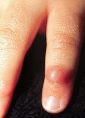
Firm, nontender, erythematous nodule on the fifth finger of a 17-month-old boy.
-
A dermal nodule extending into the subcutaneous fat.


