| Patient Education |
|
Click here for patient education.
|
|
You are in: eMedicine Specialties >
Dermatology > METABOLIC DISEASES
Amyloidosis, Nodular Localized Cutaneous
Article Last Updated: Jan 23, 2007
AUTHOR AND EDITOR INFORMATION
Section 1 of 10
Author: Marion C Miethke, MD, Clinical Assistant Professor, Department of Internal Medicine, Section of Dermatology, University of Washington
Marion C Miethke is a member of the following medical societies: Phi Beta Kappa
Coauthor(s):
Gregory J Raugi, MD, PhD, Professor, Department of Internal Medicine, Division of Dermatology, University of Washington at Seattle; Chief, Dermatology Section, Primary and Specialty Care Service, Veterans Administration Medical Center of Seattle
Editors: C Lisa Kauffman, MD, FACP, Professor, Chief, Division of Dermatology, Departments of Medicine and Pathology, Georgetown University Medical Center; Richard P Vinson, MD, Assistant Clinical Professor, Department of Dermatology, Texas Tech University School of Medicine; Consulting Staff, Mountain View Dermatology, PA; Warren R Heymann, MD, Head, Division of Dermatology, Professor, Department of Internal Medicine, University of Medicine and Dentistry of New Jersey; Catherine Quirk, MD, Clinical Assistant Professor, Department of Dermatology, Brown University; Dirk M Elston, MD, Director, Department of Dermatology, Geisinger Medical Center
Author and Editor Disclosure
Synonyms and related keywords:
amyloidosis cutis nodularis atrophicans, nodular localized cutaneous amyloidosis
Background
Localized cutaneous amyloidosis (LCA) refers to a condition characterized by the deposition of amyloid or amyloidlike proteins in the dermis. LCA encompasses several conditions characterized by amyloid deposition, including macular amyloidosis and lichen amyloidosis (see Amyloidosis, Macular and Amyloidosis, Lichen). Nodular localized cutaneous amyloidosis (NLCA) is the rarest type of LCA and is distinct from the other two.
Gottron first reported NLCA in 1950. Since then, approximately 50 patients have been reported in the North American, European, and Asian literature. This entity also is termed amyloidosis cutis nodularis atrophicans. By definition, NLCA describes a primary disease of the skin, although lesions occasionally appear similar to the skin manifestations of systemic amyloidosis.
Pathophysiology
As a term, "amyloid" was used historically to define proteins that shared similar microscopic characteristics and affinity for certain stains. Research has revealed that "amyloid" proteins are heterogeneous. The various diseases characterized by deposition of "amyloid" proteins are similarly heterogeneous but have in common the deposits of fibrillar proteins characterized as "amyloid" in the dermis. In NLCA, the amyloid is believed to derive from local plasma cells, in contrast to lichenoid or macular amyloidosis, which have keratinocyte-derived amyloid.
In NLCA, plasma cells produce immunoglobulin light chains that are precursors to the amyloid fibril protein(s) termed amyloid L. Reports differ regarding the clonality of this population of plasma cells. In some instances, plasma cells have been monoclonal, suggesting that NLCA is a neoplastic disorder; however, in another instance, plasma cells demonstrated polyclonality, which usually is a feature of a more reactive process.
Frequency
United States
Incidence and prevalence of LCA in the United States are not known; however, the scarcity of reported patients with LCA indicates that the condition may be rare.
International
Despite a paucity of reported patients, LCA, although rare, is represented in the American, Asian, and European literature.
Mortality/Morbidity
NLCA typically is benign and limited to the skin. Reported rates of progression to systemic disease are derived from case series with small numbers of patients; these rates vary from 7% to nearly 50%. Several occurrences have been associated with Sjögren syndrome.
Race
Epidemiologic data can be difficult to establish when so few patients are reported. No specific racial, ethnic, or geographic group appears more prone than another to developing NLCA.
Sex
Of the first 13 patients described in the Japanese literature, 12 were women; however, this disproportionate ratio has not been seen consistently. In a subsequent series of 12 patients, the male-to-female ratio was equal.
Age
Patients reportedly range in age from 33-86 years. Although numbers are small, reports indicate that NLCA is likely to occur during adulthood.
History
- NLCA lesions usually are asymptomatic.
- Patients can present with single or multiple lesions.
- In some reports, lesions were present for several years before patients sought medical attention.
- Troublesome aspects of NLCA primarily result from patient concerns about appearance, although plaques eventually fissured in one patient in whom the plantar aspects of the feet were affected.
- Several cases of this rare disease have been reported in patients with Sjögren syndrome.
Physical
- Firm nodules can present anywhere on the skin, including the face, scalp, extremities, trunk, and genitalia.
- Nodules vary from a few millimeters to a few centimeters.
- Nodules appear pink to brown or red.
- Overlying epidermal atrophy has been described.
- Other terms that describe the various lesions of NLCA include waxy, purpuric, yellowish, or bullous.
- Lesions tend not to ulcerate.
- NLCA lacks extracutaneous findings by definition; however, one patient reported to have NLCA had amyloid deposits in the rectum.
- Macroglossia, a typical feature of systemic amyloidosis, is not seen in NLCA.
Causes
The cause of NLCA is not known, although amyloid is derived from a localized infiltrate of plasma cells.
Colloid Milium
Leiomyoma
Pretibial Myxedema
Pseudolymphoma, Cutaneous
Lab Studies
- Normal serum protein electrophoresis and urine protein electrophoresis studies exclude multiple myeloma.
- Positive antinuclear, anti-Ro, and anti-La antibodies indicate Sjögren syndrome.
- Laboratory studies, such as CBC, serum chemistry profile, and liver function tests often were part of a general workup in several case reports of patients with NLCA. NLCA does not cause any abnormal findings in these studies.
- Urinalysis or 24-hour urine testing can be performed to check for protein. Proteinuria is not a feature of localized cutaneous disease but can be seen in systemic amyloidosis.
Imaging Studies
- In some patients, imaging studies have included chest radiography, ECG, and abdominal ultrasonography.
- Screening for amyloid within organs can be accomplished using scintigraphy with radioiodinated serum amyloid P component (ie, SAP scanning).
Procedures
- Skin biopsy provides the definitive diagnosis. No special tissue preparation or handling is required before delivering the specimen to the laboratory. Special stains and immunohistochemistry are helpful.
- Sites for an optimal biopsy include the epidermis, papillary dermis, and reticular dermis. The amyloid in NLCA is located in the reticular dermis and clearly differentiates NLCA from other forms of amyloidosis. A shave biopsy or other superficial sample may not include enough reticular dermis to complete the diagnosis.
- Consider bone marrow biopsy with gene rearrangement studies (if available) to exclude multiple myeloma.
Histologic Findings
Despite their biochemical heterogeneity, all "amyloid" deposits demonstrate a similar light microscopic appearance. They are eosinophilic and homogeneous when stained with hematoxylin and eosin and viewed with standard optics. When stained with Congo red and viewed with polarized light, deposits exhibit a characteristic and diagnostic green birefringence. Clinical morphology reflects underlying histology. Amyloid is not limited to the papillary dermis but is present in the entire dermis and in subcutaneous fat (unlike lichenoid or macular types) and may be seen within the walls of small blood vessels (see Media Files 1-3). Plasma cells, which most likely produce the amyloid, occur within an adjacent and intermingled inflammatory infiltrate. They can be sparse or numerous (similar plasma cell infiltrate occurs in nodular pulmonary amyloidosis but usually is absent in cutaneous lesions of primary systemic amyloidosis). When eosinophilic amyloid material is exposed to potassium permanganate prior to staining with Congo red, the amyloid retains its congophilia, similar to systemic amyloidosis but in contradistinction to secondary amyloidosis. When viewed with a transmission electron microscope, the apparently homogeneous deposits of amyloid are composed of loosely interwoven 6- to 10-nm–thick straight filaments. The amino acids of the filament proteins are arranged in a characteristic beta-pleated sheet tertiary structure. Amyloid deposits in the skin also contain small amounts of a plasma-derived, nonfibrillar, amyloid-P protein.
Medical Care
Various methods attempt to improve the appearance of the lesions, including topical and intralesional corticosteroids, cryotherapy, dermabrasion, shaving, curettage and electrodesiccation, carbon dioxide laser, and pulsed dye laser. Topical and intralesional corticosteroids and cryotherapy usually are not helpful. One attempt at cryotherapy produced pinpoint bleeding.
Surgical Care
- Procedures such as excision and curettage and electrodesiccation have provided satisfactory cosmetic results.
- Laser treatment has been described.
- Excessive tissue friability and difficulty with intraoperative hemostasis were described while treating one nasal lesion with carbon dioxide laser; however, a good cosmetic result was achieved.
- A patient treated with a tunable dye laser had a good result, and clinical improvement was maintained over 6 months.
- None of these treatment methods totally eradicates lesions, which can recur.
Complications
- Rarely, patients with NLCA develop multiple myeloma years later, suggesting the need for follow-up care.
Prognosis
- NLCA typically is benign and limited to the skin. Progression to systemic disease is unlikely. A recent review article reports that fewer than 15% of patients later developed systemic amyloidosis.
Medical/Legal Pitfalls
- Since NLCA is rare, recognizing that the disease exists is key. Perform or refer patients for adequate biopsy. Once the diagnosis is established, testing for multiple myeloma with serum and urine protein electrophoresis is prudent. Do not confuse generally localized disease with systemic amyloidosis, although the possibility of developing systemic disease is remote.
| Media file 1:
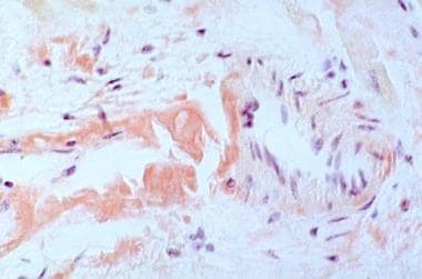
The bright pink homogeneous-appearing material seen is amyloid stained with Congo red. A distinguishing feature of amyloid in the skin is an affinity to take up Congo red stain. |
 | View Full Size Image | |
Media type: Photo
|
| Media file 2:
Amyloid shows apple green when examined with polarized light. |
 | View Full Size Image | |
Media type: Photo
|
| Media file 3:
This transmission electron micrograph of amyloid deposited in the tissue shows loosely interwoven straight filaments. |
 | View Full Size Image | |
Media type: Photo
|
- Alster TS, Manaloto RM. Nodular amyloidosis treated with a pulsed dye laser. Dermatol Surg. Feb 1999;25(2):133-5. [Medline].
- Bozikov K, Janezic T. Excision and split thickness skin grafting in the treatment of nodular primary localized cutaneous amyloidosis. Eur J Dermatol. May-Jun 2006;16(3):315-6. [Medline].
- Breathnach SM. Amyloid and amyloidosis. J Am Acad Dermatol. Jan 1988;18(1 Pt 1):1-16. [Medline].
- Carroll CB, Collison DW, Rodman OG Jr. Atrophic outpouchings of abdominal skin. Nodular cutaneous amyloidosis. Arch Dermatol. Feb 1996;132(2):223-4, 226-7. [Medline].
- Grunewald K, Sepp N, Weyrer K, et al. Gene rearrangement studies in the diagnosis of primary systemic and nodular primary localized cutaneous amyloidosis. J Invest Dermatol. Oct 1991;97(4):693-6. [Medline].
- Hagari Y, Mihara M, Hagari S. Nodular localized cutaneous amyloidosis: detection of monoclonality of infiltrating plasma cells by polymerase chain reaction. Br J Dermatol. Oct 1996;135(4):630-3. [Medline].
- Hamzavi I, Lui H. Excess tissue friability during CO2 laser vaporization of nodular amyloidosis. Dermatol Surg. Sep 1999;25(9):726-8. [Medline].
- Helm TN, Danziger J, Helm KF. Bilateral plantar amyloidosis: a unique presentation of localized cutaneous amyloidosis. Cutis. Mar 1997;59(3):142-4. [Medline].
- Hicks BC, Weber PJ, Hashimoto K, et al. Primary cutaneous amyloidosis of the auricular concha. J Am Acad Dermatol. Jan 1988;18(1 Pt 1):19-25. [Medline].
- Horiguchi Y, Takahashi C, Imamura S. A case of nodular cutaneous amyloidosis. Amyloid production by infiltrating plasma cells. Am J Dermatopathol. Feb 1993;15(1):59-63. [Medline].
- Huilgol SC, Ramnarain N, Carrington P,et al. Cytokeratins in primary cutaneous amyloidosis. Australas J Dermatol. May 1998;39(2):81-5. [Medline].
- Inazumi T, Hakuno M, Yamada H, et al. Characterization of the amyloid fibril from primary localized cutaneous nodular amyloidosis associated with Sjogren''s syndrome. Dermatology. 1994;189(2):125-8. [Medline].
- Lien MH, Railan D, Nelson BR. The efficacy of dermabrasion in the treatment of nodular amyloidosis. J Am Acad Dermatol. Feb 1997;36(2 Pt 2):315-6. [Medline].
- Masuda C, Hayashi M, Kameda Y, et al. Protein AL origin in amyloidosis cutis nodularis atrophicans. J Dermatol. Aug 1986;13(4):280-4. [Medline].
- Masuda C, Mohri S, Nakajima H. Histopathological and immunohistochemical study of amyloidosis cutis nodularis atrophicans--comparison with systemic amyloidosis. Br J Dermatol. Jul 1988;119(1):33-43. [Medline].
- Mun KS, Pailoor J, Reddy SC. Primary localised deep cutaneous amyloidosis of the eyelid. Malays J Pathol. Dec 2005;27(2):113-5. [Medline].
- Northcutt AD, Vanover MJ. Nodular cutaneous amyloidosis involving the vulva. Case report and literature review. Arch Dermatol. Apr 1985;121(4):518-21. [Medline].
- Srivastava M. Primary cutaneous nodular amyloidosis in a patient with Sjogren's syndrome. J Drugs Dermatol. Mar 2006;5(3):279-80. [Medline].
- Touart DM, Sau P. Cutaneous deposition diseases. Part I. J Am Acad Dermatol. Aug 1998;39(2 Pt 1):149-71; quiz 172-4. [Medline].
- Trau H, Shpiro D, Schewach-Millet M, et al. Nodular cutaneous amyloidosis. Am J Dermatopathol. Aug 1991;13(4):414-7. [Medline].
- Truhan AP, Garden JM, Roenigk HH Jr. Nodular primary localized cutaneous amyloidosis: immunohistochemical evaluation and treatment with the carbon dioxide laser. J Am Acad Dermatol. Jun 1986;14(6):1058-62. [Medline].
- Vestey JP, Tidman MJ, Mclaren KM. Primary nodular cutaneous amyloidosis--long-term follow-up and treatment. Clin Exp Dermatol. Mar 1994;19(2):159-62. [Medline].
- Yoneyama K, Tochigi N, Oikawa A, et al. Primary localized cutaneous nodular amyloidosis in a patient with Sjogren's syndrome: a review of the literature. J Dermatol. Feb 2005;32(2):120-3. [Medline].
Amyloidosis, Nodular Localized Cutaneous excerpt Article Last Updated: Jan 23, 2007
|