Practice Essentials
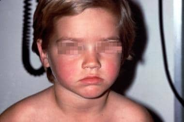
Erythema infectiosum (also known as fifth disease) is usually a benign childhood condition characterized by a classic slapped-cheek appearance (see the image below) and lacy exanthem. [1] It results from infection with human parvovirus (PV) B19, an erythrovirus. [2, 3]
See 15 Back-to-School Illnesses You Should Know, a Critical Images slideshow, to help identify conditions that may occur in young patients after they return to the classroom.
Signs and symptoms
Mild prodromal symptoms begin approximately 1 week after exposure to PV-B19 and last 2-3 days. They include the following:
-
Headache
-
Fever
-
Sore throat
-
Pruritus
-
Coryza
-
Abdominal pain
-
Arthralgias
These symptoms precede a symptom-free period of about 7-10 days, after which the infection progresses through the following stages:
-
Phase 1 - The exanthem begins with the classic slapped-cheek appearance, which typically fades over 2-4 days [1]
-
Phase 2 - This phase occurs 1-4 days later and is characterized by an erythematous maculopapular rash that fades into a classic lacelike reticular pattern as confluent areas clear [4]
-
Phase 3 - Frequent clearing and recurrences for weeks or occasionally months may occur due to stimuli such as exercise, irritation, stress, or overheating of the skin from sunlight or bathing in hot water
See Clinical Presentation for more detail.
Diagnosis
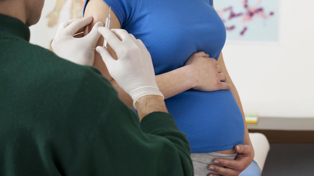
The diagnosis of erythema infectiosum usually is based on clinical presentation alone, and a workup for patients with the classic presentation is not necessary. For patients with other signs or symptoms associated with human parvovirus (PV) B19 or for exposure in a woman who is pregnant, confirmation of infection may be helpful and can be accomplished with the following specialized tests [5, 6, 7] :
-
IgM assays - Enzyme-linked immunosorbent assay (ELISA), radioimmunoassay (RIA)
-
Dot blot hybridization
-
Polymerase chain reaction (PCR) assay
-
Loop-mediated isothermal amplification
See Workup for more detail.
Management
Because erythema infectiosum most often is a benign, self-limited disease, reassuring the parents of children with the condition often is the only intervention necessary. [8] Symptomatic relief of erythema infectiosum may be provided using nonsteroidal anti-inflammatory drugs (NSAIDs) to relieve fever, malaise, headache, and arthralgia, along with topical antipruritics and antihistamines (which also relieve pruritus). Treatment also includes plenty of fluids and rest.
See Treatment and Medication for more detail.
Background
Erythema infectiosum (also known as fifth disease) is usually a benign childhood condition characterized by a classic slapped-cheek appearance and lacy exanthem. [1] It results from infection with human parvovirus (PV) B19, an erythrovirus. Human PV-B19 also is associated with other hematologic, rheumatologic, and neurologic conditions, including polyarthropathy, aplastic anemia, and hydrops fetalis. (See Etiology, Pathophysiology, and History and Physical Examination.)
In erythema infectiosum, a classic 3-phased cutaneous eruption follows a rarely noticed prodrome (see the image below). [9]
Pathophysiology
Human PV-B19, a member of the family Parvoviridae, is a heat-stable, single-stranded DNA virus [10] ; it is the only parvovirus known to cause disease in humans.
The development of erythema infectiosum in children is a normal response to infection by PV-B19. Acute infection in a host who is immunocompetent leads to a Th-1–mediated cellular immune response, with the production of specific immunoglobulin M (IgM) antibodies and subsequent formation of immune complexes. Clinical signs and symptoms of erythema infectiosum probably result from the deposition of the immune complexes in the skin and joints of individuals with this condition and not from the circulating virus. [11] The incubation period is usually 7-10 days, but it can be 4-21 days. Extremely high viral loads are noted in patients with aplastic crisis, but low-level persistent polymerase chain reaction positivity in tissues is also common among adults in a variety of tissues. [12, 13]
Arthropathy
Arthropathy is observed most commonly in adult women and occurs in fewer than 10% of children. It is a symmetrical polyarthritis, usually involving finger joints. The onset of joint symptoms occurs 2-3 weeks after exposure.
Aplastic anemia
The association of human PV-B19 with aplastic anemia is thought to be due to the affinity and cytotoxicity of the virus for erythroid progenitor cells. This complication is primarily observed in patients with underlying hemolytic anemias (eg, sickle cell disease, thalassemia) or immunodeficiency states (eg, leukemia, HIV). [14] These disease states depend on high red blood cell (RBC) production, due to shortened cell life span. The hematocrit of these patients may drop as much as 10-15% per day during acute infection. Most patients have a human PV-B19 aplastic crisis only once, and a rash following aplastic crisis is rare.
Bone marrow suppression
Previously healthy patients also develop transient (and usually clinically insignificant) bone marrow suppression and reticulocytopenia. In rare cases, mild lymphopenia, neutropenia, and thrombocytopenia also may occur.
In-utero exposure
Fetal transmission may result in severe anemia with resultant congestive heart failure and fetal hydrops. This occurs in fewer than 10% of primary maternal infections. Studies report a 1-9% risk of fetal death in pregnant women exposed to active human parvovirus infection, with a greater risk of fetal loss in early pregnancy. [15] Approximately one half of women of childbearing age are seropositive; therefore, they are immune and are of no risk to the fetus. No evidence suggests specific congenital malformations due to in-utero exposure to human PV-B19. [16, 17]
Additional morbidities
Associations between human PV-B19 infection and encephalitis, neuropathies, myocarditis, nephritis, systemic lupus erythematosus, Henoch-Schönlein purpura, pseudoerysipelas-like eruption, and rheumatoid arthritis have been described. Many of these associations, however, have not been validated. [18, 19]
Etiology
Erythema infectiosum is caused by infection with PV-B19, a member of the Parvoviridae family that dates back 7000 years in human dental and skeletal genomic studies. [20] PV-B19, the virus with the smallest DNA known to cause illness in humans, consists of a single-stranded DNA core surrounded by an unenveloped icosahedral capsid. PV-B19 requires mitotically active cells and a globoside cellular receptor for propagation, thus making erythroid cell lines a prime target. Erythroid cell line suppression usually lasts up to 2 weeks, although in some cases it is chronic, lasting months to years. [21]
The tropism for human erythroid progenitor cells and other rare sites of the globoside receptor (eg, endothelial cells, placental cells) is responsible for the more serious complications associated with the viral infection. [22, 23]
Transmission of human PV-B19 occurs through respiratory secretions, possibly through fomites, and parenterally via vertical transmission from mother to fetus and by transfusion of blood or blood products.
PV-B19 and HIV
In a Taiwanese study of the transmission of PV-B19 in men infected with HIV-1/AIDS, Lee et al found that HIV-1 ̶ seropositive men who were injection drug users (IDUs) were significantly more likely to be infected with PV-B19 than were seropositive men who had sex with men (MSM). Using serum samples from 553 male IDUs and 231 MSM, a portion of whom had HIV-1/AIDS, the investigators found PV-B19 infection in 35.4% of the HIV-1 ̶ positive members of the MSM group and in 78.8% of the HIV-1 ̶ positive members of the IDU group. [24]
Epidemiology
Occurrence in the United States
Although sporadic cases of erythema infectiosum occur, outbreaks are more common. Up to 60% of the population is seropositive for anti ̶ human PV-B19 IgG by age 20 years. The incidence peaks in winter and early spring. [7] Human PV-B19 epidemics appear to occur in a cyclical fashion every 4-7 years and are estimated to affect 30-50% of US households. Community epidemics usually last 3-6 months. Subclinical infections are common. Adults with occupational exposures to children, such as teachers or daycare workers, have a higher risk for PV-B19 seroconversion compared with the general population. [25]
International occurrence
Erythema infectiosum occurs worldwide, especially in temperate climates, with various strains implicated. [26] Antiparvovirus IgG is found equally among Americans, Asians, and Europeans. [27]
Worldwide, epidemics of erythema infectiosum tend to occur in the late winter or early spring, with incidence peaking cyclically every 4-7 years. Approximately 60% of adults are seropositive for PV-B19 by age 20 years. Infection rates vary from 20-50% in schools and households during outbreaks. [28, 29, 30]
Sex-related demographics
Males and females are infected equally by erythema infectiosum, although arthropathy is more common in women. In addition, women may be affected by complications from erythema infectiosum during pregnancy. [31, 32]
Age-related differences in incidence
Approximately 70% of erythema infectiosum cases occur in children aged 5-15 years, but the disease can develop at any age. [33] PV-B19 infection can lead to the classic symptoms of erythema infectiosum in adults but more often manifests as an acute arthropathy without cutaneous eruption.
Prognosis
Erythema infectiosum (fifth disease) is a self-limited illness that resolves without complications or sequelae in its classic childhood form. Infection in adults, hosts who are immunocompromised, and patients who are anemic or pregnant can result in more significant morbidity.
Aplastic crisis
Human PV-B19 infects erythroid cells, causing a reticulocytopenia that lasts 7-10 days. A healthy host experiences no consequences, since the normal life span of an RBC is 120 days. In patients with a background of shortened RBC survival, such as hemolytic anemia, an acute aplastic crisis ensues. At-risk patients include those with any of the following conditions:
-
Sickle cell anemia
-
Hereditary spherocytosis
-
Thalassemia
-
Glucose-6-phosphate dehydrogenase deficiency
-
Pyruvate kinase deficiency
-
Autoimmune hemolytic anemia
The incidence of human PV-B19–induced aplastic crisis in patients with chronic hemolytic anemia is 2-5% per year. Patients with aplastic crisis continue to be viremic and infectious until RBC recovery occurs.
Chronic bone marrow failure
In patients who are immunocompromised and have little defense against PV-B19, a prolonged viremia may occur, affecting all cell lines of the bone marrow. Immunodeficient states that can lead to bone marrow failure include the following:
-
HIV infection
-
Congenital immunodeficiency syndromes
-
Acute lymphocytic leukemia [14]
-
Immunosuppressive or cytotoxic therapy
Congenital infection
PV-B19 can cross the placenta during pregnancy and have a direct cytotoxic effect on fetal RBCs. [34] Infection may lead to the following:
-
Severe anemia
-
Congestive heart failure
-
Hydrops fetalis - PV-B19 is responsible for 10-15% of cases
-
Intrauterine death (miscarriage or stillborn) - In 3-10% of mothers who are infected
Rash
The rash of erythema infectiosum usually is self-resolving but may last several weeks or months with exacerbations from heat or sunlight. The onset of erythema infectiosum rash usually indicates that reticulocytosis has returned and aplastic crisis will not occur.
Papular-purpuric gloves-and-socks syndrome
This is an acute, self-limited exanthem with fine, palpable purpura usually located on the hands and feet, with sharp demarcation at the wrists and ankles. The eruption may be accompanied by fever and aphthous ulcers and occurs more commonly in adults. Rarely, in an acropetechial variant, this eruption can involve the perioral and chin area. [35, 36, 37, 38]
Arthralgias/arthropathies
These occur in up to 10% of pediatric patients and up to 50% of adult patients. Arthropathy usually lasts 2-4 weeks, but on rare occasions it can last months to years.
Additional morbidities
Other illnesses that occasionally may be linked to or triggered by PV-B19 include the following:
-
Viral-associated hemocytophagia
-
Rheumatoid arthritis/chronic polyarthritis mimicking rheumatoid arthritis [39]
-
Systemic sclerosis
-
Systemic lupus erythematosus
-
Autoimmunelike pulmonary disease [40]
-
Idiopathic thrombocytopenic purpura
-
Diamond-Blackfan–like anemia
-
Acute vasculitic syndromes
-
Atypical and nonspecific erythematous exanthem
-
Myocarditis/pancarditis [41]
-
Uveitis
-
Glomerulonephritis/nephrotic syndrome [46]
-
Sepsislike syndrome [47]
-
Bullous eruption [48]
Patient Education
Infected children with hemolytic disease or immunosuppression may be quite infectious. Therefore, respiratory isolation, especially from pregnant, chronically anemic, or immunosuppressed individuals, should be observed. Good handwashing and infection control techniques should be encouraged.
Emphasize in discussion with parents that otherwise healthy patients with erythema infectiosum are not infectious once the rash appears; therefore, they do not need to be isolated or restricted from school/day care.
For patient education information, see the Children's Health Center, as well as Fifth Disease and Skin Rashes in Children.
-
Classic slapped-cheek appearance of fifth disease.
-
Pathognomonic reticulated, lacy-appearing eruption of fifth disease.