Background
Erythema gyratum repens (EGR) is a figurate erythema that is believed to be a paraneoplastic process. [1, 2, 3, 4] In addition to other features, characteristic concentric erythematous bands forming a wood-grain appearance (see the image below) help distinguish erythema gyratum repens from other figurate erythemas, such as erythema annulare centrifugum, erythema migrans, and erythema marginatum. [5]
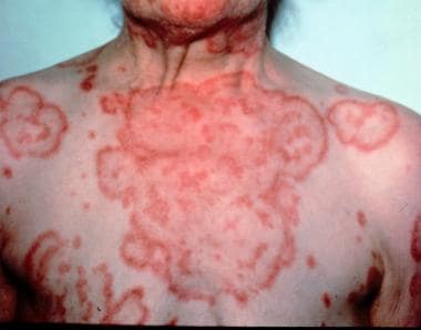 Erythema gyratum repens. The characteristic wood-grain pattern is composed of annular, erythematous concentric bands lined by a trailing edge of scale.
Pathophysiology
The pathogenesis of erythema gyratum repens remains unknown. The following hypotheses have been proposed:
-
First, tumor antigens may form and cross-react with endogenous skin antigens.
-
Second, tumor products may alter endogenous skin antigens making them susceptible to autoimmune recognition.
-
Third, tumor antigens may form immune complexes with antibodies, which are then deposited into cutaneous tissue.
A mechanism explaining the clinically apparent migrating erythema also has been proposed. This model involves a localized ground substance phenomenon. Granulocytes release factors that stimulate proliferating fibroblasts, producing ground substance with increased viscosity. This viscous ground substance serves to impede or "wall off" the tissue spread of inflammatory mediators. In erythema gyratum repens, the advancing erythema may represent the advancement of inflammatory mediators through stroma that is unable to keep them walled off.
Another hypothesis suggests that glutamine contributes to the unusual skin patterns seen in erythema gyratum repens, given the evidence of elevated glutamine levels in the body as a consequence of tumor activity and glutamine self-assembled ring formations when in aqueous solutions. [6]
Etiology
Erythema gyratum repens is associated with malignancy in as many as 80% of patients. Among visceral malignancies, the lung is the most common site, [7, 8, 9] followed by the breast, urinary bladder, uterus and/or cervix, GI tract (stomach), and prostate. [10] Erythema gyratum repens has also been associated with anal cancer. [11] Most patients with erythema gyratum repens develop the eruption before the symptoms of tumor. The time interval between the eruption of erythema gyratum repens and the detection of the tumor can range from simultaneous presentation to up to 6 years after the rash. Erythema gyratum repens also has occurred up to 7 months after the detection of malignancy.
Erythema gyratum repens is associated with some nonneoplastic conditions, such as pulmonary tuberculosis, cryptogenic organizing pneumonia, [12] lupus erythematosus, CREST (calcinosis, Raynaud phenomenon, esophageal motility disorder, sclerodactyly, and telangiectasia) syndrome, virginal breast hypertrophy, pityriasis rubra pilaris, [13, 14, 15] psoriasis, [16] and as a drug reaction to azathioprine in a patient with type I autoimmune hepatitis. [17] In a few patients, no associated conditions exist. [18]
Epidemiology
Frequency
Erythema gyratum repens is believed to be rare. A clinical review in 1992 by Boyd cited 49 patients reported in the literature. [19] A current literature search yielded a handful of additional case reports.
Race
Erythema gyratum repens reportedly occurs predominantly in white persons.
Sex
Male-to-female ratio is 2:1.
Age
Erythema gyratum repens usually occurs in patients older than 40 years, with a mean age of 63 years, but it has been reported to occur from age 16-77 years. [20]
Prognosis
The prognosis for erythema gyratum repens depends on the underlying illness. In most patients, symptoms disappear with the resolution of underlying disease. No specific complications are associated with the skin manifestations of erythema gyratum repens, and the condition alone does not lead to death. Symptoms include intense pruritus, and morbidity and mortality may occur related to the underlying condition.
-
Erythema gyratum repens. Courtesy of Jeffrey P. Callen, MD.
-
Erythema gyratum repens. The characteristic wood-grain pattern is composed of annular, erythematous concentric bands lined by a trailing edge of scale.


