Background
Erythema ab igne (EAI) is characterized as localized areas of reticulated erythema and hyperpigmentation due to chronic and repeated exposure to infrared radiation. Patients with erythema ab igne have a history of repeated exposures to heat at a lower level than that which causes a thermal burn. [1, 2] Other terms used to describe erythema ab igne include toasted skin syndrome and fire stains. [3]
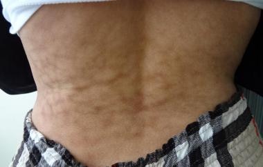
Initially, the skin in erythema ab igne patients is often mildly erythematous; however, after repeated heat exposures, the classic blue, purple, or brown reticulated hyperpigmentation develops. [1] See the image below.
Pathophysiology
Erythema ab igne (EAI) is a reticulate hypermelanosis with erythema resulting from repeated heat exposure that induces injury to the epidermis and superficial vascular plexus. The exposure, which need not be of long duration, results in cutaneous hyperthermia in the range of 43-47°C. Erythema ab igne results in histopathologic changes similar to those seen in solar-damaged skin. Although the pathogenic mechanisms in erythema ab igne are poorly understood, one study has shown that moderate heat acts synergistically with ultraviolet radiation to denature DNA in squamous cells in vitro. [4]
Etiology
Open fires reportedly result in erythema ab igne. Typically, erythema ab igne affects the legs of women aged 40-70 years who use indoor fire as a heat source. Erythema ab igne reportedly affects the face and/or palms of cooks who work over an open fire.
Some patients use a heat source (eg, heating pad, hot water bottle, heated recliner, heated blanket) to relieve chronic pain. [5] In these patients, determine the etiology of the pain. In the case of heating pads and/or hot water bottles, erythema ab igne can occur in patients with pain associated with either primary or metastatic malignancy, as well as with pain associated with chronic pancreatitis. [6, 7, 8] Heated recliners (reclining chairs) have been reported to cause erythema ab igne in patients with chronic lower back pain. [9] The application of heated popcorn kernels applied to the skin to reduce arthritic pain caused erythema ab igne in one patient. [10] One case report describes erythema ab igne in a patient with diabetic neuropathy. [11]
Other heat sources may be involved. Erythema ab igne has been described subsequent to sauna belt usage for abdominal obesity. [12] A car heater reportedly caused erythema ab igne in one patient. [13] More recently, using laptop computers while they are propped on the legs has resulted in the development of erythema ab igne. Some laptop computers can generate significant heat that can result in erythema ab igne when placed on the lap for prolonged periods. [14, 15, 16, 17, 18]
The following is a summary of heat sources reported to cause erythema ab igne [3, 19, 20] :
-
Heating pads
-
Hot water bottles
-
Electric stoves/heater
-
Open fires
-
Coal stoves
-
Peat fires
-
Wood stoves
-
Steam radiators
-
Car heaters
-
Heated reclining chairs
-
Heating or electric blanket
-
Hot bricks
-
Infrared lamps
-
Microwave popcorn
-
Laptop computer
-
Automobile seat heater
-
Hot bathing
Epidemiology
Frequency
United States
Erythema ab igne is rare. Because of the general availability of central heating, erythema ab igne is less common in the United States than in countries where open fires are commonly used for heating.
Historically, erythema ab igne was often seen on the inner thighs and legs of women who sat in front of a stove or open fire. [1, 21] Now erythema ab igne is more commonly related to heating pads or laptop computer use.
International
Currently, erythema ab igne is most commonly seen internationally following repeated use of hot water bottles, infrared lamps, and heating pads. [22, 23] Additionally, chronic pain in the lumbosacral region and consequent repeated and prolonged use of localized heat to relieve those symptoms has led to an increased incidence of erythema ab igne in this area. [24, 25, 26]
Studies have shown that physiotherapeutic treatments use ultrasound and short-wave diathermy to promote (via high-frequency mechanical waves) an extremely rapid vibration in the tissues in order to generate heat and consequent dilation of the local veins to provide pain relief. [27]
Race
Erythema ab igne has no overt racial predisposition.
Sex
Women, in particular those who are overweight, are affected by erythema ab igne more often than men. [1]
Age
Erythema ab igne primarily occurs in adults, usually of middle age (40-70 y).
Prognosis
The prognosis is good, except those cases associated with internal disease or metastatic malignancy. Early changes, such as erythema and little or no hyperpigmentation, may resolve within several months. Chronic and repeated exposure to heat may result in permanent changes such as hyperpigmentation and atrophy. In addition, thermal keratosis, squamous cell carcinoma in situ, and squamous cell carcinoma have been reported within the lesions of erythema ab igne.
Mortality/morbidity
Chronic repeated exposure to infrared radiation may result in changes similar to those seen with chronic repeated ultraviolet radiation. Carcinoma can develop from dysplastic keratinocytes harbored within the reticulated hyperpigmentation. Thermal keratosis, squamous cell carcinoma in situ, and squamous cell carcinoma have been reported in patients after chronic exposure to infrared radiation. [28] In one 90-year-old woman with erythema ab igne, Merkel cell carcinoma developed adjacent to squamous cell carcinoma. Occasionally, the first sign of splenomegaly, pancreatitis, pancreatic cancer, and other cancers is erythema ab igne resulting when patients apply external heat to relieve the underlying pain. [21]
Work exposure to heat (eg, for bakers, silversmiths, and boiler operators) may cause erythema ab igne. [1] Additionally, erythema ab igne has been reported to appear on the legs following prolonged, daily exposure to a car heater.
Patient Education
Explain the etiology of the disorder to patients, and emphasize that the cessation of heat exposure is paramount. Inform patients with erythema ab igne about the possibility of malignant degeneration in the affected areas. Educate patients about detection and the need for prompt treatment.
-
Red rash resembling lacework or a fishing net on leg.
-
Red rash resembling lacework or a fishing net on back.


