Background
In 1962, Headington and French first described trichilemmoma as a benign neoplasm with differentiation toward pilosebaceous follicular epithelium, [1] or outer root sheath. Although unusual, at times, a central zone of desmoplasia may develop and thus be termed desmoplasia trichilemmoma. [2] While benign in nature, the significance of trichilemmoma resides in the association with Cowden disease (ie, multiple hamartoma syndrome), nevus sebaceous, and the need to differentiate trichilemmomas from other more aggressive cutaneous tumors, such as trichilemmal carcinoma.
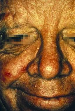
Clinically, trichilemmomas present as well-defined, smooth, asymptomatic papules or verrucoid growths. They may appear as a solitary or multiple lesions, and they usually are found on the head and face (see the image below). These papules often mimic a basal cell carcinoma or a verruca.
Trichilemmomas are often reported in association with several other neoplasms. Trichilemmoma is most commonly found secondary to nevus sebaceous of Jadassohn. [3] . Nevus sebaceous is classified as a congenital cutaneous hamartoma that presents on the scalp or face sometime between birth and childhood development. [2] Several articles have reported trichilemmomas also appearing alongside trichoblastoma, sebaceous adenoma, and syringocystadenoma papilliferum. [4, 5]
When multiple trichilemmomas are present, Cowden disease should be suspected, especially if associated with oral fibromas, goiter, gastrointestinal polyposis, thyroid disease, or a family history of breast cancer.
Pathophysiology
The underlying cause of trichilemmomas is unknown. Because of its histologic similarity to a wart, some researchers have investigated a viral etiology. [6, 7]
Johnson et al [8] performed histologic and ultrastructural analyses of 10 hyperkeratotic lesions on the extremities and 2 keratotic lesions on the face in a patient with Cowden disease. They were unable to find evidence of a viral infection in the tissues examined. Leonardi et al [9] performed a study on 25 trichilemmomas, revealing no evidence of human papillomavirus DNA in the lesions. Most trichilemmomas, therefore, appear to represent a benign tumor with differentiation towards the follicular outer root sheath (trichilemma). [7]
Stierman et al [10] were unable to detect human papillomavirus (HPV)–1, HPV-2, or mixed genital-type HPV infection in trichilemmomas using in situ hybridization. It was suggested that the association between HPV infection and the hair follicle is both one of the permanent sources of HPV DNA and the site of origin for trichilemmomas. [11] The development of trichilemmomas may be independent factors associated with increasing age, rather than HPV being an oncogenic stimulus for trichilemmomas.
Conversely, Rohwedder et al used the polymerase chain reaction (PCR) technique to show 11 cases of HPV-positive trichilemmoma. So far, HPV types 6b, 15, 17, 23, 27, and 28 have been identified in both solitary trichilemmoma lesions and in patients with Cowden syndrome. [11, 12]
Cowden syndrome is a rare autosomal dominant condition characterized by the formation of multiple types of hamartomas and neoplastic growths, which may be found throughout different body systems. This syndrome is thought to be due to the PTEN mutation, a germline mutation in exon 8 of the phosphatase and tensin homolog deleted on chromosome 10. [13] It is a point mutation of C to T at codon 1003 (CGA → TGA, arginine → stop codon). This defect demonstrates incomplete genetic penetrance with variable expressivity. Loss of heterozygosity may lead to tumor formation. [14]
The syndrome is allelic to other PTEN -related syndromes such as Lhermitte-Duclos disease (dysplastic cerebellar gangliocytoma) and Bannayan-Riley-Ruvalcaba syndrome, which is characterized by genital lentigines and hamartomatous growths. Families or patients with overlapping features have been described. [15, 16]
Etiology
The cause of a trichilemmoma is unknown. Because trichilemmoma shares some morphologic and histologic features with a verruca, some researchers have postulated that a virus may induce these lesions. Increased risk has also been found when subjects have a history of long exposure to sunlight. [17]
Epidemiology
Frequency
United States
Trichilemmomas are relatively common benign neoplasms of the follicular epithelium. Their true incidence is hard to determine and is probably underestimated. Approximately 40 cases per 100,000 consecutive skin biopsies may be found every year in any given dermatopathologic laboratory. Unlike isolated trichilemmomas, multiple trichilemmomas associated with Cowden disease are very rare.
International
The international frequency is unknown.
Race
Trichilemmomas may occur in any race. They are most common in white females, but they have also been reported in Chinese, Japanese, and black patients.
Sex
The male-to-female ratio of trichilemmomas is 1:1; however, Cowden disease has a female predominance, with a male-to-female ratio of 1:3.
Age
Trichilemmomas predominantly occur in adult patients aged 20-80 years. However, onset may occur as early as age 4 years, with a median age of onset at 30 years. [14]
Desmoplastic trichilemmomas (a histologic subtype of trichilemmoma) predominantly occur in white men over a wide age range, and the highest frequency is in the fifth decade.
One rare case of a trichilemmoma following a blaschkoid pattern has been noted in a 13-year-old boy. It is the first case seen as such. [18]
Prognosis
Trichilemmomas are benign follicular epithelial neoplasms. Of themselves, trichilemmomas are associated with minimal morbidity and no mortality. These tumors usually need to be differentiated clinically from a verruca or a basal cell carcinoma. The only morbidity associated with these tumors occurs if they are treated as a basal cell carcinoma before histologic confirmation is obtained.
Patient Education
Educate each patient with a trichilemmoma regarding the benign nature of this epithelial neoplasm. Further education regarding its association with Cowden disease may also be provided, particularly if other clinical evidence for Cowden disease exists. Patients should be informed that they are at higher risk for the development of trichilemmoma the more they are subjected to sun exposure. In addition, if nevus sebaceous remains untreated, it has a 10-20% chance of transforming into a malignant form. Appropriate plans and treatment should always be instituted. [17]
-
A patient with trichilemmoma papules on the face.
-
Low-power histologic view of a trichilemmoma.
-
Mid-power histologic view of a trichilemmoma.
-
Low-power histologic view of a desmoplastic trichilemmoma.
-
High-power histologic view of a desmoplastic trichilemmoma.
-
Tumor of the follicular infundibulum.
-
Clinical image of the face of a patient with Cowden syndrome.
-
Clinical image of the oral mucosa of a patient with Cowden syndrome.
-
Clinical image of palmar keratoses in a patient with Cowden syndrome.
-
Clinical image of sclerotic fibroma in a patient with Cowden syndrome.
-
Clinical image of multiple trichilemmomas in a patient with Cowden syndrome.


