Practice Essentials
Acne fulminans (AF), also known as acne maligna, was originally described as acute febrile ulcerative acne conglobata (AC). In 1958, at a meeting of the Detroit Dermatological Society, Burns and Colville presented a 16-year-old white boy with acute febrile disease and acne conglobata. Many similar cases have been reported since then. [1] The primary features of this disease include sudden onset, severe and often ulcerating acne, fever, polyarthritis, [2] and failure to respond to antibacterial therapy; the response to debridement in combination with steroid therapy is good. It can be the dermatologic manifestation of the synovitis-acne-pustulosis-hyperostosis-osteitis (SAPHO) syndrome. [3] Acne fulminans is a syndrome of fulminant, necrotizing acne associated with bone lesions, constitutional symptoms, and laboratory abnormalities. See the image below.
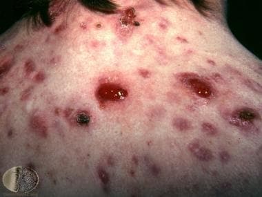 Granulomas and crusted acne lesions in acne fulminans. Courtesy of DermNet New Zealand (http://www.dermnetnz.org/assets/Uploads/doctors/follicular/images/fulmin2.jpg).
See Acne Conglobata, Acne Keloidalis Nuchae, Acne Vulgaris, and Acneiform Eruptions for complete information on these topics.
Prognosis
Recurrent acne fulminans is extremely rare. Bone lesions typically resolve with treatment, but residual radiographic changes, such as sclerosis and hyperostosis, may remain. Scarring and fibrosis may result from this acute inflammatory process.
Diagnostics
Laboratory studies
Findings in patients with acne fulminans include the following:
-
Leukocytosis (characteristic finding)
-
Increased percentage of polymorphonuclear leukocytes
-
Anemia
-
Leukemic-type reaction
-
Elevated erythrocyte sedimentation rate
-
Circulating immune complexes
-
Proteinuria
-
Sterile blood culture results
Imaging studies
Bone involvement is common. Approximately 50% of patients have lytic bone lesions demonstrated on radiographs, and 70% of patients show increased uptake using technetium scintillography ("hot spots"). Destructive lesions resembling osteomyelitis are demonstrated on radiographs in 25% of patients. Multifocal osteolytic cysts may be evident as tender bones and can be detected as hot spots by technetium scintillography.
Histologic findings
Analysis of biopsy specimens of the bony lesions shows reactive changes only.
Treatment
See Oral Steroids and Isotretinoin, Guidelines, and Medication.
Pathophysiology
Acne fulminans is an uncommon, immunologically induced, systemic disease characterized by aggressive inflammation in which the triggering antigen is believed to be from Cutibacterium acnes (formerly Propionibacterium acnes); it is a severe variant of acne vulgaris. Some authors note that elevated blood levels of testosterone may play an important role in the pathogenesis of acne fulminans. High levels of testosterone and anabolic steroids cause an increase in sebum excretion and in the population density of C acnes (formerly P acnes). Acne fulminans could be induced by anabolic steroid use in a male bodybuilder. [4] The trigger for acne induction seemed to be a testosterone therapy in a transgender boy [5] and a patient with Marfan syndrome. [6] . The increase in the amount of C acnes (formerly P acnes) or related antigens may trigger the immunologic reaction in some individuals and lead to an occurrence of acne fulminans. [7] In addition to testosterone, isotretinoin may also precipitate acne fulminans, possibly related to highly increased levels of C acnes (formerly P acnes) antigens in the patient's immune system. [8, 9]
Another theory postulates that acne fulminans may be an autoimmune complex disease because circulating immune complexes have been demonstrated in some patients with acne fulminans. Immunologically, the reaction is a type III or IV hypersensitivity reaction.
Genetic factors may play an important role in some patients; 4 sets of identical twins who developed an identical pattern of acne fulminans have been documented. [10]
Acne may be the only clinical sign of androgen excess in men, and one report is available about a boy with acne fulminans and androgen excess due to late-onset congenital adrenal hyperplasia. [11]
Acne fulminans has also been observed in patients with measles infection. [12]
Chronic inflammation in acne fulminans, characteristic to acne lesions, can also be accompanied by angiogenesis. By specific immunostaining for CD34+ endothelial cells, research revealed the presence of blood capillaries around the pilosebaceous follicles within the inflammatory or pericystic infiltrate. [13]
A young 13-year-old patient who developed acne fulminans after isotretinoin therapy at a dose of 0.1mg/kg/day without associated systemic symptoms was presented [14] .
Consensus recommendations on classification
In 2017, a panel of experts made the recommendation that acne fulminans be classified into 4 distinct clinical variants:
-
Acne fulminans with systemic symptoms (AF-SS)
-
Acne fulminans without systemic symptoms (AF-WOSS)
-
Isotretinoin-induced acne fulminans with systemic symptoms (IIAF-SS)
-
Isotretinoin-induced acne fulminans without systemic symptoms (IIAF-WOSS) [15]
Epidemiology
Acne fulminans is a rare disease. Over the past several years, fewer cases of this disease have occurred, possibly because of earlier and better treatment of acne. Approximately 200 patients with acne fulminans have been described. [16, 17, 14]
Acne fulminans predominantly affects young males aged 13-22 years with a history of acne.
-
Granulomas and crusted acne lesions in acne fulminans. Courtesy of DermNet New Zealand (http://www.dermnetnz.org/assets/Uploads/doctors/follicular/images/fulmin2.jpg).


