Continually Updated Clinical Reference
 
 
  All Sources     eMedicine     Medscape     Drug Reference     MEDLINE
 
eMedicine - Aspergillosis : Article by

Quick Find
Authors & Editors
Introduction
Clinical
Differentials
Workup
Treatment
Medication
Follow-up
Miscellaneous
Acknowledgments
Multimedia
References

Related Articles
Cellulitis

Ecthyma

Ecthyma Gangrenosum


Patient Education
Click here for patient education.


Author: Annie Chiu, MD, Staff Physician, Department of Dermatology, Cedars-Sinai Medical Group

Annie Chiu is a member of the following medical societies: American Academy of Dermatology and Women's Dermatologic Society

Coauthor(s): Alexa F Boer Kimball, MD, MPH, Associate Professor of Dermatology, Harvard University School of Medicine; Director of Clinical Unit for Research Trials in Skin, Associate Dermatologist, Department of Dermatology, Massachusetts General and Brigham and Women's Hospitals

Editors: Peter Fritsch, MD, Chair, Department of Dermatology and Venereology, University of Innsbruck, Austria; David F Butler, MD, Professor of Dermatology, Texas A&M University College of Medicine; Director, Division of Dermatology, Scott and White Clinic; Director Dermatology Residency Training Program, Scott and White Clinic; Jeffrey Meffert, MD, Assistant Clinical Professor of Dermatology, University of Texas Health Science Center-San Antonio; Catherine Quirk, MD, Clinical Assistant Professor, Department of Dermatology, Brown University; Dirk M Elston, MD, Director, Department of Dermatology, Geisinger Medical Center

Author and Editor Disclosure

Synonyms and related keywords: cutaneous aspergillosis, fungal infection, Aspergillus fumigatus, A fumigatus, Aspergillus flavus, A flavus, Aspergillus terreus, A terreus, Aspergillus chevalieri, A chevalieri, Aspergillus niger, A niger, Aspergillus ustus, A ustus

Background

Cutaneous aspergillosis is most commonly a cutaneous manifestation of disseminated infection with the fungus Aspergillus, although primary cutaneous disease can rarely occur, especially in the setting of burns or trauma. Aspergillosis typically begins as a pulmonary infection subsequent to inhalation of fungal spores. In the immunocompromised host, hematogenous dissemination and invasion of other organ systems, including the skin, can then follow the initial pulmonary infection.

Dermatologic manifestations of disseminated aspergillosis include single or multiple erythematous-to-violaceous plaques or papules, often characterized by a central necrotic ulcer or eschar. Skin lesions occur in 5-10% of patients with disseminated aspergillosis.  Infrequently, they may be the presenting sign of systemic infection. Primary cutaneous aspergillosis occurs much less commonly in the absence of hematogenous disease. In these instances, the most typical presentation is implantation of the fungus following trauma, including infections at the site of intravenous cannulas, or venipuncture wounds, especially those that have been covered with occlusive dressings. Aspergillus is a frequent contaminant found in cultures of dystrophic nails, but it can occasionally cause a true onychomycosis.

Pathophysiology

Cutaneous aspergillosis is caused by infection with ubiquitous soil- and water-dwelling saprophytes of the Aspergillus genus. Typically, infection of the pulmonary system occurs via inhalation of fungal spores, which then leads to disseminated hematogenous infection with the organism. Aspergillus fumigatus is the most common pathogen associated with disseminated disease with cutaneous involvement, whereas Aspergillus flavus1 or Aspergillus terreus most often causes the less frequent primary infections of the skin. Aspergillus niger and Aspergillus ustus have also been cultured from cutaneous lesions. Infections of the sinuses, the lungs, or the skin can lead to disseminated disease, especially in patients who are immunocompromised.

Frequency

United States

Aspergillosis is the second most common opportunistic fungal infection in patients who are immunocompromised, accounting for as many as 20% of fungal infections in patients who have received bone marrow or solid organ transplants. Key risk factors include neutropenia from hematologic malignancy or chemotherapy, immunosuppressive therapy, AIDS, and cytomegalovirus infection. 

Mortality/Morbidity

Disseminated aspergillosis is associated with a mortality rate of greater than 90%. In contrast, multiple case reports have documented resolution of primary cutaneous aspergillosis after surgical excision followed by, in some cases, systemic antifungal therapy.

Sex

No clear sexual predilection is reported.

Age

Neonates occasionally develop disseminated disease, presumably because of their immature immune mechanisms.


History

Patients with disseminated aspergillosis often present with febrile illness, pneumonia, or sinusitis unresponsive to antibiotics. Other possible presentations include the following:

    • Gradual onset of central nervous system symptoms
    • Endocarditis
    • Myocarditis
    • Esophageal and intestinal ulcers

Physical

The pertinent physical findings below are limited to the skin examination.

  • Cutaneous presentations of systemic aspergillosis most frequently begin as solitary or multiple erythematous or violaceous indurated papules or plaques. The lesions are often tender, but they can be asymptomatic. These manifestations evolve rapidly into centrally placed pustules, hemorrhagic vesicles, and, ultimately, black eschars. Involved areas can be large or small, can be localized or diffuse, and most often arise on the limbs and head. Truncal lesions are much less common.
  • Primary cutaneous infections usually develop at the sites of an intravenous catheter or a venipuncture. Reports have also associated the use of occlusive dressings, armboards, nonsterile gauze, plaster casts, and adhesive tape with primary cutaneous aspergillosis.
  • Skin lesions often initially appear as a localized cellulitis that develops into the typical necrotic ulcer or a black eschar.
  • Aspergillus chevalieri has been reported to cause a distinct skin lesion that is more hyperkeratotic and vesiculopapular in nature.

Causes

Aspergillosis is an uncommon disease in patients who are not immunocompromised because normal neutrophilic and macrophagic functions prevent infection; however, any deficiency in these immunologic parameters increases the risk of aspergillosis. For example, systemic corticosteroid therapy is a known risk factor for cutaneous aspergillosis.

Some environmental risk factors have also been implicated; these factors include construction sites and contaminated ventilation systems, presumably caused by effects on spore distribution.


Cellulitis
Ecthyma
Ecthyma Gangrenosum

Other Problems to be Considered

Fusarium infection
Mucormycosis
Cryptococcosis
Phaeohyphomycosis


Lab Studies

Findings from skin biopsy with special staining for fungus, such as with periodic acid-Schiff or methenamine silver stain, can be supportive or suggestive of Aspergillus infection, but other fungi may appear nearly identical in histopathologic sections.

Tissue, sputum, or blood culturing is usually performed, but the results may be negative or unreliable because Aspergillus is a common laboratory contaminant. If truly present in tissue, Aspergillus is a rapidly growing fungus that can be isolated in culture within 1-3 days, but longer incubation times may be required if the inoculum is very small.

In disseminated disease, the serum galactomannan assay can be used in conjunction with cultures and/or histologic examination. Galactomannan, an Aspergillus cell wall constituent, can be detected by enzyme-linked immunosorbent assay with an approximate sensitivity of 81% and specificity of 89%.2

Imaging Studies

Chest radiography can help in diagnosing a primary pulmonary infection, or it can confirm the presence of a fungal ball. CT scanning or MRI may help reveal fungal abscesses in the brain.

Histologic Findings

In tissue sections, narrow septate hyphae with delicate chitinous walls, bubbly blue cytoplasm, and acute-angle branching can be demonstrated, especially with special staining. Keep in mind that on sectioning, other fungi may appear to have acute-angle branching and Aspergillus species may appear to have more of a right-angle branching. Primary cutaneous infection often involves a granulomatous reaction in the dermis and the formation of multinucleated giant cells. Hematogenous disease is usually characterized by numerous branching hyphae surrounding and, occasionally, eroding into blood vessel lumens.


Medical Care

In both disseminated and limited cutaneous disease, high-dose intravenous amphotericin B has been the traditional antifungal used to eradicate the underlying organism. However, voriconazole is also approved as a first-line agent and is being used with increased frequency. Other treatment options include itraconazole and caspofungin.3, 4 For Aspergillus-induced onychomycosis, treatment is with oral itraconazole because topical medications have very limited efficacy in eradicating fungus from the nail.

Surgical Care

Several case reports have documented the effectiveness of surgical excision in the treatment of primary cutaneous aspergillosis. Some of the patients also received concurrent or subsequent systemic antifungal therapy.

Consultations

  • Consult a dermatologist for diagnosis, excision, and wound care.
  • Consult an infectious diseases specialist for treatment recommendations in the setting of systemic disease.


High-dose intravenous amphotericin B has traditionally been used to eradicate the underlying organism. However, voriconazole has been approved as a first-line agent in the treatment of invasive aspergillosis and is available in both parenteral and oral formulations. A multicenter, randomized, open-label trial comparing the efficacy of voriconazole to amphotericin B demonstrated better response rates, improved survival, and fewer severe adverse effects in those receiving voriconazole therapy.5

Itraconazole, posaconazole, caspofungin, terbinafine, and micafungin have also been reportedly effective in some cases. The length of treatment varies in the literature, but treatment is likely to be most effective if prolonged. Recent studies show posaconazole as an alternative treatment for invasive aspergillosis in patients previously resistant to or intolerant of other antifungal therapies.6 Combination antifungal therapies have also been used in patients with disease that is associated with a greater degree of treatment resistance.

Drug Category: Antifungal agents

The mechanism of action may involve an alteration of RNA and DNA metabolism or an intracellular accumulation of peroxide that is toxic to the fungal cell.

Drug NameAmphotericin B (Amphocin, Fungizone)
DescriptionPolyene antibiotic produced by a strain of Streptomyces nodosus; can be fungistatic or fungicidal. Binds to sterols (eg, ergosterol) in fungal cell membrane, causing intracellular components to leak with subsequent fungal cell death.
Adult Dose3-5 mg/kg/d IV of liposomal amphotericin B over 120 min; increase as tolerated
Pediatric DoseAdminister as in adults
ContraindicationsDocumented hypersensitivity
InteractionsAntineoplastic agents may enhance the potential for renal toxicity, bronchospasm, and hypotension; corticosteroids, digitalis, and thiazides may potentiate hypokalemia; the risk of renal toxicity is increased with cyclosporine, aminoglycosides, tacrolimus, cisplatin, and acetazolamide; in vitro and animal studies have suggested the development of fungal resistance to amphotericin B concurrently administered with imidazoles; when administered with amphotericin B, zidovudine leads to an increased risk of nephrotoxicity and hematologic toxicity via an unknown mechanism
PregnancyB - Fetal risk not confirmed in studies in humans but has been shown in some studies in animals
PrecautionsMonitor renal function, serum electrolyte levels (eg, magnesium, potassium), liver function, CBC count, and hemoglobin concentrations; resume therapy at lowest level (eg, 0.25 mg/kg) when therapy is interrupted for >7 d; hypoxemia, acute dyspnea, and interstitial infiltrates may occur in neutropenic patients receiving leukocyte transfusions (separate time of amphotericin infusion from time of leukocyte transfusion); fever and chills are not uncommon after first few administrations of drug; rare acute reactions may include hypotension, bronchospasm, arrhythmias, and shock

Drug NameItraconazole (Sporanox)
DescriptionFungistatic activity. Synthetic triazole antifungal agent that slows fungal cell growth by inhibiting CYP-450–dependent synthesis of ergosterol, a vital component of fungal cell membranes.
Adult Dose200 mg PO qd; not to exceed 400 mg/d; increase in 100-mg increments if no improvement (administer >200 mg/d in divided doses)
200 mg IV bid for 4 doses, followed by 200 mg/d
Pediatric DoseNot established; suggested dose 100 mg/d for systemic fungal infections
ContraindicationsDocumented hypersensitivity; breastfeeding
InteractionsAntacids may reduce absorption; edema may occur with coadministration of calcium channel blockers (eg, amlodipine, nifedipine); hypoglycemia may occur with sulfonylureas; may increase tacrolimus and cyclosporine plasma concentrations when high doses are used; rhabdomyolysis may occur with coadministration of HMG-CoA reductase inhibitors (eg, lovastatin, simvastatin); coadministration with cisapride can cause cardiac rhythm abnormalities and death; may increase digoxin levels; coadministration may increase plasma levels of midazolam or triazolam; phenytoin and rifampin may reduce levels (phenytoin metabolism may be altered)
PregnancyC - Fetal risk revealed in studies in animals but not established or not studied in humans; may use if benefits outweigh risk to fetus
PrecautionsCaution in hepatic insufficiencies

Drug NameVoriconazole (VFEND)
DescriptionUsed for primary treatment of invasive aspergillosis and salvage treatment for infection with Fusarium species or Scedosporium apiospermum. A triazole antifungal that inhibits fungal CYP450–mediated 14 alpha-lanosterol demethylation, which is essential in fungal ergosterol biosynthesis.
Adult DoseLoading dose: 6 mg/kg IV q12h infused over 2 h for 2 doses
Maintenance: 4 mg/kg IV q12h infused over 2 h; when able to tolerate PO, may switch to 200 mg PO q12h (administer PO 1 h ac or pc)
Note: For inadequate response, may increase to 300 mg PO q12h; <40 kg administer oral maintenance dose of 100 mg PO q12h (may increase to 150 mg PO q12h)
Pediatric Dose<12 years: Not established
>12 years: Data limited; administer as in adults
ContraindicationsDocumented hypersensitivity; do not administer IV form with CrCl <50 mL/min (decreased excretion of IV vehicle); coadministration with rifampin, rifabutin, carbamazepine, barbiturates, sirolimus, pimozide, quinidine, cisapride, or ergot alkaloids
InteractionsCYP450 2C19 (highest affinity), CYP2C9, and CYP3A4 (minor) substrate and inhibitor; CYP450 inducers (eg, rifampin) have shown to decrease steady state peak plasma levels by as much as 93%; may increase serum levels of drugs metabolized by CYP450 2C19 or CYP2C9, of which some are contraindicated (eg, sirolimus, pimozide, quinidine, cisapride, ergot alkaloids), others may need more frequent monitoring (eg, cyclosporine, tacrolimus, warfarin, HMG-CoA inhibitors, benzodiazepines, calcium channel blockers)
PregnancyD - Fetal risk shown in humans; use only if benefits outweigh risk to fetus
PrecautionsDecrease maintenance dose with hepatic dysfunction; common adverse effects include visual disturbances, fever, rash (including Stevens-Johnson syndrome and phototoxicity), vomiting, nausea, diarrhea, headache, sepsis, peripheral edema, abdominal pain, and respiratory disorder; rare cases of severe hepatotoxicity have been reported

Drug NameCaspofungin (Cancidas)
DescriptionUsed to treat refractory invasive aspergillosis. First of a new class of antifungal drugs (glucan synthesis inhibitors). Inhibits synthesis of beta-(1,3)-D-glucan, an essential component of fungal cell wall.
Adult Dose70 mg IV over 1 h on day 1; 50 mg IV qd thereafter
Pediatric DoseNot established
ContraindicationsDocumented hypersensitivity
InteractionsCoadministration with cyclosporine may increase risk of hepatotoxicity; carbamazepine, nelfinavir, efavirenz, or dexamethasone may decrease levels; may decrease levels of tacrolimus; rifampin decreases caspofungin levels by 30% (ie, adjust dose to 70 mg/d)
PregnancyC - Fetal risk revealed in studies in animals but not established or not studied in humans; may use if benefits outweigh risk to fetus
PrecautionsCaution in moderate hepatic dysfunction (ie, decrease dose to 35 mg/d); may exacerbate preexisting renal dysfunction or myelosuppression

Drug Category: Antifungal Agent, Systemic

Drug NameMicafungin (Mycamine)
DescriptionMember of new class of antifungal agents, echinocandins, that inhibit cell wall synthesis. Inhibits synthesis of 1,3-beta-D-glucan, an essential fungal cell wall component not present in mammalian cells.

Adult DoseDosages vary

Pediatric DoseNot established
ContraindicationsDocumented hypersensitivity
InteractionsIncreases sirolimus and nifedipine AUC approximately 20%
PregnancyC - Fetal risk revealed in studies in animals but not established or not studied in humans; may use if benefits outweigh risk to fetus
PrecautionsCommon adverse effects may include headache, nausea, vomiting, and abdominal pain; other adverse effects include rash, delirium, phlebitis, shock, leukopenia, and hyperbilirubinemia; rare cases of elevated hepatic enzyme, BUN, and creatine levels have been reported; transient acute intravascular hemolysis and hemoglobinuria may occur; do not mix or infuse in same IV line with other medications because precipitate forms with other commonly used medications (flush existing IV line with 0.9% sodium chloride solution before and after infusion); protect from light following dilution

Drug NamePosaconazole (Noxafil)
DescriptionTriazole antifungal agent. Blocks ergosterol synthesis by inhibiting the enzyme lanosterol 14-alpha-demethylase and sterol precursor accumulation. This action results in cell membrane disruption. Available as oral susp (200 mg/5 mL). Indicated for prophylaxis of invasive Aspergillus infections in patients at high risk because of severe immunosuppression.
Adult Dose200 mg (5 mL) PO tid with food or liquid nutritional supplement to enhance absorption
Pediatric Dose<13 years: Not established
>13 years: Administer as in adults
ContraindicationsDocumented hypersensitivity; coadministration with ergot alkaloids; coadministration with CYP3A4 substrates likely to result in serious toxicities (eg, terfenadine, astemizole, cisapride, pimozide, halofantrine, quinidine)
InteractionsMetabolized via UDP glucuronidation; P-gp efflux substrate; CYP3A4 inhibitor UDP-G inducers (eg, rifabutin, phenytoin) and drugs that increase gastric pH (eg, cimetidine) decrease serum levels (avoid concomitant use unless benefit outweighs risk)
Inhibits CYP3A4 and may elevate serum levels of cyclosporine, tacrolimus, sirolimus, rifabutin, midazolam, phenytoin, calcium channel blockers (eg, nifedipine, bepridil), HMG-CoA reductase inhibitors (eg, lovastatin, pravastatin), ergot alkaloids, terfenadine, astemizole, cisapride, pimozide, halofantrine, quinidine, or vinca alkaloids (eg, vincristine, vinblastine)
PregnancyC - Fetal risk revealed in studies in animals but not established or not studied in humans; may use if benefits outweigh risk to fetus
PrecautionsCommon adverse effects include nausea, vomiting, diarrhea, rash, hypokalemia, thrombocytopenia, and elevated liver enzyme levels; closely monitor patients with severe diarrhea or vomiting for breakthrough fungal infections; rare adverse events include arrhythmias caused by QTc prolongation, bilirubinemia, or liver function impairment; caution with preexisting cardiac risk factors (eg, history of arrhythmia, hypokalemia, hypomagnesemia); food improves absorption and provides optimal serum concentration; shake well before use; administer with measuring spoon provided in package; avoid if breastfeeding


Deterrence/Prevention

Laminar airflow protection and high-efficiency particulate air filters have been reported as effective ways to prevent nosocomial pulmonary infection in patients who are immunocompromised. To prevent primary cutaneous disease, use sterile dressings at catheter sites or other susceptible areas.

Prognosis

When cutaneous involvement occurs in the setting of systemic aspergillosis, the prognosis is poor.


Medical/Legal Pitfalls

Because of the potentially serious nature of these infections, prompt medical diagnosis is important.


The authors and editors of eMedicine gratefully acknowledge the contributions of previous Editor-in-Chief, William James, MD, to the development and writing of this article.


Media file 1:  
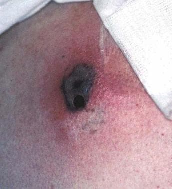
Primary cutaneous aspergillosis at a site of an intravenous catheter in a boy with leukemia.
Click to see larger pictureClick to see detailView Full Size Image
Media type:  Photo


  1. Zhang QQ, Li L, Zhu M, Zhang CY, Wang JJ. Primary cutaneous aspergillosis due to Aspergillus flavus: a case report. Chin Med J (Engl). Feb 5 2005;118(3):255-7. [Medline].
  2. Wheat LJ. Rapid diagnosis of invasive aspergillosis by antigen detection. Transpl Infect Dis. Dec 2003;5(4):158-66. [Medline].
  3. Cooke FJ, Terpos E, Boyle J, Rahemtulla A, Rogers TR. Disseminated Aspergillus terreus infection arising from cutaneous inoculation treated with caspofungin. Clin Microbiol Infect. Dec 2003;9(12):1238-41. [Medline].
  4. Koss T, Bagheri B, Zeana C, Romagnoli MF, Grossman ME. Amphotericin B-resistant Aspergillus flavus infection successfully treated with caspofungin, a novel antifungal agent. J Am Acad Dermatol. Jun 2002;46(6):945-7. [Medline].
  5. Herbrecht R, Denning DW, Patterson TF, Bennett JE, Greene RE, Oestmann JW, et al. Voriconazole versus amphotericin B for primary therapy of invasive aspergillosis. N Engl J Med. Aug 8 2002;347(6):408-15. [Medline].
  6. Walsh TJ, Raad I, Patterson TF, Chandrasekar P, Donowitz GR, Graybill R, et al. Treatment of invasive aspergillosis with posaconazole in patients who are refractory to or intolerant of conventional therapy: an externally controlled trial. Clin Infect Dis. Jan 1 2007;44(1):2-12. [Medline].
  7. Chakrabarti A, Gupta V, Biswas G, Kumar B, Sakhuja VK. Primary cutaneous aspergillosis: our experience in 10 years. J Infect. Jul 1998;37(1):24-7. [Medline].
  8. Elder D, Elenitsas R, Jaworsky C, eds. Lever's Histopathology of the Skin. Philadelphia, Pa: Lippincott-Raven; 1997:525-6.
  9. Freedberg I, Eisen AZ, Wolff K, et al, eds. Fitzpatrick's Dermatology in General Medicine. 5th ed. New York, NY: McGraw-Hill; 1999:1436-7.
  10. Klein KC, Blackwood RA. Topical voriconazole solution for cutaneous aspergillosis in a pediatric patient after bone marrow transplant. Pediatrics. Aug 2006;118(2):e506-8. [Medline].
  11. Larkin JA, Greene JN, Sandin RL, Houston SH. Primary cutaneous aspergillosis: case report and review of the literature. Infect Control Hosp Epidemiol. Jun 1996;17(6):365-6. [Medline].
  12. Naidu J, Singh SM. Aspergillus chevalieri (Mangin) Thom and Church: a new opportunistic pathogen of human cutaneous aspergillosis. Mycoses. Jul-Aug 1994;37(7-8):271-4. [Medline].
  13. Odom RB, James WD, Berger TG, eds. Andrews' Diseases of the Skin. Philadelphia, Pa: WB Saunders; 2000:415.
  14. Richardson MD, Warnock DW, eds. Fungal Infection. In: Diagnosis and Management. 2nd ed. Blackwell Science: Oxford, England; 1997:113-30.

Aspergillosis excerpt

Article Last Updated: Dec 17, 2007