Practice Essentials
Some of the major causes of acute and chronic low back pain (LBP) are associated with radiculopathy. However, radiculopathy is not a cause of back pain; rather, nerve root impingement, disc herniation (see the image below), facet arthropathy, and other conditions are causes of back pain. [1, 2, 3, 4]
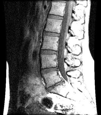 Sagittal magnetic resonance image showing loss of intervertebral disc height at L5/S1. Herniations of the nucleus pulposus are noted at L4/5 and L5/S1. Courtesy of Barton Branstetter, MD.
Lumbosacral radiculopathy, like other forms of radiculopathy, results from nerve root impingement and/or inflammation that has progressed enough to cause neurologic symptoms in the areas that are supplied by the affected nerve root(s).
Epidemiology
United States statistics
Lumbosacral radiculopathy occurs in approximately 3-5% of the population, and men and women are affected equally, although men are most commonly affected in their 40s, whereas women are most commonly affected between ages 50-60. [5] Of those who have this condition, 10-25% develop symptoms that persist for more than 6 weeks.
Functional Anatomy
The anatomy of the lumbar epidural space is the key to understanding the mechanism of lumbosacral radiculopathic pain. The sinuvertebral nerves innervate structures in the lumbar epidural space; these nerves originate distal to the dorsal root ganglion, then run back through the intervertebral foramen to supply the arteries, venous plexi, and lymphatics. At the inner aspect of the intervertebral foramen, the sinuvertebral nerves divide into ascending and descending branches that freely communicate with corresponding branches from the segment above, from the segment below, and from the opposite side.
The sinuvertebral nerve supplies the posterior longitudinal ligament, superficial annulus fibrosus, epidural blood vessels, anterior dura mater, dural sleeve, and posterior vertebral periosteum. The 2 structures capable of transmitting neuronal impulses that result in the experience of pain are the sinuvertebral nerve and the nerve root. The posterior rami of the spinal nerves supply the apophyseal joints above and below the nerve as well as the paraspinous muscles at multiple levels.
Herniation of the intervertebral disc can cause impingement of the above neuronal structures, thus causing pain. The presence of disc material in the epidural space is thought to initially result in direct toxic injury to the nerve root by chemical mediation and then exacerbation of the ensuing intraneural and extraneural swelling, which results in venous congestion and conduction block. Notably, the size of the disc herniation has not been found to be related to the severity of the patient's pain.
Pain is also believed to be mediated by inflammatory mechanisms that involve substances such as phospholipase A2, nitric oxide, and prostaglandin E. These mediators are all found in the nucleus pulposus itself. Phospholipase A2 has been found in high concentrations in herniated lumbar discs; this substance acts on cell membranes to release arachidonic acid, a precursor to other prostaglandins and leukotrienes that further advance the inflammatory cascade. Additionally, leukotriene B4 and the substance thromboxane B2 have been found to have direct nociceptive stimulatory roles.
From a biomechanical standpoint, the lumbar intervertebral discs are highly susceptible to herniation because they are exposed to tremendous forces, principally by the magnification of the forces that result from the lever effect of the human arm in lifting; the forces generated by the upper trunk mechanics with rotation, flexion/extension, and side-bending on the discs below; and by the vertical forces associated with the upright position. Because each intervertebral disc is a fluid system, hydraulic pressure is generated whenever a load is placed on the axial skeleton. The hydraulic pressure mechanisms then multiply the force on the annulus fibrosus of the intervertebral disc to make it 3-5 times that which is exerted on the axial skeleton.
Sport-Specific Biomechanics
Dancers are prone to both acute and chronic back problems, including lumbosacral radiculopathy, which develop secondary to the combination of 2 factors that are required in most dance routines: extreme physical flexibility and exposure of the spine to the extremes of its range of motion. [1] Additionally, female dancers are predisposed to disc herniation secondary to the positioning that is required in certain movements, such as the pas de deux (in which excess lumbar lordosis is present), as well as the large jumps that these dancers often perform.
Golfers are also very susceptible to disc disease and lumbosacral radiculopathy because of the repetitive torsional motion that is used in the sport. [6] The golf swing can produce up to an estimated 7500 N of compressive force across L3-L4. Competitive weight lifters and football linemen have been noted to experience even larger compressive loads.
Patient Education
Individuals with lumbosacral radiculopathy need to have an understanding of the likely etiology of their pain. The examination findings of patients with acute LBP can often be suggestive, although no clinical or historical findings have been found to significantly correlate with confirmed pain generators.
Review the basic anatomy and biomechanics of the spine with the patient. Discuss the etiology of the patient's symptoms. Also discuss the treatment plan, including a description of the recommended imaging studies, medications, injections, and therapeutic exercises. Review proper posture, the biomechanics of the spine in the activities of daily living, and simple methods to reduce the patient's symptoms. These early and simple instructions enable the patient to become an active participant in the treatment as he or she progresses to a more comprehensive home exercise program.
Patients must understand that they are making a lifelong commitment to their maintenance exercise program, because the single most important risk factor for future episodes of back pain is a previous episode. Patient education should be considered an ongoing process that must be continually refined. Directed education should continue until the patient is independent in his or her maintenance exercise program. In cases in which pain does not improve with conservative management, patient education should include all potential surgical and alternative treatment options.
For excellent patient education resources, visit eMedicineHealth's Osteoporosis Center. Also, see eMedicineHealth's patient education articles Low Back Pain and Slipped Disk.
-
Sagittal magnetic resonance image showing loss of intervertebral disc height at L5/S1. Herniations of the nucleus pulposus are noted at L4/5 and L5/S1. Courtesy of Barton Branstetter, MD.
-
Discogram showing examples of an intact disc and a disrupted disc at the lumbar level.
-
Magnetic resonance image demonstrating extension of the nucleus pulposus to the right paracentral region of the spinal cord. The disc is adjacent to the inflamed right L5 nerve root. Courtesy of Barton Branstetter, MD.


