Practice Essentials
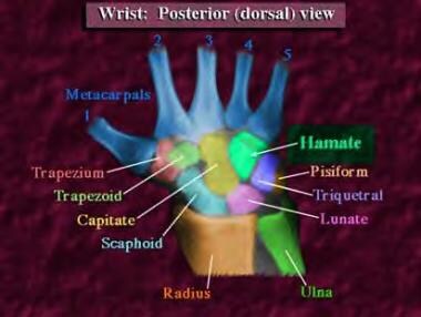
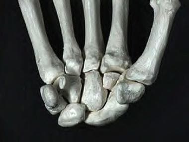
Although hamate fractures are increasing in incidence secondary to the popularity of sports activities involving racquets, bats, and clubs, these injuries remain relatively rare. [1] Estimates suggest hamate fractures constitute 2% of all carpal fractures. The hamate bone is a roughly triangular-shaped bone composed of both a body and a hook (see images below). Hamate fractures are thus classified as type I fractures involving the hook and type II fractures involving the body. Type I fractures are more common than type II fractures. [2, 3, 4]
Signs and symptoms
Physical examination findings are usually nonspecific and may even be absent. If symptoms are present, physical examination typically reveals discrete point tenderness with palpation over the hook of the hamate, diminished grip strength, and, secondary to the proximity of hamate fractures to the ulnar nerve, paresthesia may be present in the fourth and fifth fingers.
See Presentation for more detail.
Diagnosis
Fractures to the hamate may not be readily evident on radiographic images. In cases in which clinical findings suggest a fracture but the radiographic evidence is questionable, a computed tomography (CT) scan should be ordered.
See Workup for more detail.
Management
Consultation with an orthopedist or hand surgeon is recommended for all patients with hamate fractures because of the high risk of nonunion with conservative treatment.
See Treatment and Medication for more detail.
Related Medscape Reference topics:
Wrist Fracture in Emergency Medicine
Wrist Fractures and Dislocations
Related Medscape resources:
Resource Center Exercise and Sports Medicine
Resource Center Fracture
Resource Center Trauma
Epidemiology
United States statistics
Hamate fractures account for 2% of all carpal fractures. [5] Of the 2%, one third are hamate hook fractures due to repetitive swinging by golfers. Among hand and wrist injuries in professional baseball players, hook of hamate fracture is the most common indication for surgery. [6]
In hamate body fractures, the most frequent mechanisms of injury are striking an unyielding object with a clenched fist (52%) and falls (22%). Most hamate body fractures occur in males (96%). [1]
Functional Anatomy
The hamate is a triangular bone located in the distal carpal row farthest to the ulnar side (see the images below). The hamate is bordered proximally by the pisiform and the lunate in the proximal carpal row, radially by the capitate, and distally by the bases of the fourth and fifth metacarpals.
A roughly circular projection or hook on the volar surface of the hamate is the inferolateral border of the Guyon canal. The roof (superficial) of the canal is formed by the palmar carpal ligament, and the floor (deep) is formed by the flexor retinaculum. The canal carries the ulnar artery and nerve, and, for this reason, hook fractures should suggest a high probability of ulnar artery and nerve damage. [7] In addition, the hamate hook has a dual blood supply, with vessels entering from both the ulnar tip and radial base. These vessels often have a poor anastomosis, which clinically can result in nonunion due to insufficient blood supply.
Sport-Specific Biomechanics
Hamate fractures are generally associated with sports activities that use a racquet, bat, or club.
Type I fractures involving the hook of the hamate are the most common and can occur via several different mechanisms. [2, 3, 8, 9, 10, 11] First, repeated microtrauma to the hook during sports involving swinging clubs, bats, or racquets can result in a hook stress fracture. These usually occur in the nondominant hand and account for approximately one third of hamate fractures. Second, direct trauma can be applied during sports when the butt of the club rests on the hamate and the force of the swing is then transmitted directly to the bone. In addition, indirect trauma can be applied to the hook through its muscular and ligamentous attachments. This can occur either when falling on a hyperextended wrist or during power grips.
Type II fractures involving the body of the hamate are less common than type I fractures and always require direct force. [8] Most commonly, these fractures occur with a punch-press injury or dorsopalmar compression of the wrist between heavy weights. [5]
Prognosis
The prognosis of hamate fractures depends on the degree of injury encountered and the patient's effort in the physical therapy program. In a retrospective review of 29 cases, the patient's functional recovery was indirectly related to the degree of soft-tissue damage at the time of the injury (an increase in soft-tissue damage results in a decrease in functional recovery). [8] For most isolated hamate fractures treated soon after the injury, the prognosis is excellent.
Complications
The most frequent complication is nonunion. [7, 12, 13] This can follow conservative treatment in more than 50% of patients. Often, these patients present with continued palmar pain, especially with grip. Conventional radiographs can miss this diagnosis in 30-50% of patients. Therefore if the clinical suspicion is high and radiographic findings are negative, CT scanning should be performed. The treatment of nonunion involves either excision of the hamate hook or ORIF (see Surgical Intervention).
In cases in which internal fixation has been tried and has failed, excision of the fragment is the recommended treatment. These fragments may be small, and full range of motion is often preserved. Pathologic fractures due to cyst formation in the hamate may also occur. These types of fractures are treated best with bone packing, using tissue from the iliac crest, and external fixation. In addition, there has been one case report that described avascular necrosis occurring in the hamate hook. [13]
Patient Education
Patient education is an important part of the rehabilitation program for patients recovering from hamate fractures. Patients need to have a good understanding of the healing process and must adhere to recommendations provided by their physician and physical therapist to recover full strength and functional abilities.
-
Posterior (dorsal) view of the wrist.
-
Anterior palmar view.
-
Anteroposterior view of the wrist.
-
Lateral view of the wrist.
-
Oblique view of the wrist.
-
Computed tomography scan of the wrist.
-
Lateral computed tomography scan of the wrist.
-
Reconstruction of the hamate fracture.


