Continually Updated Clinical Reference
 
 
  All Sources     eMedicine     Medscape     Drug Reference     MEDLINE
 
eMedicine - Posterior Cruciate Ligament Injury : Article by

Quick Find
Authors & Editors
Introduction
Clinical
Differentials
Workup
Treatment
Medication
Follow-up
Miscellaneous
Multimedia
References

Related Articles
Anterior Cruciate Ligament Injury

Lateral Collateral Knee Ligament Injury

Medial Collateral Knee Ligament Injury

Meniscus Injuries

Talofibular Ligament Injury


Patient Education
Foot, Ankle, Knee, and Hip Center

Knee Injury Overview

Knee Injury Causes

Knee Injury Symptoms

Knee Injury Treatment

Knee Pain Introduction


Author: Charles S Peterson, MD, Consulting Staff, Arizona Sports Medicine Center

Charles S Peterson is a member of the following medical societies: American Academy of Family Physicians, American College of Sports Medicine, and American Medical Society for Sports Medicine

Coauthor(s): Thomas Agesen, MD, Assistant Clinical Professor, UMDNJ, New Jersey Medical School; Consulting Staff, Department of Physical Medicine and Rehabilitation, Mountainside Hospital, Summit Overlook Hospital; Janos P Ertl, MD, Clinical Assistant Professor, Department of Orthopedic Surgery, Chief of Orthopedic Trauma, University of California at Davis; Director of Amputee Clinic, Kaiser Hospital; Gyorgy Kovacs, MD, Department of Orthopedic Surgery, Consulting Surgeon, GOC Clinic

Editors: Gerard A Malanga, MD, Associate Professor, Department of Physical Medicine and Rehabilitation, New Jersey Medical School; Director of Pain Management, University of Medicine and Dentistry at New Jersey, Overlook Hospital; Director of Sports Medicine, Mountainside Hospital; Francisco Talavera, PharmD, PhD, Senior Pharmacy Editor, eMedicine; Russell D White, MD, Professor of Medicine, Department of Community and Family Medicine, University of Missouri-Kansas City School of Medicine, Truman Medical Center Lakewood; Jon B Whitehurst, MD, Clinical Instructor of Surgery, University of Illinois College of Medicine; Consulting Staff, Rockford Orthopedic Associates; Craig C Young, MD, Medical Director of Sports Medicine, Sports Medicine Fellowship Director, Professor, Departments of Orthopedic Surgery and Community and Family Medicine, Medical College of Wisconsin

Author and Editor Disclosure

Synonyms and related keywords: posterior cruciate ligament, PCL injury, PCL tear, posterior knee instability, posterior laxity of the knee

Background

The posterior cruciate ligament (PCL) is described as the primary stabilizer of the knee by many authors. PCL injuries are less common than anterior cruciate ligament (ACL) injuries, and they often go unrecognized. The PCL is broader and stronger than the ACL and has a tensile strength of 2000 N. Injury most often occurs when a force is applied to the anterior aspect of the proximal tibia when the knee is flexed. Hyperextension and rotational or varus/valgus stress mechanisms also may be responsible for PCL tears. Injuries may be isolated or combined with other ligamentous injuries. A PCL tear can result in varying degrees of disability, from no impairment to severe impairment.  PCL injury has been overly simplified, and the functional disability of PCL injury may be underestimated (Wang, 2002). 

The primary function of the PCL is to prevent posterior translation of the tibia on the femur. The PCL also plays a role as a central axis controlling and imparting rotational stability to the knee. This injury has received little attention in the past, compared with the ACL; however, this emphasis on the ACL has stimulated increased interest in the treatment of PCL injuries. Controversy regarding treatment of isolated PCL injuries exists in the literature, with recommendations supporting both operative and nonoperative therapy. Current management of PCL injuries unfortunately can yield relatively poor clinical outcomes, whether surgically or conservatively treated (Margheritini, 2002).

Frequency

United States

True incidence in the United States is unknown. In National Football League predraft physical examinations, a 2% incidence of isolated, asymptomatic, and unknown PCL injuries was found; operated, isolated, and combined PCL injuries were reported at an incidence of 3.5-20%. On the KT-1000 stress test examination, a 7% incidence of PCL injuries was found, of which 40% were isolated and unidirectional and 60% were multidirectional.

Functional Anatomy

The PCL originates from the intercondylar notch of the femur on the roof of the medial femoral condyle. The insertion is central on the posterior aspect of the tibial plateau, on a depression between the tibial plateaus, extending 1 cm below the articular surface (Sheps, 2005). The ligament is composed of a larger anterolateral bundle and a smaller posteromedial bundle. The anterior component is tightest in the midarc of flexion and the posterior fibers are tight in extension and deep flexion.

In addition, variable anterior and posterior meniscofemoral ligaments of Humphrey and Wrisberg attach distally and proximally to the PCL, respectively. The meniscofemoral ligaments attach distally to the posterior horn of the lateral meniscus, in a slanting orientation, providing resistance to the tibial posterior drawer (Amis, 2006). The PCL is an extrasynovial structure that lies behind the intra-articular portion of the knee. The primary function of the PCL is to resist posterior displacement of the tibia in relation to the femur; its secondary function is to prevent hyperextension and limit internal and varus/valgus rotation.

Sport Specific Biomechanics

Disruption may occur with forced hyperextension while the foot is planted in dorsiflexion. A force applied to the anteromedial aspect of the knee, as during a football tackle, results in a posteriorly directed force and a varus hyperextension force, leading to PCL and posterolateral capsular ruptures.


History

  • Knowledge of the mechanism of injury is helpful. The following 4 mechanisms of PCL injury are recognized:
    • A posteriorly directed force on a flexed knee, eg, the anterior aspect of the flexed knee striking a dashboard, may cause PCL injury.

    • A fall onto a flexed knee with the foot in plantar flexion and the tibial tubercle striking the ground first, directing a posterior force to the proximal tibia, may result in injury to the PCL.

    • Hyperextension alone may lead to an avulsion injury of the PCL from the origin. This kind of injury may be amenable to repair.

    • An anterior force to the anterior tibia in a hyperextended knee with the foot planted results in combined injury to the knee ligaments along with knee dislocation.

  • In chronic PCL tears, discomfort may be experienced with the following positions or activities:
    • A semiflexed position, as with ascending or descending stairs or an incline

    • Starting a run

    • Lifting a load

    • Walking longer distances

  • Retropatellar pain symptoms may be reported as a result of posterior tibial sagging.
  • Swelling and stiffness may be reported in cases of chondral damage.
  • Individuals may describe a sensation of instability when walking on uneven ground
  • Medial joint line pain may be reported.

Physical

In the acute stage of isolated PCL injuries, symptoms usually are vague and minimal. The following physical examination findings are common in individuals who have sustained PCL injuries:

  • Minimal to no pain
  • Minimal hemarthrosis
  • Usually full or functional range of motion (ROM)
  • Contusion over the anterior tibia
  • Posterior tibial sag
    • To observe posterior tibial sag, place patient supine and put 90° of flexion at the knee and hip. In such a position, gravity pulls posteriorly on the tibia, and in the case of PCL disruption, the tibia falls even or behind the femoral condyles. Comparison should be made to the opposite knee.

    • Grade I injury is indicated when side-to-side asymmetry exists but the tibial plateau is anterior to the femoral condyles. Grade II injury occurs when the tibial plateau is even with the femoral condyles, and grade III injury occurs when the tibial plateau falls behind the femoral condyles.

  • Posterior sag sign during extension
    • The patient is supine on the examining table, with the examiner at the end of the table. The examiner supports both of the patient's heels simultaneously with legs in full extension.

    • If a posterior sag can be seen on the injured side compared to the other side, there usually is an injury to the PCL and some secondary restraint (ie, medial collateral ligament [MCL], lateral collateral ligament [LCL], posterolateral corner).

  • Positive quadriceps active test
    • During the quadriceps active test, the patient is placed supine with the knee flexed to 90° and the foot placed flat on the examining table.

    • If an individual with an intact PCL is in such a position with the quadriceps relaxed, the tibia is 10 mm anterior to the femoral condyles. If there is a PCL disruption, gravity pulls the tibia even or behind the femoral condyles, with the quadriceps relaxed. The examiner restrains the ankle from moving, and the patient is asked to contract the quadriceps. In individuals who have a deficient PCL, the tibia moves forward; if the tibia moves forward more than 2 mm, the quadriceps active test is positive.

  • Findings of the posterior drawer test
    • The posterior drawer test is considered the most useful for documenting PCL injury.

    • The patient is placed supine with both knees flexed to 90° and the feet in neutral rotation placed flat on the table (examiner must compare side-to-side difference). As mentioned previously, in such a position the tibial plateau should be about 10 mm anterior to the femoral condyles.

    • The examiner imparts a posterior force to the proximal tibia, and if the tibia can be displaced 0-5 mm or if there is side-to-side asymmetry, a grade I injury is indicated. If the tibia can be displaced 5-10 mm or the tibial plateau can move posteriorly even with femoral condyles, a grade II injury is indicated. If the tibia can be moved more than 10 mm posteriorly or the tibial plateau moves behind the femoral condyles, a grade III injury is indicated.

    • The internal and external rotation of the foot during the posterior drawer test can assess different structures. If the foot is placed in internal rotation, the PCL and tibial collateral ligaments are tested. If the foot is placed in external rotation, the PCL, LCL, and posterolateral corner are tested. Assessment of the posterolateral corner is paramount with PCL injuries because isolated PCL injuries have a very good prognosis. However, a PCL injury combined with posterolateral corner injury has a less favorable prognosis. The external rotation recurvatum test and the reverse pivot shift test (described below) are used to assess the posterolateral corner.

  • Findings of the external rotation recurvatum test: This test is the same as the posterior sag sign described above, except the examiner notices significant subluxation of the lateral tibial plateau.
  • Findings of the posterolateral drawer test in 90° of flexion: This test is performed with the patient sitting with thighs supported by the examining table and legs lying off the end of the examining table. In such a position, the knees are at 90° of flexion. The examiner performs a posterior drawer test. If the posterolateral structures are injured, the lateral tibial plateau rotates posteriorly around the axis of the PCL as the posterior force is applied.

  • False-positive Lachman test: The Lachman test is performed to assess the integrity of the ACL. In a knee with a deficient PCL, the starting position of the tibial plateau is posterior to normal. Since the starting point is posterior, there seems to be increased anterior laxity. This results in a false-positive Lachman test. The endpoint of the Lachman test is still firm with PCL disruption.

Causes

Possible causes of PCL injuries include the following:

  • Football injuries
  • Running injuries
  • Motor vehicle accidents
  • Falls onto a flexed knee


Anterior Cruciate Ligament Injury
Lateral Collateral Knee Ligament Injury
Medial Collateral Knee Ligament Injury
Meniscus Injuries
Talofibular Ligament Injury

Other Problems to be Considered

Multiligamentous injury
Knee dislocation
Posterolateral instability


Lab Studies

  • Perform laboratory studies as indicated per patient age.

Imaging Studies

  • Radiographs
    • Acute injuries

      • Routine radiographs usually are negative in acute injuries.


      • Bony avulsions may be evident on lateral radiographs.


      • New radiological view: Axial radiograph as described by Puddu et al. Patient is supine with knees flexed to 70°, feet are plantigraded in moderate plantar flexion, and the tibia is in neutral rotation. With patient holding cassette, the radiographic beam is directed distal to proximal and parallel to the longitudinal axis of the patella. The length of the perpendicular line between the anterior tibial profile and the femoral groove is measured. If the side-to-side difference is >3 mm, posterior laxity is indicated.
         
    • Chronic lesions

      • Weight-bearing anteroposterior (AP) or Rosenberg view (posteroanterior [PA] with knees flexed) may demonstrate early medial joint arthrosis.


      • Tangential patellofemoral view (Laurin/Merchant view) may demonstrate patellofemoral arthrosis.


      • Long-film AP weight-bearing views are essential for preoperative alignment.
         
  • MRI
    • If physical examination reveals multiple injured ligaments, or if degree of injury is in question, an MRI may be justified.


    • MRI helps identify and confirm the location of the lesion; in addition, occult osteochondral lesion/fractures and meniscal lesions may be identified. MRI of acute injury is more accurate than for chronic injury, as the ligament may appear healed but be functionally deficient (Servant, 2004).

Other Tests

  • Occasionally, a patient with a posterior knee injury may present with calf pain and signs of an impending compartment syndrome, possibly due to soft tissue injury or signs of an occult vascular injury. The calf symptoms may be more pronounced than the knee symptoms. Compartment pressures need to be measured, and vascular surgery consultation must be considered for an arteriogram.

Procedures

  • Perform an arteriogram in patients with suspected vascular injury, such as an injury that might be seen with knee dislocation.


Acute Phase

Rehabilitation Program

Physical Therapy

The course of rehabilitation for a PCL injury is dependent on the type of treatment rendered (ie, operative vs nonoperative). The key to nonoperative treatment is to control the swelling, instability, and pain. The natural history of sports-related PCL injuries that are treated nonoperatively is quite good. In one study with a mean follow-up of 6.2 years, 80% of patients were satisfied with their knees and 84% had returned to their previous sport (68% at the same level of performance, 16% at a decreased level of performance).

A more recent prospective study with a mean follow-up of 5.4 years found that 50% of athletes with isolated posterior laxity returned to the same sport at the same or higher level of performance and that 33% returned to the same sport at a lower level of performance (no patient had greater than grade II injuries). In addition, the grade of laxity noted on physical examination did not change over the course of follow-up, and the grade of laxity did not correlate with radiographic joint-space narrowing.

Another study involving MRI follow-up imaging found that all low-grade and midgrade PCL injuries healed with continuity, and 19 of 22 high-grade injuries healed (4 healed with normal contour; 15 healed with continuity and altered morphology). In many cases that involve less severe PCL tears, patients are recommended to undergo conservative therapy with a progressive rehabilitation program. However, if the patient continues to experience chronic pain and instability despite therapy and functional bracing, a PCL reconstruction may be required. The choice of which route of treatment depends on the severity of the specific injury, whether the PCL injury is isolated or in combination with other ligamentous or meniscal damage, the activity level and goals of the patient, and the individual physician preference. The stages of physical therapy are discussed here and are broken into the following 2 types of rehabilitation: nonoperative and operative (ie, PCL reconstruction).

Nonoperative rehabilitation (day 1 to week 2)

Many isolated PCL injuries are missed at the time of the initial injury. The patient often cannot remember injuring the knee and often seeks medical attention at a later time. The pain, degree of swelling, and disability associated with ACL and MCL injuries is often missing from the patient’s history. Many are able to walk with normal gait immediately after the injury, and the soft endpoint of the posterior drawer test is firm by 2-3 weeks after injury (though more laxity is noted when compared to the uninjured knee). With higher-grade injuries, usually grade III +/- other ligamentous injury, the patient typically seeks medical attention immediately. In such cases, the physician should order an MRI to evaluate all the knee ligaments and assess for subchondral injury or further intra-articular pathology.

The goal of the rehabilitation for individuals undergoing a conservative program is to control the initial inflammatory phase and regain ROM with muscle function as quickly as possible.

  • Apply the rest, ice, compression, and elevation (RICE) method several times a day, in addition to any other modalities incorporated by the physical therapist to control pain and swelling (eg, electrical stimulation, cold whirlpool).


  • Patients with grade I and grade II injuries can bear weight as tolerated immediately, though some may require axillary crutches initially. Axillary crutches and a long leg brace are recommended for grade III injuries and with other associated ligamentous laxity (ie, posterolateral corner injury) or intra-articular damage.


  • Functional electrical stimulation (FES) may be used to stimulate the quadriceps muscle, but it is probably necessary only if the quadriceps muscle is shut down secondary to pain.


  • The physical therapist should instruct the patient in exercises for quadriceps and hip strengthening (eg, quadriceps sets, straight leg raises, hip abduction/adduction, multiangle quadriceps isometrics).


  • At this time, all open kinetic chain (OKC) hamstring exercises should be avoided since they impart posterior tibial translation at the knee.

Operative rehabilitation (day 1 to week 2)

Several different techniques may be used to reconstruct the PCL, so the treatment protocol is determined by the individual physician and the type of graft used in surgery.

  • Postoperatively, it is very important to control pain and swelling through the use of cold therapy, compression, and elevation.


  • The patient may bear weight as tolerated on the operated limb with the use of 2 crutches and a long leg brace.


  • Patellar mobility is important, and the patient should be instructed in self-mobilization exercises for the patella, scar, and soft tissues surrounding the kneecap to prevent fibrosis.


  • ROM should be initiated (0-90°), emphasizing full passive knee extension. Other examples of exercises that may be initiated include quadriceps sets, ankle pumps, straight leg raises, and upper body strengthening.

Surgical Intervention

Historically, controversy in treatment modalities exists because of the lack of knowledge of the natural history of this injury; in addition, reported surgical results are variable. When surgical reconstruction is considered, graft recommendations include the following:

  • Autograft

    • Patellar tendon


    • Quadriceps tendon


    • Hamstring tendons


    • Medial head of gastrocnemius
       
  • Allograft

    • Achilles tendon


    • Patellar tendon


    • Quadriceps tendon


    • Hamstring tendons

The results of operative reconstruction are variable and may be no better than nonoperative treatment. With improved techniques and understanding of the PCL anatomy, improved surgical results may be possible. Single bundle PCL reconstruction reduces the posterior tibial translation significantly, but it cannot restore the kinematics of the uninjured knee (Lenschow, 2006).

Bony PCL avulsion injuries are amenable to surgical repair of the avulsed bony fragment, with restoration of PCL integrity and function. Even delayed diagnosis of avulsion injuries can be repaired with screw fixation if the PCL substance is sufficient (Jung and Höher, 2006).  Surgical reconstruction of PCL tears is recommended in combined knee ligamentous injuries; however, controversy exists regarding the surgical technique and tissue used for ligament replacement.  Autograft reconstruction has shown similar success to allograft reconstruction (Ahn, 2005).  Preservation of the remnant bundle significantly improves the posterior stability and proprioception of the reconstructed knee joint, integrating graft with remnant fibers (Ahn, 2006; Jung and Jung, 2006).

Surgical reconstruction should be considered with multiple ligament injuries, posterolateral corner injury, or when persistent pain, instability, or disability remains despite conservative treatment (Wang, 2002). Surgical reconstruction typically improves laxity by at least one grade. Acute reconstruction is more successful than reconstruction of chronic injuries (Sekiya, 2005).

Consultations

Consultation with an orthopedic surgeon is needed for patients requiring operative intervention.

Recovery Phase

Rehabilitation Program

Physical Therapy

The recovery phase discussed here encompasses treatment from 2-12 weeks. The goal of this phase, in higher-grade injuries, is to advance weight bearing and achieve a normal gait pattern, along with a progression of functional strengthening and ROM. For those patients who undergo graft reconstruction, it is especially important to protect the graft during this period, as it is at its weakest state in the healing process.

Nonoperative rehabilitation (weeks 2-12)

  • Only patients with grade III injuries should continue to wear a brace (0-60°) until at least the third week of therapy. Shortly thereafter, the patient may be fitted for a functional knee brace.


  • Weight bearing may be progressed as tolerated, and the crutches may be discontinued at approximately 2-3 weeks.


  • At 2-3 weeks, the exercises performed in the acute phase should be progressed with light resistance as tolerated. Stationary bicycling may be recommended for improving ROM. Aquatic exercises may be recommended to improve ROM and strengthening. As the patient progresses into weeks 3-6, the exercises may be progressed to include closed kinetic chain (CKC) exercises (eg, leg press, mini squats, stair stepper, step-ups). Resistance may be increased on the bicycle as tolerated. At 8-12 weeks, the strengthening exercises should be progressed and a light jogging program may be initiated.

Operative rehabilitation (weeks 2-12)

  • The patient gradually should improve ROM (0-130°) during this time. Passive stretching is used as necessary to regain mobility. Patellar mobilization continues to be important. Modalities may be continued as needed for pain and swelling.


  • Weight bearing is progressed as tolerated, and crutches are discontinued at the discretion of the physician.


  • At 4-6 weeks, the patient may be fitted for a functional knee brace.


  • Strengthening exercises may be progressed to include CKC exercises. Aquatic resistance training may be initiated during the later part of this phase.

Surgical Intervention

See Surgical Intervention, Acute Phase.

Maintenance Phase

Rehabilitation Program

Physical Therapy

The maintenance phase (4 mo to 1 y) is the final phase of rehabilitation. This phase prepares the athlete for return to competition. Goals are focused on increasing strength, power, and endurance.

Nonoperative rehabilitation (4-9 mo)

  • Strengthening and proprioception exercises are continued and progressed as tolerated. Plyometrics and sport-specific training should be initiated and accelerated as tolerated.


  • A running program is developed, and agility drills are incorporated.


  • An isokinetic test and a KT-2000 test should be performed at 3-month, 6-month, 9-month, and 12-month follow-up visits.


  • The athlete may return to sporting activities when isokinetic and functional tests are satisfactory (determined by the physician), in addition to satisfactory clinical examination findings. The patient should not return to competitive sports until full quadriceps strength has been reestablished.


  • Prospective long-term follow-up studies comparing operative versus nonoperative outcomes are lacking. However, nonoperative treatment has demonstrated the following results:

    • After completing the rehabilitation program, 68% of patients return to their previous level of competitive function.


    • Radiographic signs of arthritis show up in 31% of patients.


    • At the time of arthrotomy, 64% of patients had degenerative medial compartment changes.


    • Significant degenerative changes developed in 44% of patients.


    • In patients with PCL deficiency for more than 5 years, 77.8% develop medial femoral condyle degenerative cartilage lesions and 46.7% develop patellar cartilage degeneration (Strobel, 2003).


    • Operative intervention is required in 42% of patients.

Operative rehabilitation (4 mo to 1 y)

  • Functional strengthening, as well as balance and proprioception, is progressed.


  • Light jogging may be progressed to running and various agility drills as tolerated. Advanced plyometrics and sports-specific training should be incorporated.


  • Isokinetic strength and KT-2000 testing often are performed; however, the necessity of these tests has not been validated to affect outcome.


Medications are used in cases of PCL injuries for pain control, inflammation, and swelling. Pain medications are used as indicated for acute pain. Nonsteroidal anti-inflammatory drugs (NSAIDs) are used to control inflammation and swelling.

Drug Category: Narcotic analgesics

Pain control is essential to quality patient care. Analgesics ensure patient comfort and have sedating properties, which are beneficial for patients who have sustained trauma or injuries.

Drug NameAcetaminophen and codeine (Tylenol #3)
DescriptionIndicated for the treatment of mild to moderate pain.
Adult Dose30-60 mg based on codeine PO q4-6h, not to exceed 12 tabs/24h
Pediatric Dose0.5-1 mg/kg/dose based on codeine PO q4-6h; 10-15 mg/kg/dose based on acetaminophen content; not to exceed 2.6 g/d of acetaminophen
ContraindicationsDocumented hypersensitivity to acetaminophen or codeine phosphate
InteractionsToxicity of codeine increases with CNS depressants, tricyclic antidepressants, MAOIs, neuromuscular blockers, CNS depressants, phenothiazines, and narcotic analgesics; rifampin can reduce analgesic effects of acetaminophen; coadministration with barbiturates, carbamazepine, hydantoins, and isoniazid may increase hepatotoxicity of acetaminophen
PregnancyC - Safety for use during pregnancy has not been established.
PrecautionsCaution in patients dependent on opiates since this substitution may result in acute opiate-withdrawal symptoms; caution in severe renal or hepatic dysfunction; hepatotoxicity is possible following various dose levels of acetaminophen in those with chronic alcoholism; severe or recurrent pain or high or continued fever may indicate a serious illness; acetaminophen is contained in many OTC products, and combined use with these products may result in cumulative acetaminophen doses that exceed recommended maximum dose

Drug Category: Nonsteroidal anti-inflammatory drugs

Have analgesic, anti-inflammatory, and antipyretic activities. Their mechanism of action is not known, but they may inhibit cyclo-oxygenase activity and prostaglandin synthesis. Other mechanisms may exist as well, such as inhibition of leukotriene synthesis, lysosomal enzyme release, lipoxygenase activity, neutrophil aggregation, and various cell membrane functions.

Drug NameIbuprofen (Motrin, Ibuprin)
DescriptionDOC for patients with mild to moderate pain. Inhibits inflammatory reactions and pain by decreasing prostaglandin synthesis.
Adult Dose400-800 mg PO tid with food
Pediatric Dose10 mg/kg PO tid with food
ContraindicationsDocumented hypersensitivity; peptic ulcer disease, recent GI bleeding or perforation, renal insufficiency, or high risk of bleeding
InteractionsCoadministration with aspirin increases risk of inducing serious NSAID-related side effects; probenecid may increase concentrations and, possibly, toxicity of NSAIDs; may decrease effect of hydralazine, captopril, and beta-blockers; may decrease diuretic effects of furosemide and thiazides; may increase PT when taking anticoagulants (instruct patients to watch for signs of bleeding); may increase risk of methotrexate toxicity; phenytoin levels may be increased when administered concurrently
PregnancyB - Usually safe but benefits must outweigh the risks.
PrecautionsCategory D in third trimester of pregnancy; caution in congestive heart failure, hypertension, and decreased renal and hepatic function; caution in coagulation abnormalities or during anticoagulant therapy

Drug NameKetoprofen (Actron, Oruvail, Orudis)
DescriptionFor relief of mild to moderate pain and inflammation. Small dosages initially are indicated in small and elderly patients and in those with renal or liver disease. Doses larger than 75 mg do not increase therapeutic effects. Administer high doses with caution, and closely observe patient for response.
Adult Dose25-50 mg PO q6-8h prn; not to exceed 300 mg/d
Pediatric Dose<3 months: Not established
3 months to 12 years: 0.1-1 mg/kg PO q6-8h
>12 years: Administer as in adults
ContraindicationsDocumented hypersensitivity
InteractionsCoadministration with aspirin increases risk of inducing serious NSAID-related side effects; probenecid may increase concentrations and, possibly, toxicity of NSAIDs; may decrease effect of hydralazine, captopril, and beta-blockers; may decrease diuretic effects of furosemide and thiazides; may increase PT when taking anticoagulants (instruct patients to watch for signs of bleeding); may increase risk of methotrexate toxicity; phenytoin levels may be increased when administered concurrently
PregnancyB - Usually safe but benefits must outweigh the risks.
PrecautionsCategory D in third trimester of pregnancy; caution in congestive heart failure, hypertension, and decreased renal and hepatic function; caution in coagulation abnormalities or during anticoagulant therapy

Drug NameNaproxen (Anaprox, Naprosyn, Naprelan)
DescriptionFor relief of mild to moderate pain. Inhibits inflammatory reactions and pain by decreasing activity of cyclo-oxygenase, which results in a decrease of prostaglandin synthesis.
Adult Dose500 mg PO followed by 250 mg q6-8h; not to exceed 1.25 g/d
Pediatric Dose<2 years: Not established
>2 years: 2.5 mg/kg/dose PO; not to exceed 10 mg/kg/d
ContraindicationsDocumented hypersensitivity; peptic ulcer disease; recent GI bleeding or perforation; renal insufficiency
InteractionsCoadministration with aspirin increases risk of inducing serious NSAID-related side effects; probenecid may increase concentrations and, possibly, toxicity of NSAIDs; may decrease effect of hydralazine, captopril, and beta-blockers; may decrease diuretic effects of furosemide and thiazides; may increase PT when taking anticoagulants (instruct patients to watch for signs of bleeding); may increase risk of methotrexate toxicity; phenytoin levels may be increased when administered concurrently
PregnancyB - Usually safe but benefits must outweigh the risks.
PrecautionsCategory D in third trimester of pregnancy; acute renal insufficiency, interstitial nephritis, hyperkalemia, hyponatremia, and renal papillary necrosis may occur; patients with preexisting renal disease or compromised renal perfusion risk acute renal failure; leukopenia occurs rarely, is transient, and usually returns to normal during therapy; persistent leukopenia, granulocytopenia, or thrombocytopenia warrants further evaluation and may require discontinuation of drug


Return to Play

When nonoperative treatment has been initiated, the athlete may return to play once the quadriceps strength has been regained. Yearly follow-up is recommended to monitor the knee for degenerative changes. Athletes with PCL injuries that have been operatively treated may return to sports at 9-12 months following surgery, pending the course and compliance with physical therapy and also the return of quadriceps strength. Follow-up at 2, 6, 12, 24, 36, 48, and 52 weeks following surgery is recommended.

Complications

Possible complications associated with PCL injury include the following:

  • Initial stiffness

  • Instability

  • Progressive arthritis

  • Postoperative complications

Prevention

Most cases of PCL injuries are not preventable. Athletes who participate in at-risk sports should maintain good strength and flexibility and should practice good techniques within their specific sports. Some physicians advocate the use of functional knee braces for reducing the risk of a recurring PCL injury when returning to activity.

Prognosis

See Physical Therapy, Acute Phase.

Education

Patient education is very important throughout the rehabilitation process for individuals with PCL injuries. Athletes should be informed of the benefits and risks of possible treatments and be involved in the decision-making process. To achieve their goals and be able to return to play, patients need to be compliant with their physician's instructions and physical therapy program as outlined by their therapist. As patients progress through their rehabilitation program, they should be instructed in a home exercise program for continued strengthening to decrease their risk for a recurrent injury.

For excellent patient education resources, visit eMedicine's Foot, Ankle, Knee, and Hip Center. Also, see eMedicine's patient education articles Knee Injury and Knee Pain.


Special Concerns

  • Patients who sustain isolated PCL tears may function quite well; however, they are predisposed to developing early degenerative articular changes. Combined PCL and other knee ligament injuries usually lead to chronic instability and progressive articular changes.
  • Although the natural history is developing, treatment of PCL injuries remains controversial. Further long-term studies of results comparing operative and nonoperative treatment are needed.


Media file 1:  
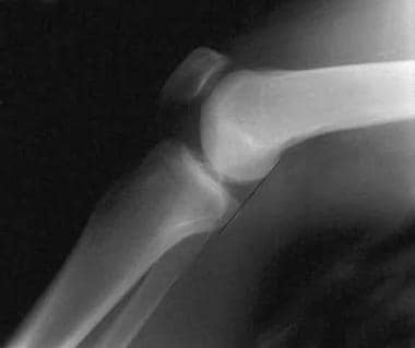
A normal lateral radiograph of a knee. In a normal knee, a line drawn along the posterior femoral condyle will not intersect the posterior tibial condyle.
Click to see larger pictureClick to see detailView Full Size Image
Media type:  X-RAY

Media file 2:  
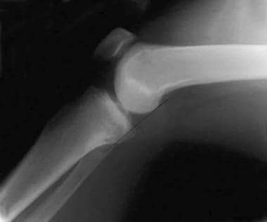
A lateral radiograph of a knee with a posterior cruciate ligament injury. Note that the same line as in Image 1 will bisect the posterior tibial condyle due to a posterior sag and an incompetent posterior cruciate ligament.
Click to see larger pictureClick to see detailView Full Size Image
Media type:  X-RAY

Media file 3:  The posterior tibia sag sign. The photo on the left demonstrates the clinical finding of the posterior tibia sag sign. A line drawn parallel to the patella accentuates the posterior tibia sag. The photo on the right demonstrates the quadriceps active drawer test described by Daniels. With the knee in 70-90° of flexion, the extensor mechanism is contracted, pulling the tibia anteriorly into a reduced position.
Click to see larger pictureClick to see detailView Full Size Image
Media type:  Photo

Media file 4:  A close-up view of a posterior tibia sag with an incompetent posterior cruciate ligament.
Click to see larger pictureClick to see detailView Full Size Image
Media type:  Photo

Media file 5:  This MRI of the knee shows a torn posterior cruciate ligament.
Click to see larger pictureClick to see detailView Full Size Image
Media type:  MRI

Media file 6:  This MRI (coronal section) shows a posterior cruciate ligament tear.
Click to see larger pictureClick to see detailView Full Size Image
Media type:  MRI

Media file 7:  This transverse MRI shows edema to the torn posterior cruciate ligament.
Click to see larger pictureClick to see detailView Full Size Image
Media type:  MRI

Media file 8:  
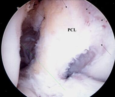
A view of the broad origin of the posterior cruciate ligament (PCL) on the medial femoral condyle of a left knee. The anterior cruciate ligament has been removed for surgical reconstruction.
Click to see larger pictureClick to see detailView Full Size Image
Media type:  Photo

Media file 9:  
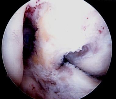
An additional view of the posterior cruciate ligament broad origin and insertion in a knee pending anterior cruciate ligament reconstruction.
Click to see larger pictureClick to see detailView Full Size Image
Media type:  Photo

Media file 10:  A right knee pending posterior cruciate ligament (PCL) reconstruction. A minimal notchplasty is completed. Two guide pins are advanced into the medial femoral condyle for tunnel placement to reconstruct the 2 bundles of the PCL.
Click to see larger pictureClick to see detailView Full Size Image
Media type:  Photo

Media file 11:  The 2 tunnels are created by reaming from outside in; 8- to 9-mm tunnels are made depending on patient size and the graft that will be used.
Click to see larger pictureClick to see detailView Full Size Image
Media type:  Photo

Media file 12:  Two red Robinson catheters are advanced through the femoral tunnels.
Click to see larger pictureClick to see detailView Full Size Image
Media type:  Photo

Media file 13:  The catheters have premade holes, which are used for suture retrieval.
Click to see larger pictureClick to see detailView Full Size Image
Media type:  Photo

Media file 14:  The catheters are advanced and threaded out the posterior knee. In this case, a posterior tibial onlay graft from an Achilles tendon allograft is used. The 2 bundles are secured to the catheters and advanced into the joint through the tunnels.
Click to see larger pictureClick to see detailView Full Size Image
Media type:  Photo

Media file 15:  The 2 Achilles tendon bundles are secured with a baseball whipstitch and threaded through the catheter holes and advanced into the femoral condyle tunnels.
Click to see larger pictureClick to see detailView Full Size Image
Media type:  Photo

Media file 16:  Additional view of the placement and advancement of the Achilles allograft.
Click to see larger pictureClick to see detailView Full Size Image
Media type:  Photo

Media file 17:  Completion and seating of the femoral allograft reconstruction. The 2 bundles are secured or stabilized by suturing over a post and washer. Note the reestablishment of the broad surface area for the reconstructed posterior cruciate ligament origin.
Click to see larger pictureClick to see detailView Full Size Image
Media type:  Photo

Media file 18:  Completion of the tibial onlay two bundle Achilles tendon allograft/posterior cruciate ligament (PCL) reconstruction. The bony calcaneus remnant is secured to the posterior tibia with 1 or 2 interfragmentary compression screws into a trough into the posterior tibia at the level of the PCL insertion. Care is taken to not penetrate the anterior tibial cortex with these screws. Note the intact original anterior cruciate ligament.
Click to see larger pictureClick to see detailView Full Size Image
Media type:  Photo


  • Ahn JH, Nha KW, Kim YC, Lim HC, Nam HW, Wang JH. Arthroscopic femoral tensioning and posterior cruciate ligament reconstruction in chronic posterior cruciate ligament injury. Arthroscopy. Mar 2006;22(3):341.e1-4. [Medline].
  • Ahn JH, Yoo JC, Wang JH. Posterior cruciate ligament reconstruction: double-loop hamstring tendon autograft versus Achilles tendon allograft--clinical results of a minimum 2-year follow-up. Arthroscopy. Aug 2005;21(8):965-9. [Medline].
  • Amis AA, Gupte CM, Bull AM, Edwards A. Anatomy of the posterior cruciate ligament and the meniscofemoral ligaments. Knee Surg Sports Traumatol Arthrosc. Mar 2006;14(3):257-63. [Medline].
  • Baker CL Jr, Norwood LA, Hughston JC. Acute combined posterior cruciate and posterolateral instability of the knee. Am J Sports Med. May-Jun 1984;12(3):204-8. [Medline].
  • Bergfeld JA, McAllister DR, Parker RD, et al. The effects of tibial rotation on posterior translation in knees in which the posterior cruciate ligament has been cut. J Bone Joint Surg Am. Sep 2001;83-A(9):1339-43. [Medline].
  • Browner BD. Skeletal Trauma: Fractures, Dislocations, Ligamentous Injuries. Philadelphia, Pa: WB Saunders Co; 1998.
  • Cain TE, Schwab GH. Performance of an athlete with straight posterior knee instability. Am J Sports Med. Jul-Aug 1981;9(4):203-8. [Medline].
  • Clancy WG Jr, Shelbourne KD, Zoellner GB, et al. Treatment of knee joint instability secondary to rupture of the posterior cruciate ligament. Report of a new procedure. J Bone Joint Surg Am. Mar 1983;65(3):310-22. [Medline].
  • Cosgarea AJ, Jay PR. Posterior cruciate ligament injuries: evaluation and management. J Am Acad Orthop Surg. Sep-Oct 2001;9(5):297-307. [Medline].
  • Cross MJ, Powell JF. Long-term followup of posterior cruciate ligament rupture: a study of 116 cases. Am J Sports Med. Jul-Aug 1984;12(4):292-7. [Medline].
  • Daniel DM, Stone ML, Barnett P, Sachs R. Use of the quadriceps active test to diagnose posterior cruciate-ligament disruption and measure posterior laxity of the knee. J Bone Joint Surg Am. Mar 1988;70(3):386-91. [Medline].
  • Delee JC. Orthopaedic Sports Medicine, Principles and Practice. Vol 2. Philadelphia, Pa: WB Saunders Co; 1994.
  • Donovan. Posterior cruciate ligament injury on artificial turf. Orthop. 1977;1:20.
  • Fowler PJ, Messieh SS. Isolated posterior cruciate ligament injuries in athletes. Am J Sports Med. Nov-Dec 1987;15(6):553-7. [Medline].
  • Harner, Miller. Complex topics in knee surgery. Clin Sports Med. 1999;18(1).
  • Janousek AT, Jones DG, Clatworthy M, et al. Posterior cruciate ligament injuries of the knee joint. Sports Med. Dec 1999;28(6):429-41. [Medline].
  • Jung TM, Höher J, Weiler A. Screw fixation of a 4 1/2-year-old PCL avulsion injury. Knee Surg Sports Traumatol Arthrosc. May 2006;14(5):469-72. [Medline].
  • Jung YB, Jung HJ, Tae SK, Lee YS, Yang DL. Tensioning of remnant posterior cruciate ligament and reconstruction of anterolateral bundle in chronic posterior cruciate ligament injury. Arthroscopy. Mar 2006;22(3):329-38. [Medline].
  • Lenschow S, Zantop T, Weimann A, Lemburg T, Raschke M, Strobel M. Joint kinematics and in situ forces after single bundle PCL reconstruction: a graft placed at the center of the femoral attachment does not restore normal posterior laxity. Arch Orthop Trauma Surg. May 2006;126(4):253-9. [Medline].
  • Margheritini F, Rihn J, Musahl V, Mariani PP, Harner C. Posterior cruciate ligament injuries in the athlete: an anatomical, biomechanical and clinical review. Sports Med. 2002;32(6):393-408. [Medline].
  • Ogata K, McCarthy JA, Dunlap J, Manske PR. Pathomechanics of posterior sag of the tibia in posterior cruciate deficient knees. An experimental study. Am J Sports Med. Nov-Dec 1988;16(6):630-6. [Medline].
  • Paddu G, Gianni E, Chambat P. The axial view in evaluating tibial translation in cases of insufficiency of the posterior cruciate ligament. Arthroscopy. 2000;16(2):217-220.
  • Parolie JM, Bergfeld JA. Long-term results of nonoperative treatment of isolated posterior cruciate ligament injuries in the athlete. Am J Sports Med. Jan-Feb 1986;14(1):35-8. [Medline].
  • Safran MR, Allen AA, Lephart SM, et al. Proprioception in the posterior cruciate ligament deficient knee. Knee Surg Sports Traumatol Arthrosc. 1999;7(5):310-7. [Medline].
  • Sekiya JK, West RV, Ong BC, Irrgang JJ, Fu FH, Harner CD. Clinical outcomes after isolated arthroscopic single-bundle posterior cruciate ligament reconstruction. Arthroscopy. Sep 2005;21(9):1042-50. [Medline].
  • Servant CT, Ramos JP, Thomas NP. The accuracy of magnetic resonance imaging in diagnosing chronic posterior cruciate ligament injury. Knee. Aug 2004;11(4):265-70. [Medline].
  • Shelbourne KD, Davis TJ, Patel DV. The natural history of acute, isolated, nonoperatively treated posterior cruciate ligament injuries. A prospective study. Am J Sports Med. May-Jun 1999;27(3):276-83. [Medline].
  • Shelbourne KD, Jennings RW, Vahey TN. Magnetic resonance imaging of posterior cruciate ligament injuries: assessment of healing. Am J Knee Surg. 1999;12(4):209-13. [Medline].
  • Sheps DM, Otto D, Fernhout M. The anatomic characteristics of the tibial insertion of the posterior cruciate ligament. Arthroscopy. Jul 2005;21(7):820-5. [Medline].
  • Siliski JM. Traumatic Disorders of the Knee. New York, NY: Springer-Verlag; 1994.
  • St Pierre P, Miller MD. Posterior cruciate ligament injuries. Clin Sports Med. Jan 1999;18(1):199-221, vii. [Medline].
  • Strobel MJ, Weiler A, Schulz MS, Russe K, Eichhorn HJ. Arthroscopic evaluation of articular cartilage lesions in posterior-cruciate-ligament-deficient knees. Arthroscopy. Mar 2003;19(3):262-8. [Medline].
  • Veltri DM, Warren RF. Isolated and combined posterior cruciate ligament injuries. J Am Acad Orthop Surg. Nov 1993;1(2):67-75.
  • Wang CJ. Injuries to the posterior cruciate ligament and posterolateral instabilities of the knee. Chang Gung Med J. May 2002;25(5):288-97. [Medline].

Posterior Cruciate Ligament Injury excerpt

Article Last Updated: Dec 22, 2006