Practice Essentials
Neutropenic enterocolitis, or typhlitis, is inflammation of the cecum. [1, 2] In 1960, Bierman and Amronin first coined the term ileocecal syndrome to describe inflammation and/or necrosis of the cecum, appendix, and/or ileum in patients with leukemia. [3, 4] Neutropenic enterocolitis (typhlitis) subsequently has been associated with aplastic anemia, lymphoma, AIDS, and immunosuppression following renal transplantation or during treatment of malignancy. [5, 6, 7, 8, 9]
A study by Cartoni et al reported a mortality of 60% due to necrotizing enterocolitis in patients with a bowel wall thickness greater than 10 mm, as compared to a mortality of 4.2% in patients with a bowel wall thickness of 10 mm or less as seen on ultrasound. [10]
Abdominal computed tomography (CT) scanning with oral and intravenous contrast is the preferred examination for neutropenic enterocolitis. The maximum normal colonic wall thickness on CT scan is 3 mm. When the colon is distended with stool, fluid, or oral contrast, the normal colonic wall is nearly imperceptible. Pericolonic fat should demonstrate homogeneous fat attenuation. Abdominal ultrasonography is an important diagnostic imaging study for neutropenic enterocolitis and is preferable to contrast enema. It may also be useful for follow-up to assess the decrease in bowel wall thickening. Ultrasonography allows follow-up imaging without repeated exposure to ionizing radiation, which is especially important in children. [2, 11, 12, 13, 14, 15, 10, 16, 17]
(See the images below.)
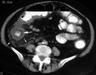 Typhlitis. Marked low-attenuation cecal wall thickening (large arrow) with moderate pericolonic inflammatory stranding (small arrows). Note thickening of transverse colon wall posteriorly.
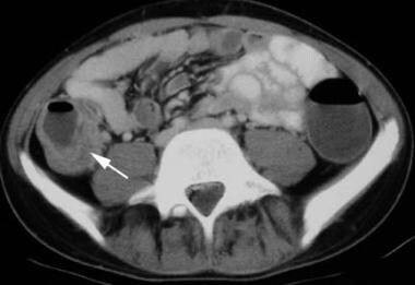 Typhlitis. Marked asymmetric cecal wall thickening (arrow) in this 64-year-old patient whose status is postchemotherapeutic for lymphoma.
Ultrasonographic findings of neutropenic enterocolitis include absent or decreased bowel peristalsis in the RLQ, thickened hypoechoic bowel wall, and markedly thickened echogenic mucosa. Color-flow imaging reveals hypervascularity of the mucosa and bowel wall. The patient may complain of pain upon palpation with the transducer. [12, 13] These findings, combined with an appropriate clinical history, indicate a high probability of neutropenic enterocolitis. CT scanning may be indicated to exclude perforation or abscess (not visualized on ultrasonography) and to establish a baseline to compare follow-up studies. RLQ small bowel loops distended with air can produce a significant ring-down artifact on ultrasonograms, thus obscuring visualization of the right colon.
Plain radiographs are nonspecific for neutropenic enterocolitis but may demonstrate a fluid-filled, masslike density in the abdominal right lower quadrant (RLQ), distention of adjacent small bowel loops, and thumbprinting. Free intraperitoneal air and pneumatosis coli rarely are observed. Barium enema and colonoscopy are contraindicated in possible neutropenic enterocolitis because of perforation risk. Confirm abnormal findings with CT scanning.
Computed Tomography
CT scanning of neutropenic enterocolitis demonstrates circumferential and occasionally eccentric low-attenuation colonic wall thickening and cecal distention. High attenuation within the thickened colonic wall may represent hemorrhage. Inflammatory pericolonic stranding of mesenteric fat is common. [11] CT scanning readily identifies complications, including pneumatosis coli, pneumoperitoneum, pericolonic fluid collections, and abscess. These complications may require urgent surgical management. CT scan findings consistent with neutropenic enterocolitis in a patient with an appropriate clinical scenario result in a high degree of confidence in the diagnosis. [2, 11]
(See the images below.)
Typhlitis. Marked low-attenuation cecal wall thickening (large arrow) with moderate pericolonic inflammatory stranding (small arrows). Note thickening of transverse colon wall posteriorly.
Typhlitis. Marked asymmetric cecal wall thickening (arrow) in this 64-year-old patient whose status is postchemotherapeutic for lymphoma.
 Typhlitis. CT of this 10-year-old patient with leukemia demonstrates fluid within the cecum, which has an asymmetrically thickened wall (arrows).
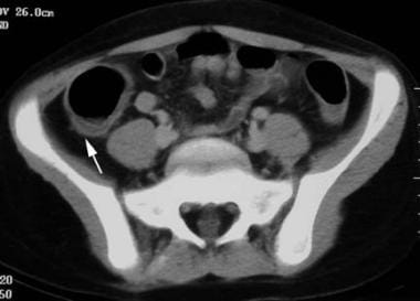 Typhlitis. Mild, asymmetrical, low-attenuation cecal wall thickening (arrow) in an 8-year-old patient with leukemia undergoing chemotherapy.
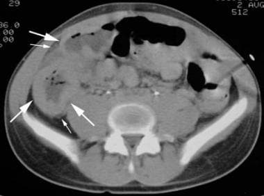 Typhlitis. Marked circumferential cecal and ascending colon wall thickening (large arrows) with mild pericolonic inflammatory stranding (small arrows).
Ultrasonography
Abdominal ultrasonography is an important diagnostic imaging study for neutropenic enterocolitis and is preferable to contrast enema. It may also be useful for follow-up to assess the decrease in bowel wall thickening. Ultrasonography allows follow-up imaging without repeated exposure to ionizing radiation, which is especially important in children. [12, 13, 14, 15, 10, 17]
Ultrasonographic findings of neutropenic enterocolitis include absent or decreased bowel peristalsis in the RLQ, thickened hypoechoic bowel wall, and markedly thickened echogenic mucosa. Color-flow imaging reveals hypervascularity of the mucosa and bowel wall. The patient may complain of pain upon palpation with the transducer. [12, 13] These findings, combined with an appropriate clinical history, indicate a high probability of neutropenic enterocolitis.
Bowel wall thickness has been suggested to be a significant prognostic factor in individuals with neutropenic enterocolitis. A retrospective study using ultrasonography showed that mortality was higher in patients with a bowel wall thickness greater than 5 mm (29%) than in those without bowel wall thickening (0%). If the bowel wall thickness cutoff was set at greater than 10 mm, mortality was 60%. [10]
-
Typhlitis. Marked low-attenuation cecal wall thickening (large arrow) with moderate pericolonic inflammatory stranding (small arrows). Note thickening of transverse colon wall posteriorly.
-
Typhlitis. Marked asymmetric cecal wall thickening (arrow) in this 64-year-old patient whose status is postchemotherapeutic for lymphoma.
-
Typhlitis. CT of this 10-year-old patient with leukemia demonstrates fluid within the cecum, which has an asymmetrically thickened wall (arrows).
-
Typhlitis. Mild, asymmetrical, low-attenuation cecal wall thickening (arrow) in an 8-year-old patient with leukemia undergoing chemotherapy.
-
Typhlitis. Marked circumferential cecal and ascending colon wall thickening (large arrows) with mild pericolonic inflammatory stranding (small arrows).


