Overview
Trauma is the leading cause of death for individuals younger than 40 years, with approximately 140,000 deaths annually in the United States alone. [1] Of these deaths, thoracic injuries are primarily responsible for 25% of cases [2] and are a major contributing factor in up to 75% of cases. [1] However, most thoracic injuries may be effectively treated with tube thoracostomy and simple fluid resuscitation. [3, 4]
Tube thoracostomy is the insertion of a tube (chest tube) into the pleural cavity to drain air, blood, bile, pus, or other fluids. [5] Whether the accumulation of air or fluid is the result of rapid traumatic filling with air or blood or an insidious malignant exudative fluid, placement of a chest tube allows for continuous, large volume drainage until the underlying pathology can be more formally addressed. The list of specific treatable etiologies is extensive (see Indications), but without intervention, patients are at great risk for major morbidity or mortality.
Editor's note: Targeted guidewire and trocar-guided placement are other techniques that are used; however, these are considered high risk for complications in the emergency department setting. Therefore, the classic technique described in this article is the highly recommended one for emergency department thoracostomy tube placement. Additionally, using ultrasound to assess tube placement is an emerging technology; however, chest radiography is the current criterion standard of care, as studies have not yet proven ultrasound to be safer or superior to radiography. For further information on other techniques see Medical Thoracoscopy and Tube Thoracostomy Management.
Also see the BTS Pleural Disease Guideline 2010. [6]
Indications
Indications are as follows:
-
Pneumothorax [7] : Open or closed; simple or tension [8]
-
Hemothorax [7]
-
Hemopneumothorax
-
Hydrothorax
-
Chylothorax [9]
-
Patients with penetrating chest wall injury who are intubated or about to be intubated
-
Considered for those about to undergo air transport who are at risk for pneumothorax
Contraindications
The need for emergent thoracotomy is an absolute contraindication to tube thoracostomy.
Relative contraindications include the following:
-
Coagulopathy
-
Pulmonary bullae
-
Pulmonary, pleural, or thoracic adhesions
-
Loculated pleural effusion or empyema
-
Skin infection over the chest tube insertion site
Equipment
Equipment is as follows:
-
Chest tube drainage device with water seal (autotransfuser unit is an option)
-
Suction source and tubing
-
Sterile gloves
-
Preparatory solution
-
Sterile drapes
-
Surgical marker
-
Lidocaine 1% with epinephrine
-
Syringes, 10-20 mL (2)
-
Needle, 25 gauge (ga), 5/8 in
-
Needle, 23 ga, 1.5 in; or 27 ga, 1.5 in; for instilling local anesthesia
-
Blade, No. 10, on a handle
-
Large and medium Kelly clamps
-
Large curved Mayo scissors
-
Large straight suture scissors
-
Silk or nylon suture, 0 or 1-0
-
Needle driver
-
Vaseline gauze
-
Gauze squares, 4 x 4 in (10)
-
Sterile adhesive tape, 4 in wide
-
Chest tube of appropriate size: Man - 28-32F; woman - 28F; child - 12-28F; infant - 12-16F; neonate - 10-12F
Positioning
The patient should be positioned supine or at a 45° angle. Elevating the patient lessens the risk of diaphragm elevation and consequent misplacement of the chest tube into the abdominal space.
The arm on the affected side should be abducted and externally rotated, simulating a position in which the palm of the hand is behind the patient's head.
A soft restraint or silk tape can be used to secure the arm in this location. If a restraint is used, make sure that good blood flow to the hand is present.
Technique
Obtain informed consent from the patient or patient’s representative except when urgent placement is required.
Assemble the drainage system and connect it to the suction source. The appearance of bubbles in the water chamber is a sign that the chest tube drainage device is functioning properly.
Position the patient as described above.
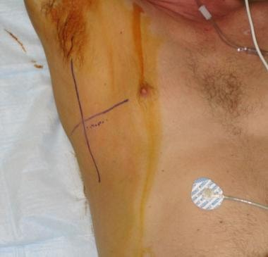
Identify the patient using two identifiers (eg, name and date of birth). If possible, match the patient's identifiers at his or her bed side with the identifiers present on a chest radiographs or computerized tomograms (CT) that was recently performed. Clearly mark the site of chest tube insertion (right or left).
Identify the fifth intercostal and the midaxillary line. The skin incision is made in between the midaxillary and anterior axillary lines over a rib that is below the intercostal level selected for chest tube insertion. A surgical marker can be used to better delineate the anatomy.
Shave excessive hair and apply a preparatory solution to a wide area of the chest wall as shown below.
Wear sterile gloves, gown, hair cover, and goggles or face shield, and apply sterile drapes to the area.
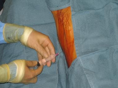
Administer analgesia. Administer a systemic analgesic (unless contraindicated). Use the 25-ga needle to inject 5 mL of the local anesthetic solution into the skin overlying the initial skin incision, as shown in the image below. Use the longer needle (23 or, preferably, 27 ga) to infiltrate about 5 mL of the anesthetic solution to a wide area of subcutaneous tissue superior to the expected initial incision. Redirect the needle to the expected course of the chest tube (following the upper border of the rib below the fifth intercostal space), and inject approximately 10 mL of the anesthetic solution into the periosteum (if bone is encountered), intercostal muscle, and the pleura. Aspiration of air, blood, pus, or a combination thereof into the syringe confirms that the needle entered the pleural cavity.
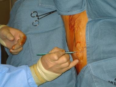
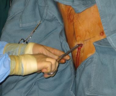
Use the No. 11 or 10 blade to make a skin incision approximately 4 cm long overlying the rib that is below the desired intercostal level of entry. The skin incision should be in the same direction as the rib itself.
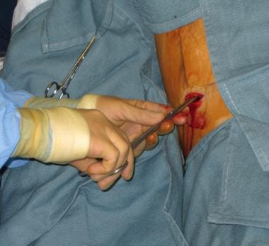
Use a hemostat or a medium Kelly clamp to bluntly dissect a tract in the subcutaneous tissue by intermittently advancing the closed instrument and opening it, as shown.
Palpate the tract with a finger as shown, and make sure that the tract ends at the upper border of the rib above the skin incision. Insertion of the chest tube as close as possible to the upper border of the rib will minimize the risks of injury to the nerve and blood vessels that follow the lower border of each rib.
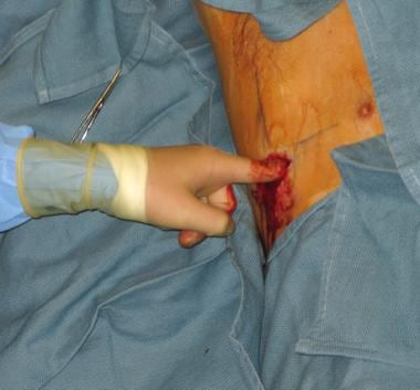 Palpation of the selected intercostal space and the superior margin of its inferior rib.
Adding more local anesthetic to the intercostal muscles and pleura at this time is recommended.
Use a closed large Kelly clamp to pass through the intercostal muscles and parietal pleura and enter into the pleural space, as shown.
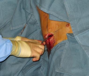 A closed and locked Kelly clamp is used to enter the chest wall into the pleural cavity. Make sure to guide the clamp over the upper margin of the rib.
This maneuver requires some force and twisting motion of the tip of the closed Kelly clamp. This motion should be done in a controlled manner so the instrument does not enter too far into the chest, which could injure the lung or diaphragm. Upon entry into the pleural space, a rush of air or fluid should occur.
The Kelly clamp should be opened (while still inside the pleural space) and then withdrawn so that its jaws enlarge the dissected tract through all layers of the chest wall as shown. This facilitates passage of the chest tube when it is inserted.
 Once the Kelly clamp enters the pleural cavity, the clamp should be opened to further enlarge the opening.
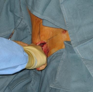
Use a sterile, gloved finger to appreciate the size of the tract and to feel for lung tissue and possible adhesions, as shown in the image below. Rotate the finger 360º to appreciate the presence of dense adhesions that cannot be broken and require placement of the chest tube in a different site, preferably under fluoroscopy (ie, by interventional radiology).
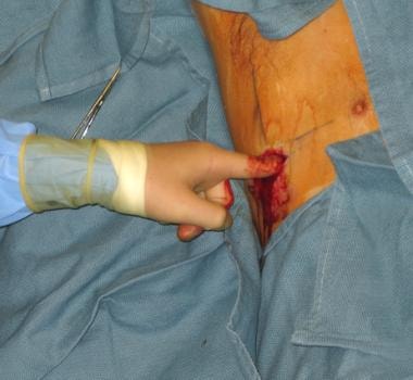 A finger is used to palpate the tract and feel for adhesions before insertion of the chest tube.
Measure the length between the skin incision and the apex of the lung to estimate how far the chest tube should be inserted. If desired, place a clamp over the tube to mark the estimated length. Some prefer to clamp the tube at a distal point, memorizing the estimated length.
Grasp the proximal (fenestrated) end of the chest tube with the large Kelly clamp and introduce it through the tract and into the thoracic cavity as shown.
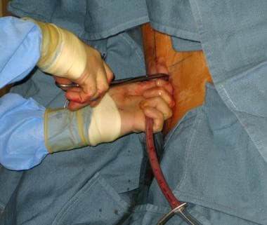 The proximal end of the chest tube is held with a Kelly clamp that is used to guide the chest tube through the tract. The distal end of the chest tube should always be clamped until it is connected to the drainage device.
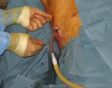
Release the Kelly clamp and continue to advance the chest tube posteriorly and superiorly. Make sure that all of the fenestrated holes in the chest tube are inside the thoracic cavity.
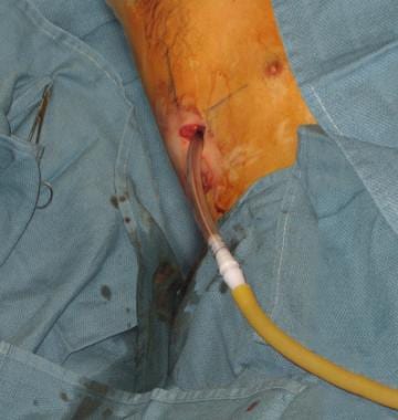
Connect the chest tube to the drainage device as shown (some prefer to cut the distal end of the chest tube to facilitate its connection to the drainage device tubing). Release the cross clamp that is on the chest tube only after the chest tube is connected to the drainage device.
Before securing the tube with stitches, look for a respiration-related swing in the fluid level of the water seal device to confirm correct intrathoracic placement.
Secure the chest tube to the skin using 0 or 1-0 silk or nylon stitches, as depicted below.
For securing sutures, two separate through-and-through, simple, interrupted stitches on each side of the chest tube are recommended. This technique ensures tight closure of the skin incision and prevents routine patient movements from dislodging the chest tube. Each stitch should be tightly tied to the skin, then wrapped tightly around the chest tube several times to cause slight indentation, and then tied again. Sealing suture: A central vertical mattress stitch with ends left long and knotted together can be placed to allow for sealing of the tract once the chest tube is removed.
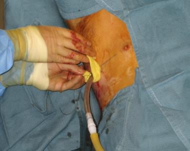
Place petrolatum (eg, Vaseline) gauze over the skin incision as shown.
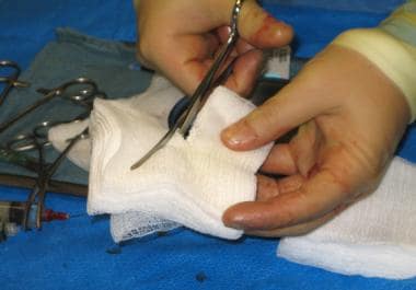
Create an occlusive dressing to place over the chest tube by turning regular gauze squares (4 x 4 in) into Y-shaped fenestrated gauze squares and using 4-in adhesive tape to secure them to the chest wall, as shown below. Make sure to provide enough padding between the chest tube and the chest wall.
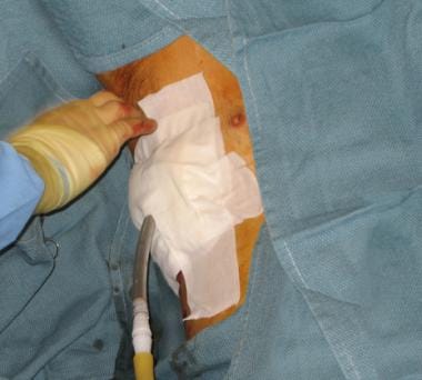 Apply support gauze dressing around the chest tube and secure it to the chest wall with 4-in adhesive tape.
Strap the emerging chest tube on to the lower trunk with a "mesentry" fold of adhesive tape, as this avoids kinking of the tube as it passes through the chest wall. It also helps reduce wound site pain and discomfort for the patient. All connections are then taped in their long axis to avoid disconnections.
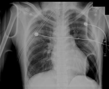
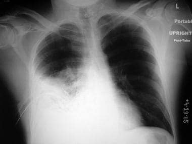
Obtain a chest radiograph, like the one below, to ensure correct placement of the chest tube.
See Tube Thoracostomy Management for removal techniques.
Pearls
In cases of high-pressure empyema or pleural effusion, removal of 50-200 mL of fluid using a syringe and a 14-ga needle, as shown below, might prevent high-pressure spraying of the accumulated fluid once the pleural space is entered with the surgical instrument.
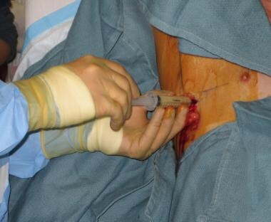 A needle and a syringe are used to decompress the pleural cavity in a case of tension empyema.
Since the intercostal vessels and nerve run on the inferior margin of each rib, incision and tunneling should be performed over the rib. Insertion of the chest tube should be performed immediately above and as close to the superior rib margin as possible in order to minimize the risks of injury to the nerve and blood vessels that follow the lower margin of each rib.
Errors that are commonly observed but easily avoidable include inadequate volume of local anesthetic, failure to wait adequate time for anesthetic to take effect, and too small an incision.
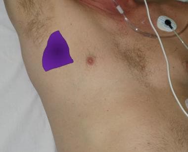
A "safe triangle" (shown below) has been described as the preferred site of insertion. This is the triangle bordered by the anterior border of the latissimus dorsi, the lateral border of the pectoralis major muscle, a line superior to the horizontal level of the nipple, and an apex below the axilla. [11]
Small-bore drains are recommended, as they are more comfortable than larger-bore tubes, but no evidence indicates that either is therapeutically superior. Large-bore drains are recommended for drainage of acute hemothorax and to monitor further blood loss. [11]
Complications
Minor complications of thoracostomy tube placement such as unresolved/reaccumulation of pneumothorax or misplacement of the tube (too deep/kinked) are common and approach approximately 30%. [12]
Improper placement is a possible complication; various placements and resolutions are as follows:
-
Horizontal (over the diaphragm) - Acceptable for hemothorax; should be repositioned for pneumothorax (See the image below.)
-
Subcutaneous - Must be repositioned
-
Placed too far into the chest (against the apical pleura) - Should be retracted
-
Placed into the abdominal space - Should be removed
Bleeding may occur, and resolution is as follows:
-
Local - Usually responds to direct pressure
-
Hemothorax (lung vs intercostal artery injury) - Might require thoracotomy if it does not resolve spontaneously
-
Hemoperitoneum (liver or spleen injury) requires emergent laparotomy.
Organ penetration usually requires surgical repair. Specific organs may include the following:
-
Stomach, colon, or diaphragm - Occurs as a result of unrecognized diaphragmatic hernia
-
Lung - Occurs as a result of pleural adhesions or use of a thoracostomy tube trocar
-
Liver or spleen – See hemoperitoneum above
Tube dislodgement is a possible complication
Empyema may occur. Chest tube (foreign object) could introduce bacteria into the pleural space.
Retained pneumothorax or hemothorax might require insertion of a second chest tube.
Re-expansion pulmonary edema is a rare and potentially fatal complication the can occur after treatment of pneumothorax or a pleural effusion. It is more common in patient with diabetes and in patients with tension or larger-size pneumothoraces and in patients with large pleural effusions. [13] The development of reexpansion pulmonary edema likely correlates with the amount of negative intrathoracic pressure, which, in turn, is related to the rate of fluid removal. The onset of cough and chest tightness is an important warning sign to end the procedure. Most experts recommend removal of 1-1.5 L at any one time. Supportive treatment is often sufficient. [14]
Periprocedural Care
Anesthesia
Systemic analgesia should be used in all conscious patients, unless contraindicated. Contraindications to use of systemic analgesia can include unstable vital signs and patient in extremis.
Procedural sedation and analgesia should be considered, unless contraindicated. For more information, see Procedural Sedation.
Local anesthesia is described in the Technique section. For more information, see Infiltrative Administration of Local Anesthetic Agents.
Follow up
For further information on other techniques and management, see Medical Thoracoscopy and Tube Thoracostomy Management.
Questions & Answers
Overview
What is the effect of tube thoracostomy on outcomes from thoracic injuries?
What are indications for tube thoracostomy?
What are contraindications for tube thoracostomy?
What equipment is required to perform a tube thoracostomy?
How is a patient positioned for a tube thoracostomy?
How is a tube thoracostomy performed?
How are high-pressure empyema or pleural effusion managed during a tube thoracostomy?
What are possible complications of a tube thoracostomy?
How is bleeding managed during tube thoracostomy?
Which organs may be penetrated during a tube thoracostomy?
What are serious complications of tube thoracostomy?
What are the indications and contraindications for anesthesia in a tube thoracostomy?
Where is information on techniques and management for a tube thoracostomy found?
-
Skin preparation and marking.
-
Local anesthesia.
-
Skin incision.
-
Blunt dissection down to the intercostal muscle.
-
Further blunt dissection down to the intercostal muscle.
-
Palpation of the selected intercostal space and the superior margin of its inferior rib.
-
A closed and locked Kelly clamp is used to enter the chest wall into the pleural cavity. Make sure to guide the clamp over the upper margin of the rib.
-
Once the Kelly clamp enters the pleural cavity, the clamp should be opened to further enlarge the opening.
-
A finger is used to palpate the tract and feel for adhesions before insertion of the chest tube.
-
The proximal end of the chest tube is held with a Kelly clamp that is used to guide the chest tube through the tract. The distal end of the chest tube should always be clamped until it is connected to the drainage device.
-
Connection of the chest tube to a drainage system.
-
A 0 or 1-0 silk or nylon suture is used to secure the chest tube to the skin.
-
Apply petrolatum (eg, Vaseline) gauze over the skin incision.
-
Preparation of a Y-shaped fenestrated drain gauze from regular gauze (4 x 4 in).
-
Apply support gauze dressing around the chest tube and secure it to the chest wall with 4-in adhesive tape.
-
The chest tube is angulated, overlying the diaphragm.
-
A needle and a syringe are used to decompress the pleural cavity in a case of tension empyema.
-
The safe triangle.
-
Chest tube in good position.


