AUTHOR AND EDITOR INFORMATION
Section 1 of 11
Author: Sharon J Gibbs, MD, Physician, Texas Back Institute
Sharon J Gibbs is a member of the following medical societies: American Academy of Physical Medicine and Rehabilitation, International Spine Intervention Society, Physiatric Association of Spine, Sports and Occupational Rehabilitation, and Texas Medical Association
Coauthor(s):
Kenneth S Dauber, MD, PA, Consulting Staff, Department of Physical Medicine and Rehabilitation, Plano Orthopedic and Sports Medicine Center
Editors: Daniel D Scott, MD, Program Director, Department of Rehabilitation Medicine, Associate Professor, University of Colorado Health Sciences Center; Francisco Talavera, PharmD, PhD, Senior Pharmacy Editor, eMedicine; Patrick M Foye, MD, FAAPMR, FAAEM, Associate Professor of Physical Medicine and Rehabilitation, Co-Director of Musculoskeletal Fellowship, Co-Director of Back Pain Clinic, Director of Coccyx Pain (Tailbone Pain, Coccydynia) Service, UMDNJ-New Jersey Medical School; Kelly L Allen, MD, Consulting Staff, Department of Physical Medicine and Rehabilitation, Lourdes Regional Rehabilitation Center, Our Lady of Lourdes Medical Center; Rene Cailliet, MD, Professor-Chairman Emeritus, Department of Rehabilitation Medicine, University of Southern California School of Medicine; Former Director, Department of Rehabilitation Medicine, Santa Monica Hospital Medical Center
Author and Editor Disclosure
Synonyms and related keywords:
golfer's elbow, medial tennis elbow, reverse tennis elbow
Background
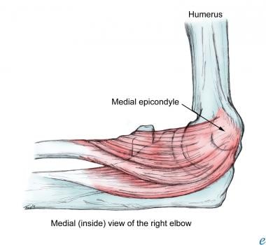
Medial epicondylitis (ME) is an overuse injury affecting the flexor-pronator muscle origin at the anterior medial epicondyle of the humerus. ME is often discussed in conjunction with lateral epicondylitis (LE), which occurs much more frequently. ME is the most common cause of medial elbow pain, although the clinician is likely to see at least 5 cases of LE for every case of ME. Patients who develop medial elbow pain appreciate their physician's knowledge of the subtle differences in the diagnosis and treatment of the 2 disorders. (See also Medial Epicondylitis [in the Orthopedic Surgery section], Medial Epicondylitis [in the Sports Medicine section], and Overuse Injury.)
Pathophysiology
ME involves primarily the flexor-pronator muscles (ie, pronator teres, flexor carpi radialis, palmaris longus) at their origin on the anterior medial epicondyle. Less often, ME also affects the flexor carpi ulnaris and flexor digitorum superficialis. Repetitive stress at the musculotendinous junction and its origin at the epicondyle leads to tendinitis in its most acute form and to tendinosis in its more chronic form. In addition, an ulnar neuropraxia caused by compression of the ulnar nerve in or around the medial epicondylar groove has been estimated to occur in up to 50% of ME cases. The tendinosis that occurs is primarily the result of failure of the damaged tendon to heal. Microscopic examination of the involved tissue shows granulation tissue, fibrovascular and fibrocartilaginous tissue, tendon microfragmentation, calcification, and necrosis. Histologically, damage to the involved tendons has been described as angiofibroblastic hyperplasia tendinosis and fibrillary degeneration of collagen. A simple, acute inflammatory reaction is noted to be a much less common finding than are the previously described tendinosis changes.
Race
No studies indicate a race predilection.
Sex
A male-to-female ratio of 2:1 has been reported.
Age
Peak incidence is in patients aged 20-49 years, but ME is also seen in teens and older adults, especially if they engage in hobbies, jobs, or sports activities that make them prone to overuse injuries. (See also the article Prevalence of Orthopaedic Maladies in People Who Flyfish: An Internet-Based Survey, on Medscape.)
History
ME is characterized by pain over the medial epicondyle. Pain worsens with wrist flexion and forearm pronation activities. Patients may report discomfort even when simply shaking hands with someone. Up to 50% of patients with ME complain of occasional or constant numbness and/or tingling sensation that radiates into their fourth and fifth fingers, suggesting involvement of the ulnar nerve. The patient's history may include the occurrence of an acute injury as a result, for instance, of taking a divot in golf, throwing a pitch in baseball, or hitting a hard serve in tennis.
Physical
Tenderness with palpation over the anterior aspect of the medial epicondyle is the most consistent finding. Other characteristics of ME include the following:
- Typically, pain is reproduced with resisted wrist flexion or resisted forearm pronation.
- Occasionally, the area of tenderness extends approximately 1 inch toward the proximal flexor-pronator muscle mass just distal to the epicondyle.
- The range of motion of the elbow and wrist is usually within normal limits.
- Patients may have symptoms of an ulnar neuropathy (eg, decreased sensation in the ulnar nerve distribution, a positive elbow-flexion test, a positive Tinel sign). In more severe cases, decreased sensation is associated with intrinsic weakness; intrinsic muscle atrophy may be noted.
Causes
The causes of ME include the following: - The condition can result from the repetitive use of flexor-pronator muscles, especially with valgus stress at the medial epicondyle.
- The onset can be related to the patient's occupation (if, for example, his/her job requires repetitive actions, such as the consistent use of a screwdriver or hammer).
- ME's onset can accompany acute injury.
- An excessive topspin in tennis, excessive grip tension, improper pitching techniques in baseball, and an improper golf swing are common sports-related causes of ME.
Lateral Epicondylitis
Olecranon Bursitis
Other Problems to Be Considered
Arthritis
Cervical radiculopathy
Flexor-pronator strain
Pronator teres syndrome
Shoulder musculoskeletal injuries
Thoracic outlet syndrome
Ulnar collateral ligament injury
Ulnar neuropathy
Lab Studies
- Laboratory studies are generally not indicated in ME.
Imaging Studies
- A radiograph of the elbow is often performed to rule out associated lesions (eg, loose bodies, bony avulsion, osteoarthritis). Typically, anteroposterior and lateral films are adequate.
- Oblique views are needed if loose bodies are suggested upon examination or because of a catching or clicking sensation described by the patient.
- Valgus stress radiographs should be obtained if medial instability is suggested.
- Magnetic resonance imaging (MRI) may be helpful in looking for the characteristic pathology that is seen in chronic cases of ME; MRI also permits assessment of the medial collateral ligament and ulnar nerve.1 Although in most cases an MRI scan is not necessary, it should be considered in individuals with atypical symptoms or in patients who are not responsive to conservative measures.
Other Tests
- If ulnar nerve involvement is suggested, a nerve conduction study and electromyography should be performed.
Rehabilitation Program
Physical Therapy
The physician may recommend that the patient with ME receive physical or occupational therapy. The discipline of therapy usually depends on the type of facility available, the accessibility of therapists, and physician preference. The proper means of treatment for ME are discussed below, in the Occupational Therapy section.
Occupational Therapy
Treatment begins with rest, ice, compression, and bracing, to decrease pain and inflammation. One to 6 weeks of relative rest of the affected muscles and tendons is typically advised, until discomfort subsides. Icing is employed for 5-10 minutes, 4-6 times per day and is particularly important if a patient presents after an acute event. Patients should be instructed to avoid icing over the ulnar nerve. Compression with a medial counterforce brace (ie, a tennis elbow splint) with a pad placed anteromedially on the proximal forearm over the flexor-pronator mass is routine (see Image 1). Discontinue if symptoms of an ulnar neuropathy worsen. In addition, if the symptoms are severe, brace with a wrist splint worn in the neutral position in order to rest the wrist flexors. In milder cases, a counterbalance brace may be used alone instead of a rigid splint; this limits extremes of motion while allowing some movement for functional activities. In the case of ulnar nerve involvement, a nighttime elbow extension splint should be considered. The splint is made in 30-45º of elbow flexion. A daytime elbow pad also may be useful, by limiting additional trauma to the nerve. After the patient's initial discomfort has subsided, a rehabilitation program with an occupational therapist should be initiated for muscle/tendon reconditioning. Begin with gentle stretching and add gradual strengthening of the flexor-pronator muscles, as the patient tolerates. Follow this with functional activities and with patient education aimed at avoiding re-injury. The patient should be advised to perform very slow stretching exercises 10-15 times to warm up muscles and increase flexibility, before doing any strengthening exercises or functional activities. Strengthening begins slowly with isometrics and progresses to eccentric exercises (see Images 2-3), with a gradual increase in resistance. Take care to cut back on exercises if they cause a recurrence of symptoms. Icing for 5-10 minutes after exercise is reasonable, especially if the patient reports pain in the affected area following exercise. Concomitant modalities may include ultrasonography, iontophoresis, phonophoresis, transcutaneous electrical nerve stimulation, and low-energy, extracorporeal shock-wave therapy. Successful relief from symptoms is variable. Shock-wave therapy has been shown to be less effective for ME than it is for LE.2
Surgical Intervention
Epicondylar debridement is rarely indicated but has proven to be effective in cases in which conservative treatment has failed. In addition, the ulnar nerve may be decompressed surgically.3
Consultations
Referral to an orthopedic surgeon is appropriate after 6-10 months if conservative treatment fails.
Other Treatment
If conservative measures fail, injection with local anesthetic and steroid to the point of maximal tenderness is appropriate. Special care should be taken to avoid injection directly into the tendon or the ulnar nerve. If concern for dislocation of the ulnar nerve exists, the injection should be performed with the elbow extended or semiflexed. The number of injections should be limited to 3 to decrease the risk of tendon atrophy or rupture. Short-term relief of discomfort with cortisone injection may be expected, but a complete rehabilitation program, as previously described, is a more effective and long-lasting way to treat ME.
Nonsteroidal anti-inflammatory drugs (NSAIDs), taken orally, are the medicines of choice for ME to help control pain and any associated inflammation. NSAIDs are used on average only for the first 7-10 days of the treatment period. For the patient, taking these medications with food may help to decrease the possible gastrointestinal side effects. The following list is not meant to be comprehensive but simply to provide examples of options.
Drug Category: Nonsteroidal anti-inflammatory drugs
NSAIDs have analgesic, anti-inflammatory, and antipyretic activities. Their mechanism of action is not known, but they may inhibit cyclo-oxygenase activity and prostaglandin synthesis. Other mechanisms may exist as well, such as inhibition of leukotriene synthesis, lysosomal enzyme release, lipoxygenase activity, neutrophil aggregation, and various cell-membrane functions.
| Drug Name | Ibuprofen (Motrin, Advil, Nuprin) |
| Description | DOC for mild to moderate pain. Inhibits inflammatory reactions and pain by decreasing prostaglandin synthesis. |
| Adult Dose | 400 mg PO q4-6h, 600 mg PO q6h, or 800 mg PO q8h; not to exceed 3.2 g/d |
| Pediatric Dose | <6 months: Not established 6 months to 12 years: 4-10 mg/kg/dose PO tid/qid >12 years: Administer as in adults (Consult pediatrician) |
| Contraindications | Documented hypersensitivity; avoid in peptic ulcer disease; recent GI bleeding or perforation; renal insufficiency; high risk of bleeding |
| Interactions | May decrease the effects of loop diuretics with coadministration; coadministration with anticoagulants may increase prothrombin time (PT); monitor and watch for signs of bleeding; may increase serum lithium levels and the risk of methotrexate toxicity; probenecid may increase the toxicity of NSAIDs |
| Pregnancy | B - Fetal risk not confirmed in studies in humans but has been shown in some studies in animals D - Fetal risk shown in humans; use only if benefits outweigh risk to fetus
|
| Precautions | Caution in congestive heart failure, hypertension, decreased renal and hepatic function, anticoagulation abnormalities, or during anticoagulant therapy |
| Drug Name | Naproxen (Naprosyn, Aleve, Naprelan, Anaprox) |
| Description | For the relief of mild to moderate pain; naproxen inhibits inflammatory reactions and pain by decreasing the activity of cyclo-oxygenase, which is responsible for prostaglandin synthesis |
| Adult Dose | 500 mg PO bid; not to exceed 1.25 g/d |
| Pediatric Dose | <2 years: Not established >2 years: 2.5 mg/kg/dose PO; not to exceed 10 mg/kg/d (Consult pediatrician) |
| Contraindications | Documented hypersensitivity; avoid in peptic ulcer disease; recent GI bleeding or perforation; renal insufficiency; high risk of bleeding |
| Interactions | May decrease the effects of loop diuretics with coadministration; coadministration with anticoagulants may increase prothrombin time (PT); monitor and watch for signs of bleeding; may increase serum lithium levels and the risk of methotrexate toxicity; probenecid may increase the toxicity of NSAIDs |
| Pregnancy | B - Fetal risk not confirmed in studies in humans but has been shown in some studies in animals D - Fetal risk shown in humans; use only if benefits outweigh risk to fetus
|
| Precautions | Acute renal insufficiency; interstitial nephritis; hyperkalemia; hyponatremia and renal papillary necrosis may occur; patients with pre-existing renal disease or compromised renal perfusion, risk of acute renal failure; leukopenia occurs rarely, is transient, and usually returns to normal during therapy; persistent leukopenia, granulocytopenia, or thrombocytopenia warrants further evaluation and may require discontinuation of the drug |
| Drug Name | Etodolac (Lodine, Lodine XL) |
| Description | For relief of mild to moderate pain; etodolac inhibits inflammatory reactions and pain by decreasing the activity of cyclo-oxygenase, which is responsible for prostaglandin synthesis. |
| Adult Dose | 200-400 mg PO q8h prn; not to exceed 1200 mg/d |
| Pediatric Dose | Not established |
| Contraindications | Documented hypersensitivity |
| Interactions | Coadministration with aspirin increases the risk of inducing serious, NSAID-related side effects; probenecid may increase the concentrations and possibly the toxicity of NSAIDs; may decrease the effect of beta-blockers, hydralazine, and captopril; may decrease the diuretic effects of furosemide and thiazides; may prolong prothrombin time when administered concurrently with anticoagulants; monitor PT closely and instruct patients to watch for signs and symptoms of bleeding; etodolac may increase the risk of methotrexate toxicity; phenytoin levels may increase when coadministered with etodolac |
| Pregnancy | C - Fetal risk revealed in studies in animals but not established or not studied in humans; may use if benefits outweigh risk to fetus D - Fetal risk shown in humans; use only if benefits outweigh risk to fetus
|
| Precautions | Acute renal insufficiency; interstitial nephritis, hyperkalemia, hyponatremia, and renal papillary necrosis may occur; patients with preexisting renal disease or compromised renal perfusion, risk of acute renal failure; leukopenia occurs rarely, is transient, and usually returns to normal during therapy; persistent leukopenia, granulocytopenia, or thrombocytopenia warrant further evaluation and may require discontinuation of the drug |
Further Outpatient Care
- The patient should be seen for follow-up about 2-3 weeks after the initial evaluation to make sure that symptoms are subsiding and to determine whether the patient is ready to begin the reconditioning phase of his/her rehabilitation.
Deterrence
- Patient education is a key element in preventing the recurrence of symptoms. Patients often have to modify the activities or the particular techniques that have led them to develop ME.
Prognosis
- Overall, the prognosis is good, with few patients needing to progress to steroid injection and even fewer (typically less than 10%) needing surgical intervention to find relief.
Patient Education
- Patient education is an essential part of the rehabilitation process and is an important means of preventing the recurrence of symptoms. Patient education includes the following elements:
- Modifying equipment (for example, increasing grip size on equipment, such as tennis rackets, golf clubs, and hammers) and using flexible shafts in golf
- Modifying activities (such as avoiding repetitive activities that cause medial elbow stress), decreasing grip pressure, decreasing topspin in tennis, and evaluating throwing techniques may be appropriate.
- For excellent patient education resources, visit eMedicine's Hand, Wrist, Elbow, and Shoulder Center. Also, see eMedicine's patient education article Tennis Elbow.
Medical/Legal Pitfalls
- Failure to recognize an acute fracture or a complete ligament tear requiring immediate orthopedic referral
Special Concerns
Waiting too long from symptom onset to treatment could make recovery much more prolonged and may increase the chance that surgical intervention will be required; thus, early identification and treatment is of great importance.
| Media file 1:
A medial counterforce brace is used in treating medial epicondylitis. The proximal extension serves to protect the medial epicondyle. Forearm braces without this extension can be used as well. Patients should be able to extend and flex fully at the elbow while wearing this brace. |
 | View Full Size Image | |
Media type: Photo
|
| Media file 2:
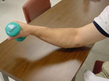
Strengthening exercises are performed once pain has subsided with active range of motion. The starting position (slight pronation) of an eccentric exercise for medial epicondylitis is shown. In order to prevent further injury, a trained therapist should instruct patients in exercises to confirm proper weight and technique. (The X indicates the medial epicondyle). |
 | View Full Size Image | |
Media type: Photo
|
| Media file 3:
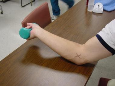
The eccentric exercise proceeds until full supination has been reached. |
 | View Full Size Image | |
Media type: Photo
|
- Banks KP, Ly JQ, Beall DP, et al. Overuse injuries of the upper extremity in the competitive athlete: magnetic resonance imaging findings associated with repetitive trauma. Curr Probl Diagn Radiol. Jul-Aug 2005;34(4):127-42. [Medline].
- Krischek O, Hopf C, Nafe B, et al. Shock-wave therapy for tennis and golfer's elbow--1 year follow-up. Arch Orthop Trauma Surg. 1999;119(1-2):62-6. [Medline].
- Gabel GT, Morrey BF. Operative treatment of medical epicondylitis. Influence of concomitant ulnar neuropathy at the elbow. J Bone Joint Surg Am. Jul 1995;77(7):1065-9. [Medline].
- Carson WG. Overuse injuries of the elbow in the throwing athlete. In: Baker CL, ed. The Hughston Clinic Sports Medicine Book. Baltimore, Md: Williams & Wilkins; 1995:324-31.
- Ciccotti MG, Ramani MN. Medial epicondylitis. Tech Hand Up Extrem Surg. Dec 2003;7(4):190-6. [Medline].
- Gabel GT, Morrey BF. Medial epicondylitis. In: Morrey BF, ed. The Elbow and Its Disorders. 3rd ed. Philadelphia, Pa: WB Saunders; 2000:537-42.
- Kraushaar BS, Nirschl RP. Tendinosis of the elbow (tennis elbow). Clinical features and findings of histological, immunohistochemical, and electron microscopy studies. J Bone Joint Surg Am. Feb 1999;81(2):259-78. [Medline].
- Ollivierre CO, Nirschl RP, Pettrone FA. Resection and repair for medial tennis elbow. A prospective analysis. Am J Sports Med. Mar-Apr 1995;23(2):214-21. [Medline].
- Stahl S, Kaufman T. The efficacy of an injection of steroids for medial epicondylitis. A prospective study of sixty elbows. J Bone Joint Surg Am. Nov 1997;79(11):1648-52. [Medline].
- Stahl S, Kaufman T. Ulnar nerve injury at the elbow after steroid injection for medial epicondylitis. J Hand Surg [Br]. Feb 1997;22(1):69-70. [Medline].
- Vangsness CT Jr, Jobe FW. Surgical treatment of medial epicondylitis. Results in 35 elbows. J Bone Joint Surg Br. May 1991;73(3):409-11. [Medline]. [Full Text].
Medial Epicondylitis excerpt Article Last Updated: Nov 26, 2007
|