Practice Essentials
Spondylolysis is a defect in the pars interarticularis that may or may not be accompanied by forward translation of one vertebra relative to another (spondylolisthesis).
Kilian, Robert, and Lambl first described spondylolysis accompanied by spondylolisthesis in the literature in the mid-1800s. The number of different spinal abnormalities contributing to development of spondylolisthesis was appreciated only after Naugebauer's anatomic studies in the late 1800s. [1] (See the image below.) Radiographic studies allow visualization and grading of spondylolisthesis but may not always reveal the presence of an isolated spondylolysis (without spondylolisthesis). Most patients with low-grade isthmic spondylolisthesis and degenerative spondylolisthesis can be treated conservatively.
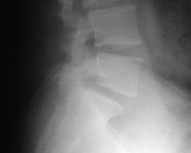 Radiograph of the lumbosacral junction showing a grade 1 spondylolytic spondylolisthesis at L5-S1.
Signs and symptoms of spondylolisthesis
Signs and symptoms of isthmic spondylolisthesis include the following
-
Hamstring tightness is observed almost universally, even in low-grade spondylolisthesis
-
Lumbar spasm may be present
-
A palpable step-off is noted with slips equal to or greater than grade 2
-
With higher degrees of spondylolisthesis, an increased lumbosacral kyphosis is seen (50% or greater), along with a compensatory thoracolumbar lordosis; truncal shortening may be present; with severe slips, the rib cage may rest on the iliac crest
-
Dermatomal weakness may be present if a radiculopathy or an element of stenosis is present
-
A waddling gait may be noted secondary to hamstring tightness producing a shortened stride length
-
If spondylolisthesis is not present, spondylolysis presents with paraspinal spasm, pain provocation with lumbar spine extension, and tight hamstrings
Signs and symptoms of degenerative spondylolisthesis include the following:
-
These patients present with less prominent physical findings; pain often is provoked with lumbar spine extension
-
If lumbar stenosis is present, then reflexes may be diminished; radicular findings also may be present
In congenital/dysplastic spondylolisthesis, physical findings are similar to those described above for isthmic spondylolisthesis.
Patients with traumatic and pathologic spondylolisthesis also present with similar findings. A good neurologic evaluation is important.
Workup in spondylosis and spondylolisthesis
Radiologic workup can include the following:
-
Radiographic studies - These allow visualization and grading of spondylolisthesis but may not always reveal the presence of an isolated spondylolysis (without spondylolisthesis)
-
Bone scanning - Bone scanning with single-photon emission computed tomography (SPECT) imaging is useful and often helps to direct management
-
Computed tomography (CT) scanning - This not only documents the presence and severity of spondylolysis but can help to rule out more serious causes for a positive bone scan
-
Magnetic resonance imaging (MRI) - MRI may visualize edema in the marrow around the sight of an acute spondylolytic defect
Management
The goals of exercise in spondylolisthesis are to improve abdominal strength and increase flexibility. Since tight hamstrings are almost always part of the clinical picture, appropriate hamstring stretching is important. Instruction in pelvic tilt exercises may help to reduce any postural component causing increased lumbar lordosis. Myofascial release may play a role in reducing pain from the surrounding soft tissues.
Surgical treatment is indicated when any type of spondylolisthesis is accompanied by a neurologic deficit. Persistent disabling back pain after conservative management may be considered an indication. High-grade slips (greater than 50%) more commonly require surgical intervention. Traumatic spondylolisthesis is rare but almost always requires surgical stabilization. [2]
Pathophysiology
Spondylolysis is a defect in the pars interarticularis that may or may not be accompanied by forward translation of one vertebra relative to another (spondylolisthesis). See the image below.
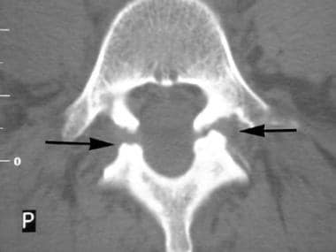 Axial computed tomography (CT) scan shows bilateral spondylolysis. Note elongation of the spinal canal at this level.
Wiltse, Macnab, and Newman developed a classification to help outline causes of vertebral translation in an anterior direction. [3, 4] Their categories include the following:
-
Type I: Congenital spondylolisthesis
-
Type II: Isthmic spondylolisthesis
-
Type III: Degenerative spondylolisthesis
-
Type IV: Traumatic spondylolisthesis
-
Type V: Pathologic spondylolisthesis
Type I: Congenital spondylolisthesis is characterized by the presence of dysplastic sacral facet joints, allowing forward translation of one vertebra relative to another. Orientation of facets in an axial or sagittal plane may allow for forward translation, producing undue stress on the pars, resulting in a fracture.
Type II: Isthmic spondylolisthesis is caused by the development of a stress fracture of the pars interarticularis.
Type III: Degenerative spondylolisthesis is commonly caused by intersegmental instability produced by facet arthropathy. This variation usually occurs in the adult population and, in most cases, does not progress beyond a grade I spondylolisthesis (see grading system below). [5]
Type IV: Traumatic spondylolisthesis can, in rare instances, result from acute stresses (trauma) to the facet or pars.
Type V: Any bone disorder may destabilize the facet mechanism producing pathologic spondylolisthesis. Iatrogenic spondylolisthesis, lastly, may occur if an overzealous surgeon performs too great of a facetectomy.
The most commonly used grading system for spondylolisthesis is the one proposed by Meyerding in 1947. The degree of slippage is measured as the percentage of distance the anteriorly translated vertebral body has moved forward relative to the superior end plate of the vertebra below. Classifications use the following grading system:
-
Grade 1: 1- 25% slippage
-
Grade 2: 26-50% slippage
-
Grade 3: 51-75% slippage
-
Grade 4: 76-100% slippage (see the image below)
-
Grade 5: Greater than 100% slippage
Epidemiology
Frequency
United States
Wiltse and Beutler each reported an incidence of 6-7% for isthmic spondylolysis. [3, 6] Up to 5% of children aged 5-7 years have been found to have spondylolysis, many of whom are asymptomatic. The incidence increases up to the 7% by age 18. Athletic activities requiring repetitive hyperextension and rotation or repetitive combined flexion-extension predispose some athletes to developing pars defects. [7, 8, 9, 10] Gymnasts, linemen in college football, weight lifters, javelin throwers, pole-vaulters, and judoists are most commonly affected. [11, 12] Approximately 82% of cases of isthmic spondylolisthesis occur at L5-S1. [13] Another 11.3% occur at L4-L5. Congenital defects, including spina bifida occulta, have been linked to occurrence of isthmic spondylolisthesis. Scoliosis has been found to occur along with spondylolysis as well. [14] Roughly 50% of all cases of spondylolysis are not associated with spondylolisthesis.
Degenerative spondylolisthesis occurs more frequently with increasing age. The L4-L5 interspace is affected 6-10 more times than any other level. Sacralization of L5 is frequently seen with L4-5 degenerative spondylolisthesis.
Mortality/Morbidity
See the list below:
-
Increased mortality is not associated with spondylolisthesis. While some patients may have persistent low back pain, significant disability is rare unless the patient has severe neurologic compromise that has not been addressed.
-
The most common morbidity is persistent low back pain or nerve impingement. Because disk degeneration is accelerated at the sight of level of the spondylolysis, diskogenic pain may occur. Degenerative spondylolisthesis produces characteristic arthritic symptoms that may worsen with age.
Race
See the list below:
-
Isthmic spondylolytic defects affect roughly 1.1% of black females. The most commonly affected group is the white male with an incidence of 6.4%. Arkara Plains Indians and Aleut people groups have a very high incidence of spondylolytic defects, due to a combination of genetic and environmental factors.
-
Degenerative spondylolisthesis affects black females more commonly than white females (and females are more commonly affected than men).
Sex
Beutler et al noted a 2:1 male-to-female ratio of occurrence in asymptomatic patients with spondylolysis. [6]
-
Females with isthmic spondylolytic lesions appear to be more prone to progressive displacement and may need surgical intervention more often than males.
-
Congenital spondylolisthesis (dysplastic type) occurs with a 2:1 female-to-male ratio with symptoms beginning around the adolescent growth spurt. These comprise about 14-21% of all cases of spondylolisthesis.
-
Degenerative spondylolisthesis occurs more commonly in females with a 5:1 female-to-male ratio. The incidence increases after age 40 years.
Age
See the list below:
-
Acute isthmic spondylolysis often occurs during the first and second decades of life. Most cases occur before the patient reaches age 15 years. In rare cases, acute spondylolysis may be seen in early adulthood. Younger patients are at higher risk than older patients for developing progressive spondylolisthesis. The risk for progression in adults is rare when the lesion is at L5. In contrast, lesions at L4-5 may progress into adulthood because of increased sagittal rotation, shear translation, and axial rotation at this segment.
-
Congenital/dysplastic spondylolisthesis has been documented in children as young as 3.5 months. More commonly, congenital spondylolistheses go undiagnosed until later in life after an individual has been ambulating for quite some time.
-
Degenerative spondylolisthesis occurs most commonly after age 40 years.
-
Radiograph of the lumbosacral junction showing a grade 1 spondylolytic spondylolisthesis at L5-S1.
-
Lumbar oblique radiograph showing the "Scottie Dog." A pars defect is seen at L5.
-
Bone scan with single-photon emission computed tomography (SPECT) imaging showing acute spondylolysis
-
Axial computed tomography (CT) scan shows bilateral spondylolysis. Note elongation of the spinal canal at this level.
-
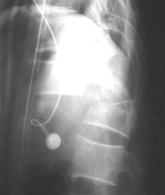
Grade 4 traumatic spondylolisthesis.
-
Diagram in the oblique projection shows the components of the vertebrae that result in the appearance of a Scottie dog with a collar.
